. 2023 Feb 21;15(5):1079.
doi: 10.3390/polym15051079.

Effect of Electrospun PLGA/Collagen Scaffolds on Cell Adhesion, Viability, and Collagen Release: Potential Applications in Tissue Engineering

Affiliations

Effect of Electrospun PLGA/Collagen Scaffolds on Cell Adhesion, Viability, and Collagen Release: Potential Applications in Tissue Engineering

Aldo Guzmán-Soria et al. Polymers (Basel). .

Abstract

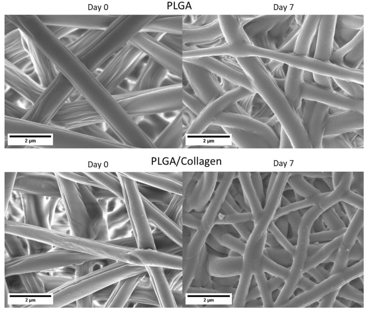
The development of scaffolding obtained by electrospinning is widely used in tissue engineering due to porous and fibrous structures that can mimic the extracellular matrix. In this study, poly (lactic-co-glycolic acid) (PLGA)/collagen fibers were fabricated by electrospinning method and then evaluated in the cell adhesion and viability of human cervical carcinoma HeLa and NIH-3T3 fibroblast for potential application in tissue regeneration. Additionally, collagen release was assessed in NIH-3T3 fibroblasts. The fibrillar morphology of PLGA/collagen fibers was verified by scanning electron microscopy. The fiber diameter decreased in the fibers (PLGA/collagen) up to 0.6 µm. FT-IR spectroscopy and thermal analysis confirmed that both the electrospinning process and the blend with PLGA give structural stability to collagen. Incorporating collagen in the PLGA matrix promotes an increase in the material's rigidity, showing an increase in the elastic modulus (38%) and tensile strength (70%) compared to pure PLGA. PLGA and PLGA/collagen fibers were found to provide a suitable environment for the adhesion and growth of HeLa and NIH-3T3 cell lines as well as stimulate collagen release. We conclude that these scaffolds could be very effective as biocompatible materials for extracellular matrix regeneration, suggesting their potential applications in tissue bioengineering.

Keywords: HeLa cells; NIH-3T3 fibroblasts; PLGA; collagen; electrospun fibers; scaffolds; tissue regeneration.


Conflict of interest statement

The authors declare no conflict of interest.

Figures

Figure 1
Figure 1
SEM images of PLGA and PLGA/collagen scaffolds.
Figure 2
Figure 2
Fiber diameter histograms of corresponding PLGA and PLGA/collagen scaffolds.
Figure 3
Figure 3
Fourier transform infrared spectroscopy (FTIR) spectra of PLGA, PLGA/collagen scaffolds, lyophilized collagen, and collagen prepared by solvent casting in HFIP.
Figure 4
Figure 4
Weight loss of PLGA and PLGA/collagen scaffolds in the time exposed to enzymatic degradation with collagenase.
Figure 5
Figure 5
SEM images of enzymatic degradation of PLGA and PLGA/collagen scaffolds after 7 days of exposure to PBS and collagenase.
Figure 6
Figure 6
FTIR spectra of PLGA/collagen scaffolds until complete 7 days of degradation in collagenase.
Figure 7
Figure 7
(A) Differential scanning calorimetry (DSC), and (B) XRD pattern of PLGA and PLGA/collagen. Thermograms of lyophilized and prepared by solvent casting in HFIP collagen were also shown for comparison.
Figure 8
Figure 8
Mechanical analysis of PLGA and PLGA/collagen electrospun fibers (a). Strain-Stress curve and (b) Statical analysis of Young’s modulus, Tensile strength, and elongation at break of PLGA and PLGA/collagen scaffolds. (* denotes a significant difference with a value p < 0.05) (n = 6).
Figure 9
Figure 9
(A) SEM images of HeLa cells and NIH 3T3 attached to the collagen hydrogel, PLGA, and PLGA/collagen scaffolds for 48 h. (B) The number of HeLa cells and NIH 3T3 adhered to scaffolds. (* denotes a significant difference with a value p < 0.05, and arrows indicate the presence of cells on scaffolds).
Figure 10
Figure 10
MTT test of cell viability using HeLa cells and NIH-3T3 fibroblasts on a positive control, collagen hydrogel, PLGA, and PLGA/collagen scaffolds at different exposure times, 1, 3, 5, and 7 days. (* denotes a significant difference with a value p < 0.05).
Figure 11
Figure 11
Collagen release by NIH-3T3 fibroblasts cultured on PLGA and PLGA/collagen scaffolds. As a control group, PLGA/collagen scaffolds without cells were incubated. (* denotes a significant difference with a value p < 0.05).

References

    1. Rahmati M., Mills D.K., Urbanska A.M., Saeb M.R., Venugopal J.R., Ramakrishna S., Mozafari M. Electrospinning for Tissue Engineering Applications. Prog. Mater. Sci. 2021;117:100721. doi: 10.1016/j.pmatsci.2020.100721. - DOI
    1. Sadeghi A., Moztarzadeh F., Aghazadeh Mohandesi J. Investigating the Effect of Chitosan on Hydrophilicity and Bioactivity of Conductive Electrospun Composite Scaffold for Neural Tissue Engineering. Int. J. Biol. Macromol. 2019;121:625–632. doi: 10.1016/j.ijbiomac.2018.10.022. - DOI - PubMed
    1. Khorshidi S., Solouk A., Mirzadeh H., Mazinani S., Lagaron J.M., Sharifi S., Ramakrishna S. A Review of Key Challenges of Electrospun Scaffolds for Tissue-Engineering Applications. J. Tissue Eng. Regen. Med. 2016;10:715–738. doi: 10.1002/term.1978. - DOI - PubMed
    1. Qu H., Fu H., Han Z., Sun Y. Biomaterials for Bone Tissue Engineering Scaffolds: A Review. RSC Adv. 2019;9:26252–26262. doi: 10.1039/C9RA05214C. - DOI - PMC - PubMed
    1. Kwak S., Haider A., Gupta K.C., Kim S., Kang I.K. Micro/Nano Multilayered Scaffolds of PLGA and Collagen by Alternately Electrospinning for Bone Tissue Engineering. Nanoscale Res. Lett. 2016;11:323. doi: 10.1186/s11671-016-1532-4. - DOI - PMC - PubMed

LinkOut - more resources