

doi: 10.1016/0092-8674(87)90593-9.
A cell that dies during wild-type C. elegans development can function as a neuron in a ced-3 mutant
Affiliations
- PMID: 3690660
- PMCID: PMC3773210
- DOI: 10.1016/0092-8674(87)90593-9
Item in Clipboard
A cell that dies during wild-type C. elegans development can function as a neuron in a ced-3 mutant
Cell.
.
Abstract
Mutations in the C. elegans gene ced-3 prevent almost all programmed cell deaths, so that in a ced-3 mutant there are many extra cells. We show that the pharyngeal neuron M4 is essential for feeding in wild-type worms, but in a ced-3 mutant, one of the extra cells, probably MSpaaaaap (the sister of M4), can sometimes take over M4's function. The function of MSpaaaaap, unlike that of M4, is variable and subnormal. One possible explanation is that its fate, being hidden by death and not subject to selection, has drifted randomly during evolution. We suggest that such cells may play roles in the evolution of cell lineage analogous to those played by pseudogenes in the evolution of genomes.
Figures
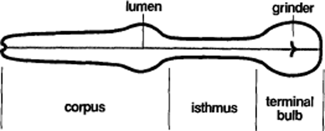
Anterior is to the left.
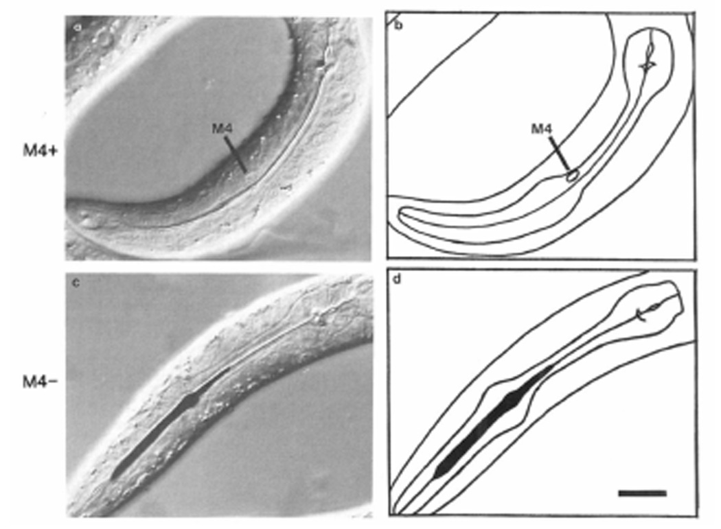
(a) Nomarski differential interference contrast micrograph of the pharynx of an intact wild-type worm. (b) Tracing of (a). Anterior is left, dorsal up. Wild-type eggs were placed on an NGM plate that had been spread with ink. After 4–5 hr, the animals were mounted in sodium azide, which anesthetizes them, and photographed. The line down the center of the pharynx is the lumen, which is closed in normal anesthetized worms. The ink eaten by intact worms like this one is passed back to the intestine (not shown). (c) Nomarski photomicrograph of the pharynx of a wild-type worm in which M4 had been killed. (d) Tracing of(c). The worm was placed on an ink plate immediately after M4 was killed, and photographed 4–5 hr later under anesthesia. The lumen of the front half of the pharynx, including the entire corpus and the front half of the isthmus, is stuffed with ink. Scale bar = 10 µm.
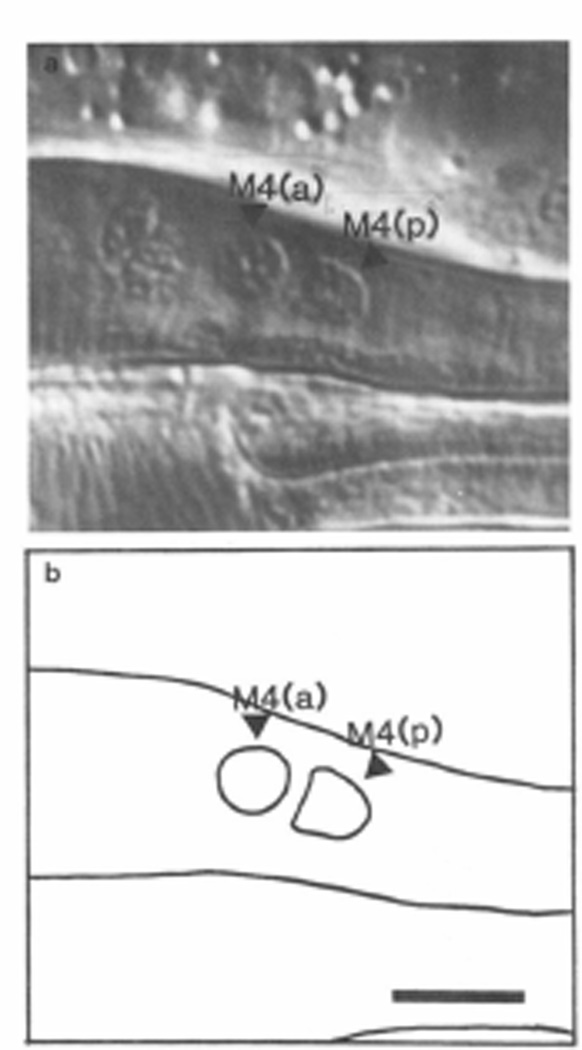
(a) Nomarski photograph of the posterior metacarpus and anterior isthmus of a ced-3 worm, showing two nuclei where wild-type has only M4. (b) Tracing of (a). Anterior left, dorsal up. Scale bar = 5 µm.
References
-
- Albertson DG, Thomson JN. The pharynx of Caenorhabditis elegans. Phil. Trans. Roy. Sot. London Ser. B. 1976;275:299–325. - PubMed
-
- Chalfie M, Horvitz HR, Sulston JE. Mutations that lead to reiterations in the cell lineages of C. elegans. Cell. 1981;24:59–69. - PubMed
-
- Croll NA. The Behavior of Nematodes. London: Edward Arnold; 1970.
-
- Ellis HM, Horvitz HR. Genetic control of programmed cell death in the nematode C. elegans. Cell. 1986;44:817–829. - PubMed
Publication types
MeSH terms
Grants and funding
LinkOut - more resources
Full Text Sources
Other Literature Sources
