. 2023 Mar 11;14(1):1338.
doi: 10.1038/s41467-023-37041-7.

Molecular mechanism of biased signaling at the kappa opioid receptor

Affiliations

Molecular mechanism of biased signaling at the kappa opioid receptor

Amal El Daibani et al. Nat Commun. .

Abstract

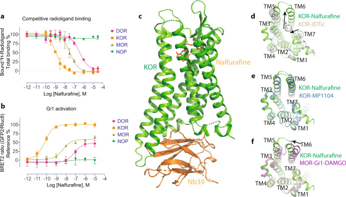
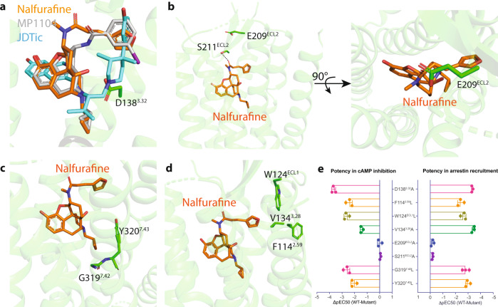
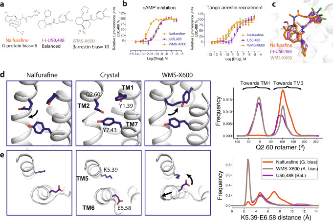
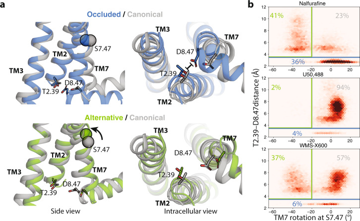
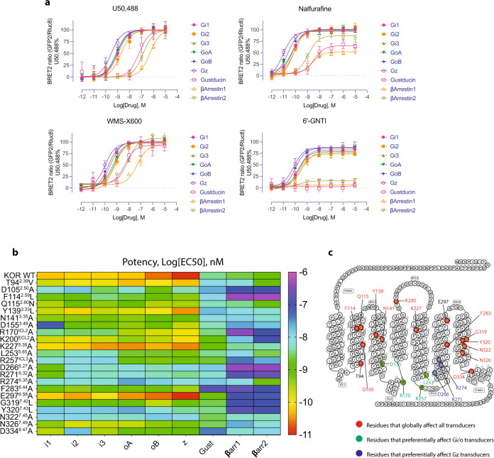
The κ-opioid receptor (KOR) has emerged as an attractive drug target for pain management without addiction, and biased signaling through particular pathways of KOR may be key to maintaining this benefit while minimizing side-effect liabilities. As for most G protein-coupled receptors (GPCRs), however, the molecular mechanisms of ligand-specific signaling at KOR have remained unclear. To better understand the molecular determinants of KOR signaling bias, we apply structure determination, atomic-level molecular dynamics (MD) simulations, and functional assays. We determine a crystal structure of KOR bound to the G protein-biased agonist nalfurafine, the first approved KOR-targeting drug. We also identify an arrestin-biased KOR agonist, WMS-X600. Using MD simulations of KOR bound to nalfurafine, WMS-X600, and a balanced agonist U50,488, we identify three active-state receptor conformations, including one that appears to favor arrestin signaling over G protein signaling and another that appears to favor G protein signaling over arrestin signaling. These results, combined with mutagenesis validation, provide a molecular explanation of how agonists achieve biased signaling at KOR.


Conflict of interest statement

The authors declare no competing interests.

Figures

Fig. 1
Fig. 1. Structure of active-state KOR bound to nalfurafine.
a The binding affinity of nalfurafine at four opioid receptors. 3H-Diprenorphine was used as radioligands for DOR, KOR, and MOR; 3H-Nociceptin was used as a radioligand for NOP. b The functional activity of nalfurafine at the opioid receptors. References were DPDPE, U50,488, DAMGO, and Nociceptin for DOR, KOR, MOR, and NOP, respectively. Data are expressed as the mean ± SEM of three independent experiments (n = 3 experiments each done in duplicate). c Structure of KOR-nalfurafine-Nb39 complex. d Comparison of active-state KOR-nalfurafine and inactive-state KOR-JDTic structures (intracellular view). e Comparison of active-state KOR-nalfurafine and KOR-MP1104 structure (intracellular view). f Comparison of KOR-nalfurafine and MOR-Gi1-DAMGO complex structures (intracellular view). Values in each plot were summarized in Supplemental Table 2.
Fig. 2
Fig. 2. Molecular signatures of nalfurafine-bound KOR.
a Alignment of the antagonist JDTic, agonist MP1104, and nalfurafine in the binding pocket of KOR. b The residue E209ECL2 forms a lid on the top of nalfurafine. c The interaction between the cyclopropyl methyl group of nalfurafine and hydrophobic pocket residues. d The furan ring of nalfurafine forms unique interaction with the ‘triad’ pocket including F1142.59/W124ECL1/V1343.28. e Mutagenesis studies confirm the role of residues that interact with nalfurafine. Data are expressed as the mean ± SEM of three independent experiments (n = 3 experiments each done in duplicate) and summarized from curves attached in Supplemental Figs. 1–3. Values in each plot were summarized in Supplemental Tables 3–5.
Fig. 3
Fig. 3. Molecular dynamics simulations of functionally selective KOR agonists.
a Chemical structure of G protein biased agonist nalfurafine, balanced agonist U50,488, and arrestin-biased agonist WMS-X600. U50,488 was used as a reference agonist that has a bias factor = 1; G protein bias factor of nalfurafine (95% confidence interval) = 6 (4.5–8.2); arrestin2 bias factor of WMS-X600 (95% confidence interval) = 10 (6.5–15.4). Calculation of bias factor was described in the “Methods”. b Functional characterization of KOR agonists in G protein-mediated cAMP inhibition and arrestin-mediated recruitment. Data are expressed as the mean ± SEM of three independent experiments (n = 3 experiments each done in duplicate). c Overlay of nalfurafine, U50,488, and WMS-X600 in the binding pocket of KOR. d Differences in Q1152.60 rotamer orientations favored by different ligands. In the left panel, Q1152.60 points towards TM3, keeping the pocket close to its starting configuration (nalfurafine). In the right panel, the rotation of Q1152.60 out of the pocket depresses Y661.39, allowing Y3207.43 to move forward and the top of TM7 to rotate counter-clockwise (WMS-X600). The Q1152.60 rotamer is quantified as the dihedral angle formed by the C, Ca, Cg, and Cd atoms of Q1152.60. e Differences in TM5 and TM6 conformation favored by different ligands. The distribution of K2275.39 amine nitrogen to E2976.58 carboxylate oxygen distances shows that the two residues generally do form a salt bridge when WMS-X600 or U50,488 is bound but do not when nalfurafine is bound. Values in each plot were summarized in Supplemental Table 6.
Fig. 4
Fig. 4. Nalfurafine, U50,488, and WMS-X600 favor different receptor conformations.
a Side and intracellular views of representations of the occluded (blue) and alternative state (green) compared to the canonical state (gray). Here the alternative and occluded states are represented by simulation frames and the canonical state is represented by the KOR-nafurafine-Nb39 crystal structure. b Density plots of the distance between the carboxylate oxygens of D3348.47 and the hydroxyl oxygen of T942.39 and the rotation of TM7 at the S3247.47 (see “Methods”). Simulation frames were classified as being in the occluded state if the T942.39–D3348.47 distance is less than 3.5 Å and otherwise in the alternative state if the TM7 rotation is less than −20° or else in the canonical active state.
Fig. 5
Fig. 5. The functional characterization of ligand-specific transducer coupling.
a The functionally selective KOR agonists display preference towards unique transducer coupling. Data are expressed as the mean ± SEM of three independent experiments (n = 3 experiments each done in duplicate). b The mutagenesis screening represented by the heatmap confirms that nalfurafine-specific transducer coupling is affected by specific residues of the receptor. c Positions of residues in nalfurafine-mediated transducer coupling. Values in each plot were summarized in Supplemental Tables 9–11.

References

    1. Seyedabadi, M., Gharghabi, M., Gurevich, E. V. & Gurevich, V. V. Structural basis of GPCR coupling to distinct signal transducers: implications for biased signaling. Trends. Biochem. Sci.47, 570–581 (2022). - PMC - PubMed
    1. Eichel K, et al. Catalytic activation of beta-arrestin by GPCRs. Nature. 2018;557:381–386. doi: 10.1038/s41586-018-0079-1. - DOI - PMC - PubMed
    1. Latorraca NR, et al. Molecular mechanism of GPCR-mediated arrestin activation. Nature. 2018;557:452–456. doi: 10.1038/s41586-018-0077-3. - DOI - PMC - PubMed
    1. DeWire SM, et al. A G protein-biased ligand at the mu-opioid receptor is potently analgesic with reduced gastrointestinal and respiratory dysfunction compared with morphine. J. Pharm. Exp. Ther. 2013;344:708–717. doi: 10.1124/jpet.112.201616. - DOI - PubMed
    1. Manglik A, et al. Structure-based discovery of opioid analgesics with reduced side effects. Nature. 2016;537:185–190. doi: 10.1038/nature19112. - DOI - PMC - PubMed

Publication types