Review
. 2023 Jun;12(2):243-260.
doi: 10.1007/s40119-023-00309-6. Epub 2023 Mar 11.

Pediatric Myocarditis

Affiliations
Review

Pediatric Myocarditis

Jason L Williams et al. Cardiol Ther. 2023 Jun.

Abstract

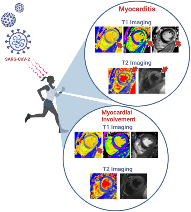
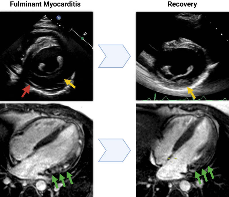
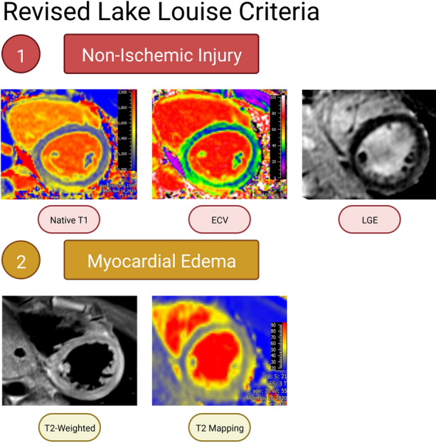
Myocarditis is a condition caused by acute or chronic inflammation of the cardiac myocytes, resulting in associated myocardial edema and myocardial injury or necrosis. The exact incidence is unknown, but is likely underestimated, with more mild cases going unreported. Diagnosis and appropriate management are paramount in pediatric myocarditis, as it remains a recognized cause of sudden cardiac death in children and athletes. Myocarditis in children is most often caused by a viral or infectious etiology. In addition, there are now two highly recognized etiologies related to Coronavirus disease of 2019 (COVID-19) infection and the COVID-19 mRNA vaccine. The clinic presentation of children with myocarditis can range from asymptomatic to critically ill. Related to severe acute respiratory syndrome-Coronavirus 2 (SARs-CoV-2), children are at greater risk of developing myocarditis secondary to COVID-19 compared to the mRNA COVID-19 vaccine. Diagnosis of myocarditis typically includes laboratory testing, electrocardiography (ECG), chest X-ray, and additional non-invasive imaging studies with echocardiogram typically being the first-line imaging modality. While the reference standard for diagnosing myocarditis was previously endomyocardial biopsy, with the new revised Lake Louise Criteria, cardiac magnetic resonance (CMR) has emerged as an integral non-invasive imaging tool to assist in the diagnosis. CMR remains critical, as it allows for assessment of ventricular function and tissue characterization, with newer techniques, such as myocardial strain, to help guide management both acutely and long term.

Keywords: CMR; COVID-19; Lake Louise Criteria; MIS-C; Myocarditis; Pediatric; SARs-CoV-2.


Conflict of interest statement

Jason L. Williams, Hannah M. Jacobs, and Simon Lee have nothing to disclose.

Figures

Fig. 1
Fig. 1
Evidence of myocarditis versus myocardial involvement after COVID-19. Note abnormalities (denoted by red arrows) in T1 and T2 imaging, which is diagnostic for myocarditis, in the above, compared to only abnormality in T1 imaging without concomitant abnormalities in T2 imaging in the myocardial involvement images
Fig. 2
Fig. 2
Clinical presentation of acute myocarditis
Fig. 3
Fig. 3
Example of fulminant myocarditis. Note the extensive lateral wall thickening seen during the acute episode that has resolved back to normal (yellow arrow), consistent with myocardial edema. This area also showed extensive delayed myocardial enhancement on MRI that has also resolved (green arrows). There is an additional pericardial effusion seen (red arrow), consistent with perimyocarditis
Fig. 4
Fig. 4
A, B H+E stain, original magnification 400×. The myocardium in these images has a prominent, predominantly lymphocytic, interstitial inflammatory infiltrate (yellow arrows) with several foci of associated myocyte injury (red stars)
Fig. 5
Fig. 5
Revised Lake Louise Criteria for cardiac MRI diagnosis of myocarditis. The patient must have abnormalities in any T1 sequence (representing non-ischemic myocardial injury) and in any T2 sequence (representing myocardial edema). Supporting criteria are evidence of pericarditis (pericardial effusion, or abnormalities in the pericardium) and/or evidence of systolic left ventricular dysfunction (regional or global wall motion abnormalities)

References

    1. Law YM, Lal AK, Chen S, Cihakova D, Cooper LT, Jr, Deshpande S, et al. Diagnosis and management of myocarditis in children: a scientific statement from the American Heart Association. Circulation. 2021;144(6):e123–e135. doi: 10.1161/CIR.0000000000001001. - DOI - PubMed
    1. Vasudeva R, Bhatt P, Lilje C, Desai P, Amponsah J, Umscheid J, et al. Trends in acute myocarditis related pediatric hospitalizations in the United States, 2007–2016. Am J Cardiol. 2021;149:95–102. doi: 10.1016/j.amjcard.2021.03.019. - DOI - PubMed
    1. Maron BJ, Doerer JJ, Haas TS, Tierney DM, Mueller FO. Sudden deaths in young competitive athletes: analysis of 1866 deaths in the United States, 1980–2006. Circulation. 2009;119(8):1085–1092. doi: 10.1161/CIRCULATIONAHA.108.804617. - DOI - PubMed
    1. Fabre A, Sheppard MN. Sudden adult death syndrome and other non-ischaemic causes of sudden cardiac death. Heart. 2006;92(3):316–320. doi: 10.1136/hrt.2004.045518. - DOI - PMC - PubMed
    1. Doolan A, Langlois N, Semsarian C. Causes of sudden cardiac death in young Australians. Med J Aust. 2004;180(3):110–112. doi: 10.5694/j.1326-5377.2004.tb05830.x. - DOI - PubMed

LinkOut - more resources