. 2023 Mar 11;6(1):261.
doi: 10.1038/s42003-023-04619-2.

SMAD2/3 signaling in the uterine epithelium controls endometrial cell homeostasis and regeneration

Affiliations

SMAD2/3 signaling in the uterine epithelium controls endometrial cell homeostasis and regeneration

Maya L Kriseman et al. Commun Biol. .

Abstract

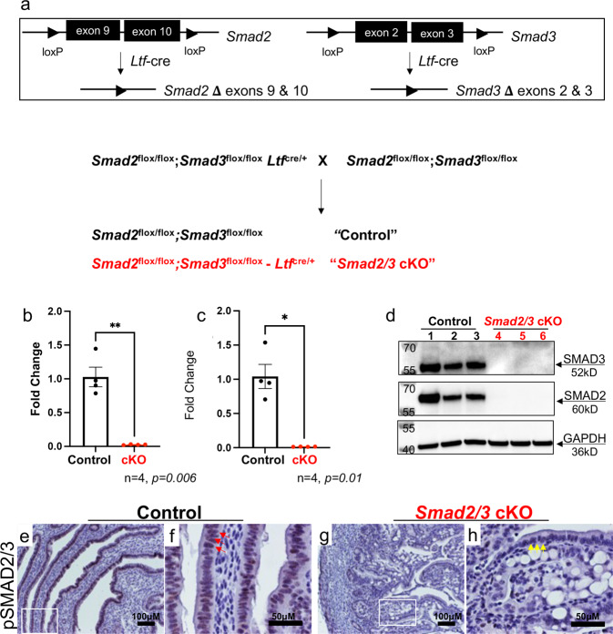
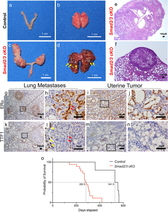
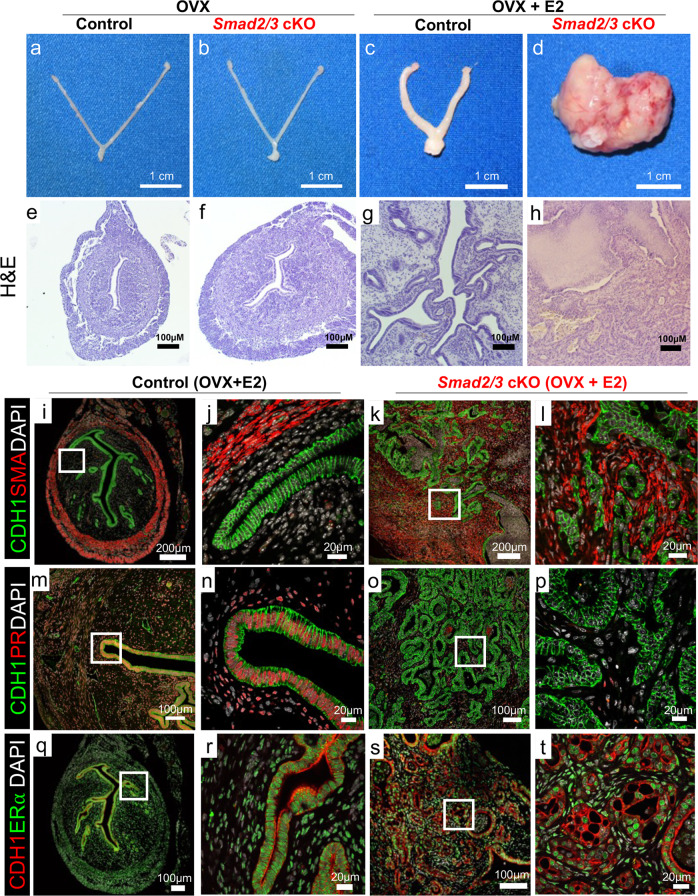
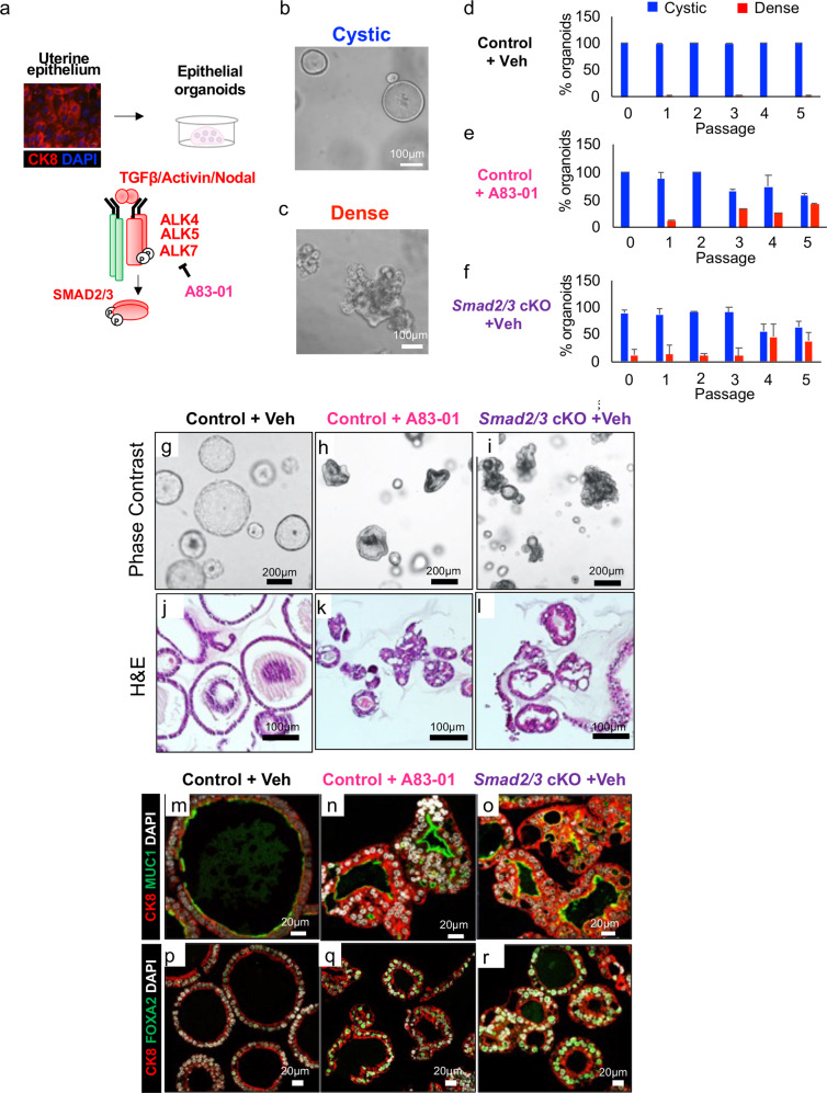
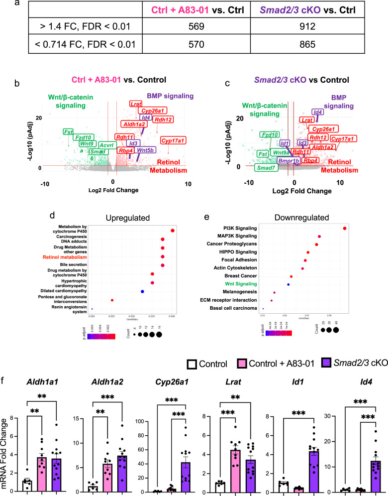
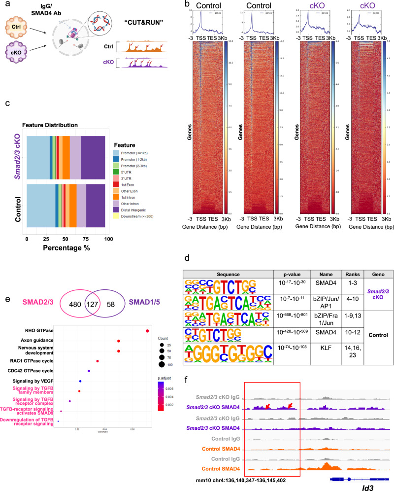
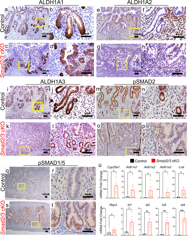
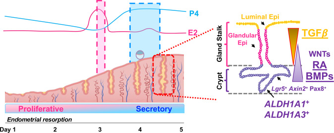
The regenerative potential of the endometrium is attributed to endometrial stem cells; however, the signaling pathways controlling its regenerative potential remain obscure. In this study, genetic mouse models and endometrial organoids are used to demonstrate that SMAD2/3 signaling controls endometrial regeneration and differentiation. Mice with conditional deletion of SMAD2/3 in the uterine epithelium using Lactoferrin-iCre develop endometrial hyperplasia at 12-weeks and metastatic uterine tumors by 9-months of age. Mechanistic studies in endometrial organoids determine that genetic or pharmacological inhibition of SMAD2/3 signaling disrupts organoid morphology, increases the glandular and secretory cell markers, FOXA2 and MUC1, and alters the genome-wide distribution of SMAD4. Transcriptomic profiling of the organoids reveals elevated pathways involved in stem cell regeneration and differentiation such as the bone morphogenetic protein (BMP) and retinoic acid signaling (RA) pathways. Therefore, TGFβ family signaling via SMAD2/3 controls signaling networks which are integral for endometrial cell regeneration and differentiation.


Conflict of interest statement

The authors declare no competing interests.

Figures

Fig. 1
Fig. 1. Generation of mice with double conditional deletion of SMAD2 and SMAD3 using Ltf-cre.
a Diagram showing the schematic used to obtain conditional deletion of SMAD2 and SMAD3 in the uterine epithelium using Ltf-cre. bh Confirmation that effective deletion of the Smad2 and Smad3 floxed exons and protein levels were decreased in the uterine epithelium of Smad2/3 cKO mice at the mRNA level (b, c, n = 4 per genotype) and in protein lysates from purified epithelium (d, n = 3 per genotype). Lanes in (d) were generated from the same blot, which was sequentially probed and stripped with each of the indicated antibodies. eh Immunohistochemical analysis of phosphorylated SMAD2 and SMAD3 (pSMAD2/3) in uterine cross-sections of control (n = 3) and Smad2/3 cKO mice (n = 3). Red arrows (f) show positively stained cells in uterine epithelial cells of controls, while yellow arrows (h) highlight the unstained epithelial cells in Smad2/3 cKO uterus. Histograms represent mean ± SEM analyzed by an unpaired two-tailed t-test.
Fig. 2
Fig. 2. Conditional deletion of SMAD2/3 results in metastatic endometrial tumors and death.
a, b Gross uterus of control (a) and Smad2/3 cKO (b) mice at 9 months of age. Smad2/3 cKO mice showed the presence of uterine masses. c, d Lungs dissected from control (c) and Smad2/3 cKO mice (d), showing that the lungs of the Smad2/3 cKO mice developed metastatic nodules (yellow arrows). e, f Cross-sections of the uterine tumor (e) and metastatic nodules (f) from Smad2/3 cKO mice stained with Hematoxylin and Eosin (H&E). gj Immunohistochemistry of ERα in the lung nodules (g, h) or uterine tumors (i, j) from Smad2/3 cKO mice. Expression of ERα is observed in the uterine tumors (i, j), and in the lung nodules (outlined by white dotted circles), but not in the adjacent normal tumor tissue. kn Immunohistochemistry of the lung cell marker, TTF1, in lung (k, l) and uterine tumor cross-sections (m, n) showing that neither the uterine tumors (m, n) nor metastatic nodules (k, l) express TTF1. However, the normal lung cells adjacent to the lung nodules do express TTF1 (l, yellow arrows). Red arrows in (h, l) show ERα positive cells in the lung nodules (h) that are TTF-negative in a sequential section (l). o Survival analysis comparing the survival of control mice (50% survival, 541.5 days) to Smad2/3 cKO mice (50% survival, 282.5 days).
Fig. 3
Fig. 3. Endometrial tumor development is estrogen dependent in Smad2/3 cKO mice.
ad Gross uteri from adult control (a, c) and Smad2/3 cKO mice (b, d) collected 3 months after ovariectomy (OVX) and treated without (a, b) or with estradiol releasing pellets (c, d). Only the Smad2/3 cKO mice that received the estradiol treatment developed tumors. eh H&E stained uterine cross-sections stained of control (e, g) and Smad2/3 cKO (f, h) that were ovariectomized and treated without (e, f) or with estradiol (g, h). it Immunostaining of control (i, j, m, n, q, r) and Smad2/3 cKO mice (k, l, o, p, s, t) cross sections following OVX + E2 treatment. il Tissue sections were stained with the epithelial cell marker, E-cadherin (CDH1, green) and smooth muscle actin (SMA, red). Compared to controls (i, j) sections from Smad2/3 cKO mice (k, l) show disordered epithelial cell and smooth muscle layers. mp Tissue sections were stained with E-cadherin (CDH1, green) and progesterone receptor (PR, red). PR can be seen in the nuclei of the control mice (m, n) but not in the epithelium of Smad2/3 cKO mice (o, p). qt Uterine cross sections were stained with E-cadherin (CDH1, red) and estrogen receptor α (ERα, green) antibodies. Cross sections from control (q, r) and Smad2/3 cKO (s, t) mice were positive for ERα. Nuclei were stained with DAPI (white). H&E and immunostaining experiments were performed in samples from at least 3 control and 3 Smad2/3 cKO mice.
Fig. 4
Fig. 4. Inhibition of SMAD2/3 signaling disrupts morphology and differentiation of endometrial epithelial organoids.
a Schematic showing the strategy used to isolate uterine epithelium from control and Smad2/3 cKO mice for the culture of epithelial organoids. Organoids from control mice were grown in the presence or absence of the TGFβ receptor (ALK4/ALK5/ALK7) inhibitor, A83-01 for 5 passages. b, c Phase contrast images of cystic (b, round-shaped) or dense (c, lobular shaped) organoids. df Quantification of cystic vs. dense organoids across the three conditions, control + vehicle (d), control + A83-01 (e), and Smad2/3 cKO (f). Values displayed as percentage of total organoids from n = 3 mice per group over 5 passages, mean ± SEM. gi Phase contrast imaging of the organoids from control mice grown in the absence (g) or presence (h) of A83-01, and from Smad2/3 cKO mice (i). jl H&E stained cross sections of organoids from control mice (j), control mice treated with the A83-01 inhibitor (k), and from Smad2/3 cKO mice (l). mr Cross sections of endometrial organoids from control mice cultured in the absence (m, p) or presence of A83-01 (n, q) or from Smad2/3 cKO mice (o, r). The organoids were immunostained with the epithelial cell marker antibody, cytokeratin 8 (CK8, red) and the mucin 1 antibody (mo, MUC1, green), or with CK8 (red) and the glandular cell marker, FOXA2 (pr, green). These experiments were performed in organoids derived from at least three mice per group.
Fig. 5
Fig. 5. Gene expression profiling of endometrial organoids reveals that inhibition of SMAD2/3 signaling increases retinoid- and BMP-signaling pathways.
RNA-sequencing of the endometrial epithelial organoids was performed to identify the gene expression differences between control and Smad2/3 cKO organoids. a Differentially expressed genes identified by RNAseq between the various organoid groups (Control + A83-01 vs. Control and Smad2/3 cKO vs. Control). b, c Volcano plots highlighting gene-level differences identified by RNAseq between Control + A83-01 vs. Control (b) and Smad2/3 cKO vs. Control (c) organoids. RNAseq data represent differentially expressed genes from four different mice per group, >1.4 fold or <0.714 fold change, FDR < 0.01. d, e Gene ontology analyses of overexpressed genes in Smad2/3 cKO organoids indicates that “Retinol Metabolism” pathway genes are overrepresented in Smad2/3 cKO organoids (d), while pathways related to “WNT/β-catenin” and are downregulated (e). f qPCR validation of differentially expressed genes identified by RNAseq in an independent set of organoids from Control, Control + A83-01, and Smad2/3 cKO mice (n = 6, n = 9 and n = 12, per group, respectively). RNAseq was performed in organoids from four mice per group. Histograms represent mean ± SEM analyzed by an ordinary One Way ANOVA with Tukey multiple comparison post test.
Fig. 6
Fig. 6. Analysis of SMAD4 binding in endometrial organoids reveals differential binding across the genome.
a Diagram outlining the procedures used to identify SMAD4-bound genes in organoids from Control and Smad2/3 cKO mice using CUT & RUN. b Heatplot showing the SMAD4 signal distribution across the transcriptional start site (TSS) and transcriptional end site (TES) in Control and Smad2/3 cKO organoids. As expected, the SMAD4 signal in the Smad2/3 cKO organoids was decreased when compared to the SMAD4 signal in Control organoids. c Feature distribution comparison between the SMAD4 binding sites in Control and Smad2/3 cKO organoids. d Motif sequence analyses in the SMAD4-bound regions in Control and Smad2/3 cKO organoids. e Differentially bound SMAD4 genes in Control (representing SMAD2/3 targets) and Smad2/3 cKO organoids (representing SMAD1/5 targets) and the gene ontology analysis of the differentially bound genes in Control organoids. f Genome track screenshot showing increased SMAD4 enrichment in the upstream promoter region of the BMP-target gene, Id3, in Smad2/3 cKO organoids when compared to Control organoids. CUT & RUN experiments were performed in the organoids from >3 mice per genotype, analyzed and sequenced as duplicates.
Fig. 7
Fig. 7. Detection of retinoid- and BMP-signaling pathways in control and Smad2/3 cKO mice.
ad Cross-sections from 17-week-old control (a, b) and Smad2/3 cKO (c, d) mice stained with ALDH1A1 antibody. ALDH1A1 is enriched in the crypts of the mouse endometrial glands. eh ALDH1A2 IHC in the uteri of control (e, f) and Smad2/3 cKO mice (g, h) ALHD1A2 is localized to the subepithelial stromal compartment. il ALDH1A3 IHC in control (I, j) and Smad2/3 cKO (k, l) mice shows enrichment in the crypts of endometrial glands. mp pSMAD2 IHC in control (m, n) and Smad2/3 cKO (o, p) uterine cross-sections. Decreased pSMAD2 is observed in Smad2/3 cKO mice. q–t pSMAD1/5 IHC in the uterine cross-sections of control (q, r) and Smad2/3 cKO (s, t) mice shows increased pSMAD1/5 reactivity in the uteri of Smad2/3 cKO mice. IHC experiments were performed in >3 mice per genotype. u Quantitative PCR analysis of uterine epithelium from control (n = 4) and Smad2/3 cKO (n = 6) for genes involved in retinoid signaling (Cyp26a1, Aldh1a1, -1a2, -1a3, Lrat, and Rbp4) or BMP signaling (Id1, Id2, Id3, Id4). Histograms represent mean ± SEM analyzed by an unpaired two-tailed t-test.
Fig. 8
Fig. 8. Schematic showing the effect of TGFβ signaling in endometrial cell regeneration and differentiation.
Diagram indicating the dynamic remodeling of the endometrium throughout the estrous cycle, transitioning from a proliferative to a secretory state under the control of the steroid hormones, estrogen (E2) and progesterone (P4). The regenerative potential of the endometrium is controlled by the presence of endometrial Lgr5+, Axin2+, and Pax8+ stem cells, likely in the crypts of the uterine glands, with growth factors such as WNTs, controlling differentiation. Our results indicate that Aldh1a1+ and Aldh1a3+ cells are putative endometrial stem cells in the uterine glands that are controlled by TGFβ, BMP and retinoic acid (RA) signaling.

References

    1. Cousins FL, Pandoy R, Jin S, Gargett CE. The Elusive Endometrial Epithelial Stem/Progenitor Cells. Front. Cell Dev. Biol. 2021;9:640319. doi: 10.3389/fcell.2021.640319. - DOI - PMC - PubMed
    1. Nguyen HPT, et al. N-cadherin identifies human endometrial epithelial progenitor cells by in vitro stem cell assays. Hum. Reprod. 2017;32:2254–2268. doi: 10.1093/humrep/dex289. - DOI - PubMed
    1. Valentijn AJ, et al. SSEA-1 isolates human endometrial basal glandular epithelial cells: phenotypic and functional characterization and implications in the pathogenesis of endometriosis. Hum. Reprod. 2013;28:2695–2708. doi: 10.1093/humrep/det285. - DOI - PubMed
    1. Tempest N, Maclean A, Hapangama DK. Endometrial Stem Cell Markers: Current Concepts and Unresolved Questions. Int. J. Mol. Sci. 2018;19:3240. doi: 10.3390/ijms19103240. - DOI - PMC - PubMed
    1. Gurung S, Werkmeister JA, Gargett CE. Inhibition of Transforming Growth Factor-beta Receptor signaling promotes culture expansion of undifferentiated human Endometrial Mesenchymal Stem/stromal Cells. Sci. Rep. 2015;5:15042. doi: 10.1038/srep15042. - DOI - PMC - PubMed

Publication types