

The Multifaceted Roles of Macrophages in NAFLD Pathogenesis
- PMID: 36907380
- PMCID: PMC10148157
- DOI: 10.1016/j.jcmgh.2023.03.002
The Multifaceted Roles of Macrophages in NAFLD Pathogenesis
Abstract
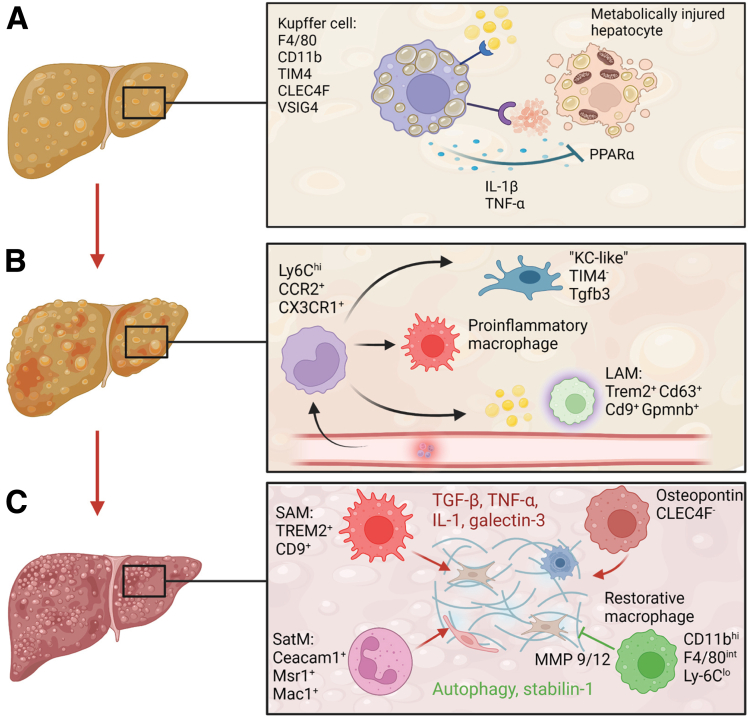
Nonalcoholic fatty liver disease (NAFLD) is the liver manifestation of the metabolic syndrome. NAFLD constitutes a spectrum of pathologies ranging from simple hepatic steatosis (nonalcoholic fatty liver) to the more progressive form of steatohepatitis and fibrosis, which can culminate in liver cirrhosis and hepatocellular carcinoma. Macrophages play multiple roles in the context of NAFLD pathogenesis by regulating inflammatory responses and metabolic homeostasis in the liver and thereby may represent an attractive therapeutic target. Advances in high-resolution methods have highlighted the extraordinary heterogeneity and plasticity of hepatic macrophage populations and activation states thereof. Harmful/disease-promoting as well as beneficial/restorative macrophage phenotypes co-exist and are dynamically regulated, thus this complexity must be taken into consideration in strategies concerning therapeutic targeting. Macrophage heterogeneity in NAFLD includes their distinct ontogeny (embryonic Kupffer cells vs bone marrow-/monocyte-derived macrophages) as well as their functional phenotype, for example, inflammatory phagocytes, lipid- and scar-associated macrophages, or restorative macrophages. Here, we discuss the multifaceted role of macrophages in the pathogenesis of NAFLD in steatosis, steatohepatitis, and transition to fibrosis and hepatocellular carcinoma, focusing on both their beneficial and maladaptive functions at different disease stages. We also highlight the systemic aspect of metabolic dysregulation and illustrate the contribution of macrophages in the reciprocal crosstalk between organs and compartments (eg, the gut-liver axis, adipose tissue, and cardiohepatic metabolic interactions). Furthermore, we discuss the current state of development of pharmacologic treatment options targeting macrophage biology.
Keywords: HCC; NASH; liver fibrosis; macrophage; obesity; type 2 diabetes.
Copyright © 2023 The Authors. Published by Elsevier Inc. All rights reserved.
Figures

References
-
- Peiseler M., Schwabe R., Hampe J., et al. Immune mechanisms linking metabolic injury to inflammation and fibrosis in fatty liver disease - novel insights into cellular communication circuits. J Hepatol. 2022;77:1136–1160. - PubMed
Publication types
MeSH terms
LinkOut - more resources
Full Text Sources
Medical
