Diagnostic imaging of adnexal masses in pregnancy
- PMID: 36907575
- PMCID: PMC10191762
- DOI: 10.5468/ogs.22287
Diagnostic imaging of adnexal masses in pregnancy
Abstract
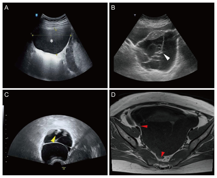
Adnexal masses detected during pregnancy require a prompt and accurate diagnosis to ensure fetal safety and good oncological outcomes. Computed tomography is the most common and useful diagnostic imaging modality for diagnosing adnexal masses; however, it is contraindicated in pregnant women because of the teratogenic effect of radiation on the fetus. Therefore, ultrasonography (US) is commonly used as the main alternative for the differential diagnosis of adnexal masses during pregnancy. Additionally, magnetic resonance imaging (MRI) can assist in the diagnosis when US findings are inconclusive. As each disease has characteristic US and MRI findings, understanding these features is important for the initial diagnosis and subsequent treatment. Thus, we thoroughly reviewed the literature and summarized the key findings of US and MRI to apply these in real-world clinical practice for various adnexal masses detected during pregnancy.
Keywords: Adnexal disease; Diagnostic imaging; Magnetic resonance imaging; Pregnant woman; Ultrasonography.
Conflict of interest statement
The authors declare no conflicts of interest relevant to this article.
Figures
References
-
- Kumari I, Kaur S, Mohan H, Huria A. Adnexal masses in pregnancy: a 5-year review. Aust N Z J Obstet Gynaecol. 2006;46:52–4. - PubMed
-
- Bernhard LM, Klebba PK, Gray DL, Mutch DG. Predictors of persistence of adnexal masses in pregnancy. Obstet Gynecol. 1999;93:585–9. - PubMed
-
- Nelson MJ, Cavalieri R, Graham D, Sanders RC. Cysts in pregnancy discovered by sonography. J Clin Ultrasound. 1986;14:509–12. - PubMed
Grants and funding
LinkOut - more resources
Full Text Sources
Research Materials