Review
. 2023 Apr;37(2):273-299.
doi: 10.1016/j.hoc.2022.12.001.

Molecular Basis and Genetic Modifiers of Thalassemia

Affiliations
Review

Molecular Basis and Genetic Modifiers of Thalassemia

Nicolò Tesio et al. Hematol Oncol Clin North Am. 2023 Apr.

Abstract

Thalassemia syndromes are common monogenic disorders and represent a significant health issue worldwide. In this review, the authors elaborate on fundamental genetic knowledge about thalassemias, including the structure and location of globin genes, the production of hemoglobin during development, the molecular lesions causing α-, β-, and other thalassemia syndromes, the genotype-phenotype correlation, and the genetic modifiers of these conditions. In addition, they briefly discuss the molecular techniques applied for diagnosis and innovative cell and gene therapy strategies to cure these conditions.

Keywords: Genetic modifiers; Genotype-phenotype; Globin genes; Hemoglobin; Hemoglobin switch; Molecular genetics; Thalassemia.


Conflict of interest statement

Disclosure The authors have nothing to disclose.

Figures
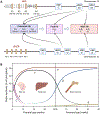
Fig. 1.
Fig. 1.
A. Schematic representation of the α- and β-globin gene clusters and the types of hemoglobin produced at different developmental stages. For simplicity, pseudogenes are not shown and the β-globin gene cluster is shown in reverse orientation with respect to conventional genomic coordinates. Regulatory elements are shown in orange, globin genes in blue, with resultant hemoglobin tetramers shown in boxes. B. Developmental stage-specific globin gene expression and site of erythropoiesis. Created with BioRender.com
Fig. 2.
Fig. 2.
Molecular machinery involved in fetal-to-adult hemoglobin switch. Displayed are some of the transcription factors, chromatin complexes and loci involved in developmental regulation of the β-globin gene cluster. Created with BioRender.com
Fig. 3.
Fig. 3.
Genotype-phenotype correlation of α-thalassemia. The severity of α-thalassemia widely ranges from the silent carrier with a single gene deletion, α-thalassemia trait with two genes deleted in trans (left) or in cis (right), three genes deleted (deletional HbH disease), two genes deleted plus one nondeletional mutation (nondeletional HbH disease), up to the most severe form with all four genes deleted (Bart’s hydrops fetalis syndrome). Created with BioRender.com
Fig. 4.
Fig. 4.
Genetic modifiers of β-thalassemia. Created with BioRender.com
Fig. 5.
Fig. 5.
A. Deletional mutations of the β-globin gene cluster, including deletional HPFH. B. Non-deletional HPFH caused by single point mutations at the level of promoters of γ-globin genes. Created with BioRender.com
Fig. 6.
Fig. 6.
Cell and gene therapy approaches for the cure of β-thalassemia. Numerous approaches could be utilized to add a globin gene, to activate a gene paralog (like fetal globin), or to directly correct the mutation. Several examples are listed applicable to lentiviral transduction or gene editing. The process of ex vivo gene modification includes collection of hematopoietic stem and progenitor cells from mobilized peripheral blood or bone marrow harvest, ex vivo genetic manipulation, treatment of patient with myeloablative conditioning regimen, and re-infusion of modified cells to peripheral blood or bone marrow. Created with BioRender.com
Fig. 7.
Fig. 7.
Descriptive analysis of β-thalassemia mutations annotated on the IthaGenes database (https://www.ithanet.eu/db/ithagenes). Pie chart shows frequency of deletions, insertions, and single nucleotide variants among the listed β-thalassemia mutations. On the left is shown the distribution of insertions and deletions causing β-thalassemia according to indel size up to 50 nucleotides. Indels longer than 50 nucleotides, which comprise 32/246 (13%), are not shown in plot. On the right is shown the distribution of β-thalassemia variants according to nucleotide substitution. Number of unique mutations and deviation from expected 8.3% for each of the 12 substitution types are shown. 58% of currently annotated β-thalassemia single nucleotide variants could be potentially corrected with C>T, C>G, or A>G base editors. Created with BioRender.com

References

    1. Kattamis A, Kwiatkowski JL, Aydinok Y. Thalassaemia. Lancet. 2022;399(10343):2310–2324. - PubMed
    1. Modell B, Darlison M. Global epidemiology of haemoglobin disorders and derived service indicators. Bull World Health Organ. 2008;86(6):480–487. - PMC - PubMed
    1. Weatherall DJ. Genetic variation and susceptibility to infection: the red cell and malaria. Br J Haematol. 2008;141(3):276–286. - PubMed
    1. Weatherall DJ. The Evolving Spectrum of the Epidemiology of Thalassemia. Hematol Oncol Clin North Am. 2018;32(2):165–175. - PubMed
    1. Kattamis A, Forni GL, Aydinok Y, Viprakasit V. Changing patterns in the epidemiology of β-thalassemia. Eur J Haematol. 2020;105(6):692–703. - PMC - PubMed

Publication types