. 2023 Mar 13;10(1):13.
doi: 10.1186/s40779-023-00442-2.

Pericytes protect rats and mice from sepsis-induced injuries by maintaining vascular reactivity and barrier function: implication of miRNAs and microvesicles

Affiliations

Pericytes protect rats and mice from sepsis-induced injuries by maintaining vascular reactivity and barrier function: implication of miRNAs and microvesicles

Zi-Sen Zhang et al. Mil Med Res. .

Abstract

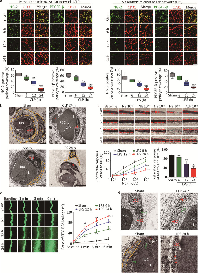
Background: Vascular hyporeactivity and leakage are key pathophysiologic features that produce multi-organ damage upon sepsis. We hypothesized that pericytes, a group of pluripotent cells that maintain vascular integrity and tension, are protective against sepsis via regulating vascular reactivity and permeability.

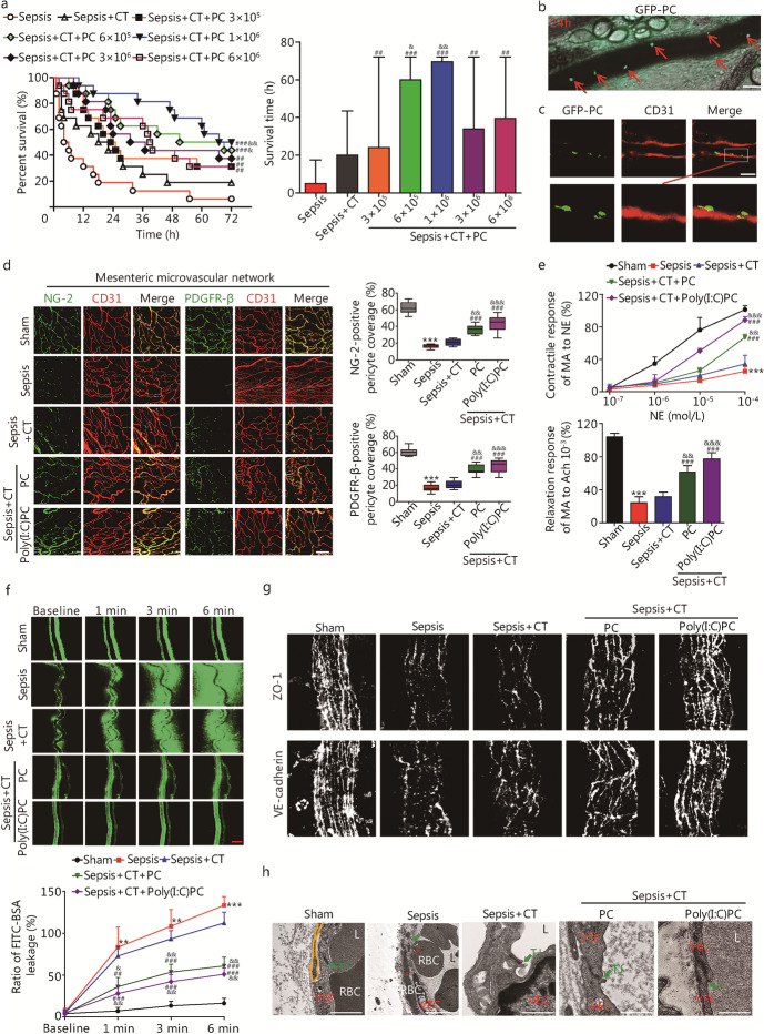
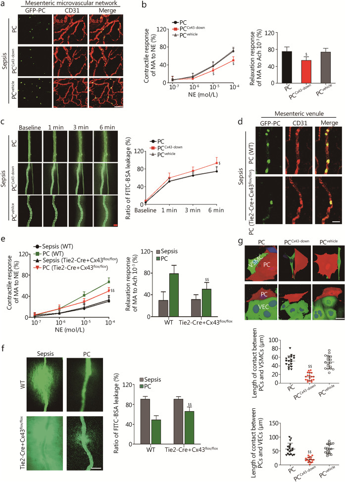
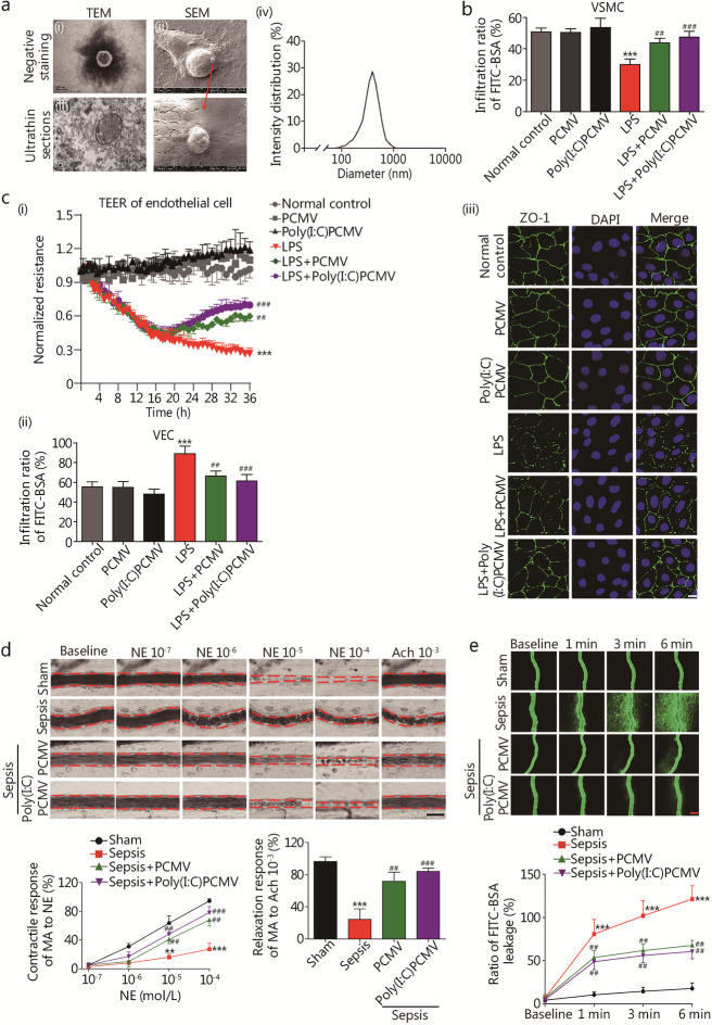
Methods: We conducted a series of in vivo experiments using wild-type (WT), platelet-derived growth factor receptor beta (PDGFR-β)-Cre + mT/mG transgenic mice and Tie2-Cre + Cx43flox/flox mice to examine the relative contribution of pericytes in sepsis, either induced by cecal ligation and puncture (CLP) or lipopolysaccharide (LPS) challenge. In a separate set of experiments with Sprague-Dawley (SD) rats, pericytes were depleted using CP-673451, a selective PDGFR-β inhibitor, at a dosage of 40 mg/(kg·d) for 7 consecutive days. Cultured pericytes, vascular endothelial cells (VECs) and vascular smooth muscle cells (VSMCs) were used for mechanistic investigations. The effects of pericytes and pericyte-derived microvesicles (PCMVs) and candidate miRNAs on vascular reactivity and barrier function were also examined.

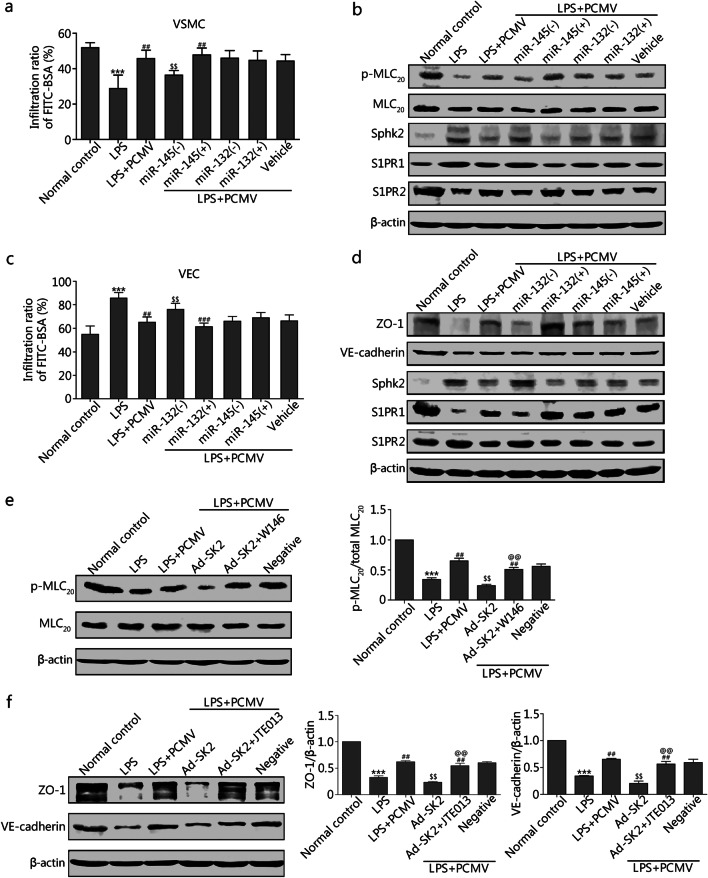
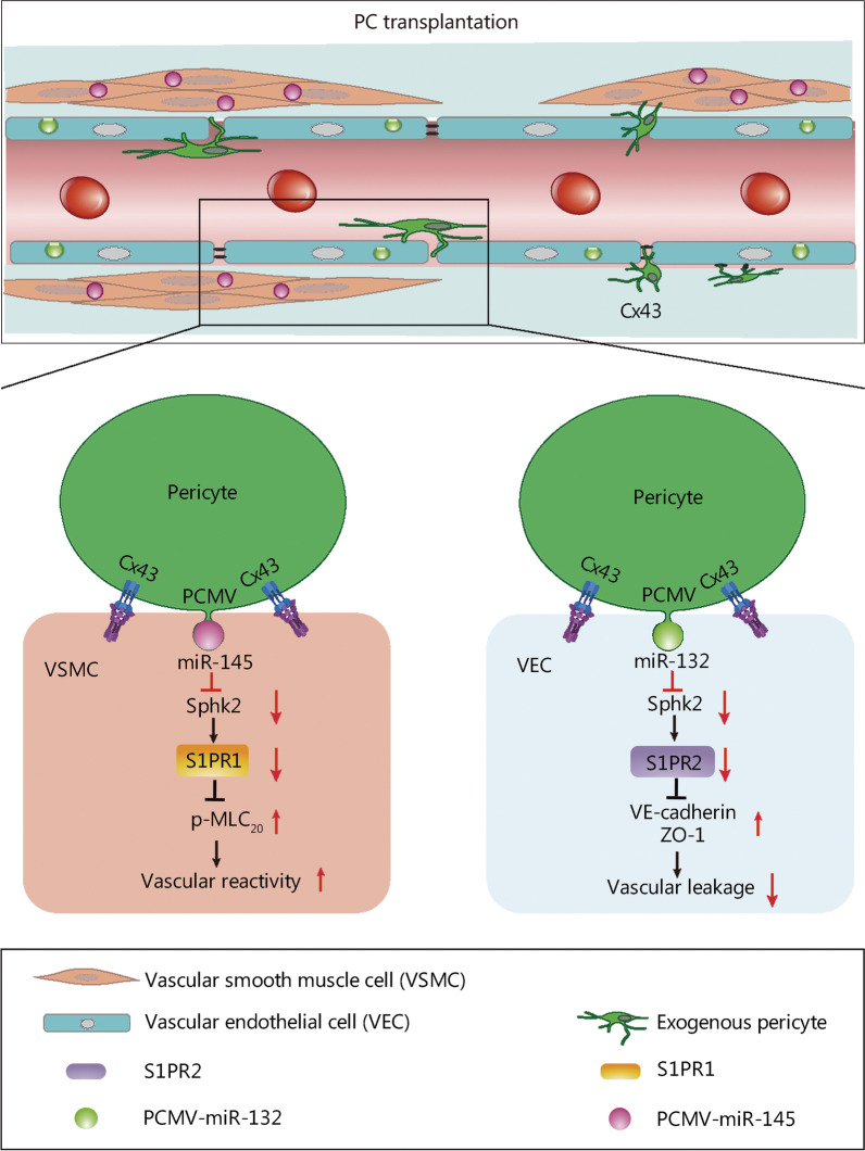
Results: CLP and LPS induced severe injury/loss of pericytes, vascular hyporeactivity and leakage (P < 0.05). Transplantation with exogenous pericytes protected vascular reactivity and barrier function via microvessel colonization (P < 0.05). Cx43 knockout in either pericytes or VECs reduced pericyte colonization in microvessels (P < 0.05). Additionally, PCMVs transferred miR-145 and miR-132 to VSMCs and VECs, respectively, exerting a protective effect on vascular reactivity and barrier function after sepsis (P < 0.05). miR-145 primarily improved the contractile response of VSMCs by activating the sphingosine kinase 2 (Sphk2)/sphingosine-1-phosphate receptor (S1PR)1/phosphorylation of myosin light chain 20 pathway, whereas miR-132 effectively improved the barrier function of VECs by activating the Sphk2/S1PR2/zonula occludens-1 and vascular endothelial-cadherin pathways.

Conclusions: Pericytes are protective against sepsis through regulating vascular reactivity and barrier function. Possible mechanisms include both direct colonization of microvasculature and secretion of PCMVs.

Keywords: Cx43; Microvesicle; Pericyte; Vascular permeability; Vascular reactivity.


Conflict of interest statement

The authors declare that they have no competing interests.

Figures

Fig. 1
Fig. 1
Sepsis induces pericyte loss, vascular hyporeactivity and leakage in rats. a Mesenteric microvascular networks from CLP and LPS (10 mg/kg)-induced sepsis at 6, 12 and 24 h were stained for NG-2 (pericyte marker; green), PDGFR-β (pericyte marker; green), and CD31 (VEC marker; red). Pericyte coverage rate of endothelium was quantified by analyzing percentage of CD31+ capillaries opposed to NG-2+ and PDGFR-β+ PCs (n = 8 rats). Scale bars: 100 μm. b TEM observation of ultrastructural changes of pericyte in mesenteric venules at 24 h after CLP and LPS administration (yellow arrowheads indicate pericyte loss and swelling, *indicate erythrocyte diapedesis). Scale bars: 2 μm. c Changes in vascular response of mesenteric arterioles to NE and Ach in vivo (n = 8 rats). d Vascular leakage of mesenteric venules measured by the appearance of intravenously injected FITC–BSA and quantitation of FITC–BSA+ vessel (n = 8 rats). Scale bars: 50 μm. e Representative TEM images of tight junctions in mesenteric venules after CLP and LPS administration at 24 h (green arrow indicate the tight junction, red arrowheads indicate the endothelial fragments and disrupted VEC junctions). Scale bars: 1 μm. PC pericyte, CLP cecal ligation and puncture, LPS lipopolysaccharides, NG-2 nerve/glial antigen 2, PDGFR-β platelet-derived growth factor receptor beta, VEC vascular endothelial cell, RBC red blood cell, L lumen, NE norepinephrine, Ach acetylcholine, MA mesenteric arteriole, TJ tight junction, TEM transmission electron microscopy. Data shown as mean ± SD. *P < 0.05, **P < 0.01, ***P < 0.001 vs. Sham (one-way ANOVA)
Fig. 2
Fig. 2
The transplanted pericytes improve the vascular hyporeactivity and leakage after sepsis. a Effects of transplanting different amount of exogenous pericytes on animal survival (n = 16 rats). Intravital microscopy (b, red arrows indicate GFP-PC) and immunofluorescence (c) by CLSM were used to monitor the GFP-PC location on mesenteric venules at 24 h after transplantation of exogenous pericytes (106). Scale bars: 50 μm. d Mesenteric microvascular networks were stained for NG-2, PDGFR-β, and CD31 at 24 h after resuscitation (n = 8 rats). Scale bars: 100 μm. e Changes in vascular response of mesenteric arterioles to NE and Ach in vivo after sepsis in rats (n = 8). f Vascular leakage of mesenteric venules measured by the appearance of intravenously injected FITC–BSA and quantitation of FITC–BSA+ vessel (n = 8 rats). Scale bars: 50 μm. g Immunohistochemistry for ZO-1 and VE-cadherin in mesenteric venules. Scale bars: 20 μm. h Representative TEM images of tight junctions in mesenteric venules (green arrows indicate the tight junction, *indicate the erythrocyte diapedesis). Scale bars: 1 μm. NG-2 nerve/glial antigen 2, PDGFR-β platelet-derived growth factor receptor beta, CT conventional treatment, CLSM confocal laser scanning microscopy, PC pericyte, Poly(I:C)PC polyinosine-polycytidylic acid pre-treatment pericyte, NE norepinephrine, Ach acetylcholine, MA mesenteric arteriole, ZO-1 zonula occludens-1, VE-cadherin vascular endothelial cadherin, VEC vascular endothelial cell, RBC red blood cell, TJ tight junction, L lumen, TEM transmission electron microscopy. Data shown as mean ± SD. **P < 0.01, ***P < 0.001 vs. Sham; ##P < 0.01, ###P < 0.001 vs. Sepsis; &&P < 0.01, &&&P < 0.001 vs. Sepsis + CT (one-way ANOVA)
Fig. 3
Fig. 3
Transplanted pericytes regulate vascular reactivity and permeability via Cx43 after sepsis. a Immunofluorescence by CLSM was used to monitor the PCCx43−down colonization on mesenteric microvascular networks in septic rats. Scale bars: 100 μm. b Changes in vascular response of mesenteric arterioles to NE and Ach in vivo after PCCx43−down transplantation (n = 8 rats). c Vascular leakage of mesenteric venules measured after PCCx43−down transplantation (n = 8 rats). Scale bars: 50 μm. d Immunofluorescence by CLSM was used to monitor the GFP-PC location on mesenteric venules at 24 h after sepsis in Tie2-Cre + Cx43flox/flox mice. Scale bars: 20 μm. e Changes in vascular response of mesenteric arterioles to NE and Ach in vivo after sepsis in Tie2-Cre + Cx43flox/flox mice (n = 8). f Vascular leakage of mesenteric venules measured after sepsis in Tie2-Cre + Cx43flox/flox mice (n = 8). Scale bars: 20 μm. g 3D projection images of contact area on 24 h in the pericytes-VSMCs/VECs culture at a 1:9 pericyte:VSMCs/VECs ratio (PC group: pericyte with no-treatment; PCCx43−down group: infection of pericytes with shRNA adenovirus targeting Cx43; PCvehicle group: infection of pericytes with control adenovirus). Pericytes, VSMCs/VECs and nuclei are shown in red, green and blue, respectively. Scale bars: 20 μm. CLSM confocal laser scanning microscopy, PC pericyte, NE norepinephrine, Ach acetylcholine, MA mesenteric arteriole, VECs vascular endothelial cells, VSMCs vascular smooth muscle cells. Data shown as mean ± SD. $P < 0.05, $$P < 0.01 vs. PC or PC (WT) (one-way ANOVA)
Fig. 4
Fig. 4
PCMVs regulate the contractile response of VSMCs and barrier function of VECs after sepsis. a Identification of PCMV. (i-ii) Representative TEM micrographs of microvesicle isolated from pericyte; (iii) Representative SEM micrographs of microvesicle observed from pericytes; (iv) PCMV diameter measured by DLS analysis. b Role of PCMVs and Poly(I:C)PCMVs (2 × 106 microvesicles/ml) on the contractile response of rat VSMC to NE at 12 h after LPS (2 μg/ml) stimulation (n = 8 cells). c Role of PCMVs and Poly(I:C)PCMVs on the barrier function of rat VECs after LPS administration. i PCMVs and Poly(I:C)PCMVs were added into rat VECs, and TEER of each group was measured (n = 3 cells); ii PCMVs and Poly(I:C)PCMVs were added into VECs, and FITC–BSA penetration of each group was measured (n = 8 cells); iii VECs treated with PCMV were analyzed by immunofluorescence for ZO-1. Scale bars: 20 μm. d Changes in vascular response of mesenteric arterioles to NE and Ach in vivo after PCMV transplantation (n = 8 rats). Scale bars: 50 μm. e Vascular leakage of mesenteric venules measured after PCMV transplantation (n = 8 rats). Scale bars: 50 μm. PC pericyte, PCMV pericyte-derived microvesicle, TEM transmission electron microscopy, SEM scanning electron microscopy, VECs vascular endothelial cells, VSMCs vascular smooth muscle cells, LPS lipopolysaccharides, TEER transendothelial electrical resistance, ZO-1 zonula occludens-1, NE norepinephrine, Ach acetylcholine, MA mesenteric arteriole. Data shown as mean ± SD. **P < 0.01, ***P < 0.001 vs. Normal control or Sham; ##P < 0.01, ###P < 0.001 vs. LPS or Sepsis (one-way ANOVA)
Fig. 5
Fig. 5
PCMVs carry miR-145 and miR-132 to play coordinated effects on the VSMCs and VECs. a Effects of different types of PCMVs on contractile response of rat VSMC to NE after LPS administration (n = 8 cells). b Western blotting analysis of p-MLC20, Sphk2, S1PR1 and S1PR2 from VSMCs treated with different types of PCMVs (n = 3 cells). c Different types of PCMVs were added into rat VECs, and FITC–BSA penetration of each group was measured (n = 8 cells). d Western blotting analysis of ZO-1, VE-cadherin, Sphk2, S1PR1 and S1PR2 from VECs treated with different types of PCMVs (n = 3 cells). e–f Western blotting analysis of p-MLC20, ZO-1 and VE-cadherin from VSMCs and VECs with S1PR1 inhibition (W146) and S1PR2 inhibition (JTE013) (n = 3 cells). LPS lipopolysaccharides, PC pericyte, PCMV pericyte-derived microvesicle, VECs vascular endothelial cells, VSMCs vascular smooth muscle cells, Sphk2 sphingosine kinase 2, S1PR1 sphingosine-1-phosphate receptor 1, p-MLC20 phosphorylation of myosin light chain 20, ZO-1 zonula occludens-1, VE-cadherin vascular endothelial cadherin, Ad-SK2 adenovirus-mediated overexpression of Sphk2, Negative infection of VSMCs or VECs with negative control adenovirus. Data shown as mean ± SD. ***P < 0.001 vs. Normal control; ##P < 0.01, ###P < 0.001 vs. LPS; $$P < 0.01 vs. LPS + PCMV; @@P < 0.01 vs. Ad-SK2 (one-way ANOVA)
Fig. 6
Fig. 6
A schematic diagram of the protective role of pericytes in sepsis. After sepsis, pericyte desquamation, increased expression of endothelial S1PR2 and decreased ZO-1 and VE-cadherin are associated with vascular endothelial barrier breakdown; increased expression of S1PR1 and decreased p-MLC20 in VSMCs are associated with the vascular hyporeactivity. After pericyte transplantation, pericytes colonize in the mesenteric vein and form direct contact with endothelial cells to form a gap junction. Pericytes also secreted microvesicles (MVs) containing miR-145/132 to VSMCs and VECs to produce additional protective effects. miR-145 mainly acts on VSMCs to improve the vascular reactivity via inhibiting the expression of Sphk2 and S1PR1, and increasing the expression of p-MLC20. miR-132 mainly acts on VECs to improve the barrier function via inhibiting the expression of Sphk2 and S1PR2, and increasing the expression of ZO-1 and VE-cadherin. PC pericyte, PCMV pericyte-derived microvesicle, VEC vascular endothelial cell, VSMC vascular smooth muscle cell, p-MLC20 phosphorylation of myosin light chain 20, Sphk2 sphingosine kinase 2, S1PR1 sphingosine-1-phosphate receptor 1, ZO-1 zonula occludens-1, VE-cadherin vascular endothelial cadherin

References

    1. Yao YM, Zhang H. Better therapy for combat injury. Mil Med Res. 2019;6(1):23. - PMC - PubMed
    1. Singh V, Akash R, Chaudhary G, Singh R, Choudhury S, Shukla A, et al. Sepsis downregulates aortic Notch signaling to produce vascular hyporeactivity in mice. Sci Rep. 2022;12(1):2941. doi: 10.1038/s41598-022-06949-3. - DOI - PMC - PubMed
    1. Ince C, Mayeux PR, Nguyen T, Gomez H, Kellum JA, Ospina-Tascon GA, et al. The endothelium in sepsis. Shock. 2016;45(3):259–270. doi: 10.1097/SHK.0000000000000473. - DOI - PMC - PubMed
    1. Auriemma CL, Zhuo H, Delucchi K, Deiss T, Liu T, Jauregui A, et al. Acute respiratory distress syndrome-attributable mortality in critically ill patients with sepsis. Intensive Care Med. 2020;46(6):1222–1231. doi: 10.1007/s00134-020-06010-9. - DOI - PMC - PubMed
    1. Senatore F, Balakumar P, Jagadeesh G. Dysregulation of the renin-angiotensin system in septic shock: mechanistic insights and application of angiotensin II in clinical management. Pharmacol Res. 2021;174:105916. doi: 10.1016/j.phrs.2021.105916. - DOI - PubMed

Publication types