. 2023 Feb 24:10:985040.
doi: 10.3389/fvets.2023.985040. eCollection 2023.

COPA3 peptide supplementation alleviates the heat stress of chicken fibroblasts

Affiliations

COPA3 peptide supplementation alleviates the heat stress of chicken fibroblasts

Sharif Hasan Siddiqui et al. Front Vet Sci. .

Abstract

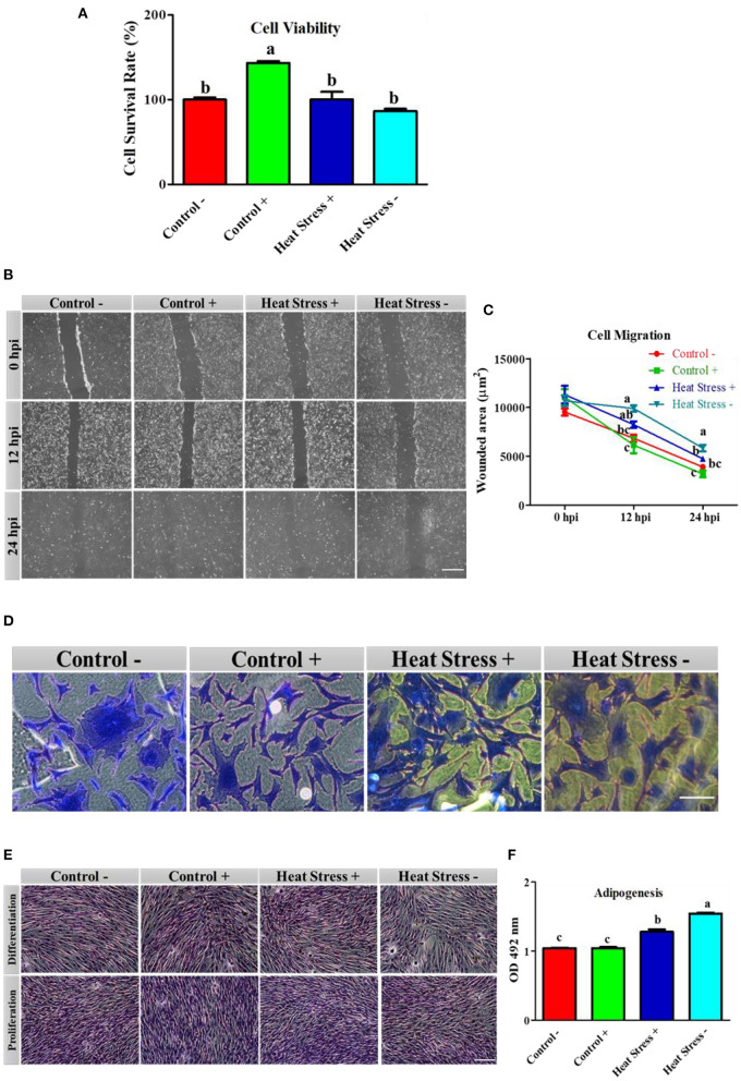
Heat stress inhibits cellular proliferation and differentiation through the production of reactive oxygen species. Under stress conditions, antioxidant drugs promote stable cellular function by reducing the stress level. We sought to demonstrate 9-mer disulfide dimer peptide (COPA3) supplementation stabilizes fibroblast proliferation and differentiation even under heat stress conditions. In our study, fibroblasts were assigned to two different groups based on the temperature, like 38°C group presented as Control - and 43°C group presented as Heat Stress-. Each group was subdivided into two groups depending upon COPA3 treatment, like 38°C + COPA3 group symbolized Control+ and the 43°C + COPA3 group symbolized as Heat Stress+. Heat stress was observed to decrease the fibroblast viability and function and resulted in alterations in the fibroblast shape and cytoskeleton structure. In contrast, COPA3 stabilized the fibroblast viability, shape, and function. Moreover, heat stress and COPA3 were found to have opposite actions with respect to energy production, which facilitates the stabilization of cellular functions by increasing the heat tolerance capacity. The gene expression levels of antioxidant and heat shock proteins were higher after heat stress. Additionally, heat stress promotes the mitogen-activated protein kinase/ extracellular signal-regulated kinase-nuclear factor erythroid 2-related factor 2 (MAPK/ERK-Nrf2). COPA3 maintained the MAPK/ERK-Nrf2 gene expressions that promote stable fibroblast proliferation, and differentiation as well as suppress apoptosis. These findings suggest that COPA3 supplementation increases the heat tolerance capacity, viability, and functional activity of fibroblasts.

Keywords: COPA3; fibroblast; heat stress; heat tolerance; proliferation; viability.


Conflict of interest statement

The authors declare that the research was conducted in the absence of any commercial or financial relationships that could be construed as a potential conflict of interest.

Figures

Figure 1
Figure 1
COPA3 protects the stressed fibroblasts' morphology by increasing heat tolerance. (A) Model experimental strategy; 38°C and 43°C temperatures indicate the control and the heat stress group, respectively; each group was subdivided into two groups based on COPA3 treatment. (B) Survival rates of fibroblasts at 38°C obtained using the CCK-8 assay; data represent mean ± SE; a−ddifferent letters indicate significant difference in mean fibroblasts in 38°C (P < 0.05). (C) Survival rate of fibroblasts at 43°C temperature obtained using the CCK-8 assay; data represent mean ±SE; a, bdifferent letters indicate significant difference in the mean number of fibroblasts at 43°C (P < 0.05). (D) Comparative survival rates of the fibroblasts at 38°C and 43°C obtained using the CCK-8 assay; data represent mean ± SE; *P < 0.05, and **P < 0.001. (E) Giemsa staining showing the morphology of the fibroblasts at 4 × and 20 × microscopic focus; scale bar = 100 μm and 20 μm, respectively. (F) Concentration of lactate dehydrogenase (LDH); data represent mean ±SE; a, bdifferent letters indicate significant difference in different groups (P < 0.05). (G) Concentration of nitric oxide (NO); data represent mean ± SE; a, bdifferent letters indicate significant difference in different groups (P < 0.05). Groups are distinguished by temperatures and COPA3 treatment, that is, fibroblasts at 38°C (Control- group), 38°C with COPA3 treatment (Control+ group), 43°C with COPA3 treatment (Heat Stress+ group), and 43°C temperature (Heat Stress- group). Each experiment was conducted in four biological replicates (n = 4).
Figure 2
Figure 2
Effects of COPA3 on stressed fibroblasts' viability, cytoskeleton structure, and functions. (A) Percentages of fibroblasts' survivability obtained using the CCK-8 assay. (B) Fibroblasts' migration activity analysis by wound healing assay; hpi indicates hour post incubation; scale bar = 100 μm. (C) Wounded area measurement; data represent mean ±SE; a−cdifferent letters indicate significant difference in different groups at specific time periods (P < 0.05). (D) Coomassie blue stain showing the cytoskeleton shape; scale bar = 20 μm. (E) Oil Red O (ORO) staining showing the lipid droplets in fibroblasts; scale bar = 100 μm. (F) Adipogenesis in fibroblasts; data represent mean ± SE; a−cdifferent letters indicate significant difference in different groups (P < 0.05). Groups are distinguished by temperatures and COPA3 treatment, that is, fibroblasts at 38°C (Control- group), 38°C with COPA3 treatment (Control+ group), 43°C with COPA3 treatment (Heat Stress+ group), and 43°C temperature (Heat Stress- group). Each experiment was conducted in four biological replicates (n = 4).
Figure 3
Figure 3
Effects of COPA3 on stressed fibroblasts' apoptosis through energy regulation. (A) Concentration of adenosine triphosphate (ATP); data represent mean ± SE; a, bdifferent letters indicate significant difference in different groups (P < 0.05). (B) Concentration of glucose 6 phosphate dehydrogenase (G6PDH); data represent mean ± SE; a, bdifferent letters indicate significant difference in different groups (P < 0.05). (C) Live and dead cell stain showing the live cells and apoptosed cells; scale bar = 50 μm. (D) Crystal violet stain showing cell attachment; scale bar = 100 μm. (E) Percentage of cell attachment; data represent mean ± SE; a, ddifferent letters indicate significant difference in different groups (P < 0.05). Groups are distinguished by temperatures and COPA3 treatment, that is, fibroblasts at 38°C (Control- group), 38°C with COPA3 treatment (Control+ group), 43°C with COPA3 treatment (Heat Stress+ group), and 43°C temperature (Heat Stress- group). Each experiment was conducted in four biological replicates (n = 4).
Figure 4
Figure 4
Effects of COPA3 on mRNA expression levels of antioxidant enzymes and heat shock proteins. (A–J) Relative mRNA expression levels corresponding to the genes of catalase, superoxide dismutase, glutathione S-transferase omega-2, glutathione S-transferase theta 1, glutathione S-transferase A3, cytochrome c oxidase subunit 5a, heat shock protein70, heat shock protein 60, heat shock protein 47, and heat shock protein 40 obtained by RT-qPCR; data represent mean ± SE; a−cdifferent letters indicate significant difference in different groups (P < 0.05). (K) Cluster analysis of mRNA expression. Groups are distinguished by temperatures and COPA3 treatment, that is, fibroblasts at 38°C (Control- group), 38°C with COPA3 treatment (Control+ group), 43°C with COPA3 treatment (Heat Stress+ group), and 43°C temperature (Heat Stress- group). Each experiment was conducted in four biological replicates (n = 4).
Figure 5
Figure 5
Effects of COPA3 on heat stressed fibroblasts' MAPK/ERK-Nrf2 related gene expression. (A–D) Relative mRNA expression levels of the genes of extracellular signal-regulated kinases, jun N-terminal kinase, p38 mitogen-activated protein kinases, and nuclear factor erythroid 2–related factor 2 involved in the MAPK/ERK-Nrf2 obtained by RT-qPCR; data represent mean ± SE; a, bdifferent letters indicate significant difference in different groups (P < 0.05). (E) Cluster analysis of the mRNA expression corresponding to the MAPK/ERK-Nrf2 components. Groups are distinguished by temperatures and COPA3 treatment, that is, fibroblasts at 38°C (Control- group), 38°C with COPA3 treatment (Control+ group), 43°C with COPA3 treatment (Heat Stress+ group), and 43°C temperature (Heat Stress- group). Each experiment was conducted in four biological replicates (n = 4).
Figure 6
Figure 6
Effects of COPA3 on the relationship between heat and MAPK/ERK-Nrf2 mRNA expression in fibroblasts. Correlations among the mRNA expression levels of the antioxidant enzymes, heat shock proteins, and the MAPK/ERK-Nrf2 related gene. (A) Control- group, (B) Control+ group, (C) Heat Stress+ group, and (D) Heat Stress- group. Principal component analysis (PCA) of the mRNA expression levels of the antioxidant enzymes, heat shock proteins, and the MAPK/ERK-Nrf2 related gene. Based on the position of the different genes on the X and Y axis predict the relation among different genes in particular group. (E) Control- group, (F) Control+ group, (G) Heat Stress+ group, and (H) Heat Stress- group. PC1 and PC2 were selected as they explained the most variation.

Similar articles

Cited by

References

    1. Siddiqui SH, Kang D, Park J, Khan M, Belal SA, Shin D, et al. . Altered relationship between gluconeogenesis and immunity in broilers exposed to heat stress for different durations. Poult Sci. (2021) 100:101274. 10.1016/j.psj.2021.101274 - DOI - PMC - PubMed
    1. Kurop MK, Huyen CM, Kelly JH, Blagg BSJ. The heat shock response and small molecule regulators. Eur J Med Chem. (2021) 226:113846. 10.1016/j.ejmech.2021.113846 - DOI - PMC - PubMed
    1. Siddiqui SH, Khan M, Kang D, Choi HW, Shim K. Meta-analysis and systematic review of the thermal stress response: gallus gallus domesticus show low immune responses during heat stress. Front Physiol. (2022) 13:809648. 10.3389/fphys.2022.809648 - DOI - PMC - PubMed
    1. Rivera RM, Kelley KL, Erdos GW, Hansen PJ. Reorganization of microfilaments and microtubules by thermal stress in two-cell bovine embryos. Biol Reprod. (2004) 70:1852–62. 10.1095/biolreprod.103.024901 - DOI - PubMed
    1. Gu ZT, Wang H, Li L, Liu YS, Deng XB, Huo SF, et al. . Heat stress induces apoptosis through transcription-independent p53-mediated mitochondrial pathways in human umbilical vein endothelial cell. Sci Rep. (2014) 4:4469. 10.1038/srep04469 - DOI - PMC - PubMed