. 2022 Nov 30;8(2):174-185.
doi: 10.1016/j.jacbts.2022.08.003. eCollection 2023 Feb.

Inflammatory Macrophage Interleukin-1β Mediates High-Fat Diet-Induced Heart Failure With Preserved Ejection Fraction

Affiliations

Inflammatory Macrophage Interleukin-1β Mediates High-Fat Diet-Induced Heart Failure With Preserved Ejection Fraction

Hong Liu et al. JACC Basic Transl Sci. .

Abstract

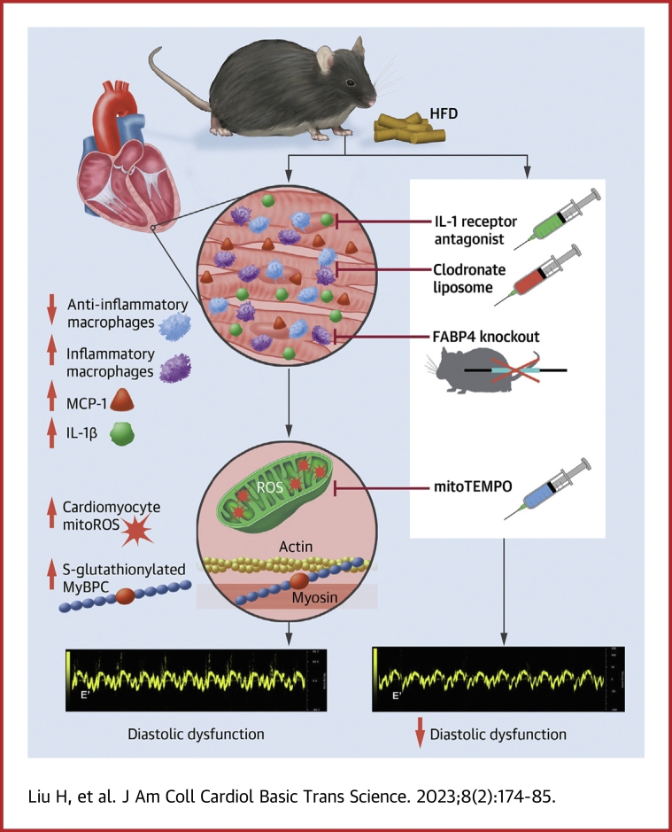
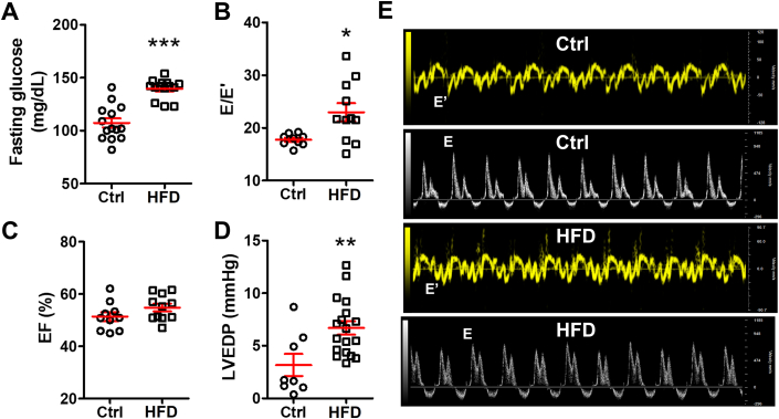
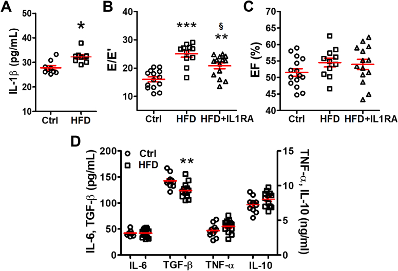
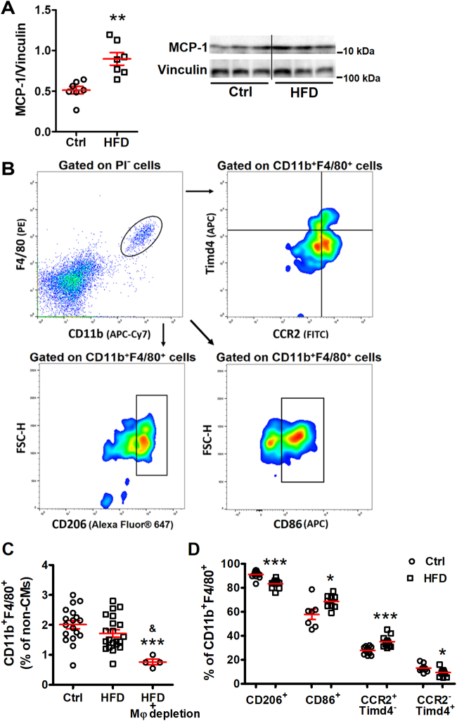
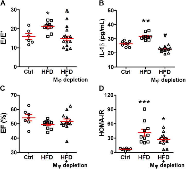
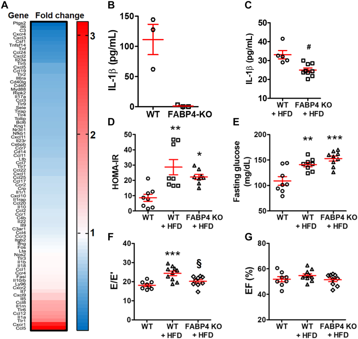
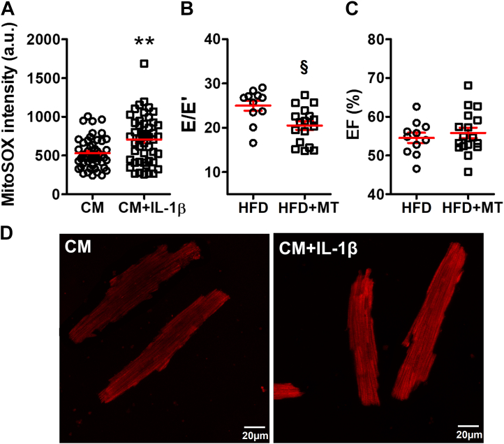
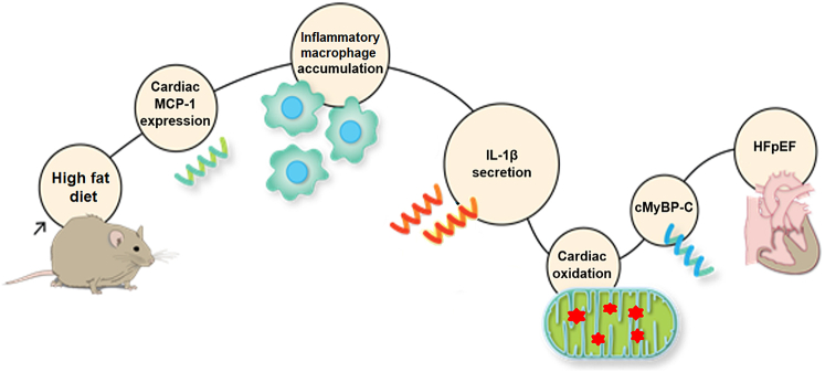
Diabetes mellitus (DM) is a main risk factor for diastolic dysfunction (DD) and heart failure with preserved ejection fraction. High-fat diet (HFD) mice presented with diabetes mellitus, DD, higher cardiac interleukin (IL)-1β levels, and proinflammatory cardiac macrophage accumulation. DD was significantly ameliorated by suppressing IL-1β signaling or depleting macrophages. Mice with macrophages unable to adopt a proinflammatory phenotype were low in cardiac IL-1β levels and were resistant to HFD-induced DD. IL-1β enhanced mitochondrial reactive oxygen species (mitoROS) in cardiomyocytes, and scavenging mitoROS improved HFD-induced DD. In conclusion, macrophage-mediated inflammation contributed to HFD-associated DD through IL-1β and mitoROS production.

Keywords: CCR2, C-C motif chemokine receptor 2; CM, cardiomyocyte; DD, diastolic dysfunction; DM, diabetes mellitus; EF, ejection fraction; FABP4, fatty acid binding protein 4; HF, heart failure; HFD, high-fat diet; HFpEF; HFpEF, heart failure with preserved ejection fraction; IL, interleukin; IL-1β; IL1RA, interleukin 1 receptor antagonist; KO, knockout; MCP, monocyte chemoattractant protein; MyBP-C, myosin binding protein C; TGF, transforming growth factor; TNF, tumor necrosis factor; Timd4, T cell immunoglobulin and mucin domain containing 4; WT, wild-type; diabetes; diastolic dysfunction; inflammation; macrophage; mitoROS, mitochondrial reactive oxygen species; mitochondria.


Conflict of interest statement

This project was supported by National Institutes of Health grants R01 HL104025 (Dr Dudley) and R01 HL106592 (Dr Dudley). All other authors have reported that they have no relationships relevant to the contents of this paper to disclose.

Figures

None
Graphical abstract
Figure 1
Figure 1
HFD Causes Cardiac Diastolic Heart Failure in Mice High-fat diet (HFD)-induced (A) hyperglycemia (n = 14 mice per group), (B) an increased ratio of transmitral Doppler early filing velocity to tissue Doppler early diastolic mitral annular velocity (E/E′) (n = 9 to 11 per group), and (D) left ventricular end-diastolic pressure (LVEDP) elevation by hemodynamic test (n = 7 to 18 mice per group) with (C) preserved ejection fraction (EF) value (n = 9 to 11 mice per group). (E) Representative echocardiographic images of tissue Doppler and pulsed wave Doppler from control (Ctrl) and HFD mice. Bars are mean ± SEM. Unpaired t-test was used. ∗P <0.05; ∗∗P < 0.01; ∗∗∗P < 0.001 vs Ctrl.
Figure 2
Figure 2
IL-1β Mediates HFD-Induced Diastolic Dysfunction (A) Cardiac interleukin (IL)-1β level tested by enzyme-linked immunosorbent assay was increased in the mice with HFD; n = 8 to 9 mice per group. (B) IL-1β antagonist improved E/E′ in HFD mice; n = 11 to 14 mice per group. (C) The EF remained unchanged after IL1RA treatment; n = 11 to 14 mice per group. (D) Other cytokines in hearts were tested by enzyme-linked immunosorbent assay; n = 10 to 12 mice per group. Bars are mean ± SEM. Unpaired t-test (A and D) or 1-way analysis of variance with Bonferroni post hoc tests (B and C) were used. ∗P < 0.05; ∗∗P < 0.01; ∗∗∗P < 0.001 vs Ctrl; §P < 0.05 vs HFD. IL1RA = interleukin 1 receptor antagonist; TGF = transforming growth factor; TNF = tumor necrosis factor; other abbreviations as in Figure 1.
Figure 3
Figure 3
Cardiac Macrophages Are Activated in HFD Hearts (A) Monocyte chemoattractant protein (MCP)-1 level was significantly higher in HFD hearts by Western blot; n = 7 mice per group. (B) Representative flow cytometry images showing the gating strategy and analysis to identify macrophages subsets. Isolated noncardiomyocyte cells were pregated on CD11b+F4/80+ as cardiac macrophages and were further divided into subsets based on their expression of CCR2, T cell immunoglobulin and mucin domain containing 4 (Timd4), CD206, or CD86. (C) Percentage of cardiac macrophages (CD11b+F4/80+) among noncardiomyocyte interstitial cells; n = 4 to 21 mice per group. (D) Percentage change of each macrophage subset; n = 8 to 12 mice per group. Bars are mean ± SEM. Unpaired t-test (A and D) or 1-way analysis of variance with Bonferroni post hoc tests (C) were used. ∗P < 0.05; ∗∗P < 0.01; ∗∗∗P < 0.001 vs Ctrl; and &P < 0.01 vs HFD. CCR2 = C-C motif chemokine receptor 2; PI = propidium iodide; other abbreviations as in Figure 1.
Figure 4
Figure 4
Cardiac Macrophages Are Required for HFD-Induced Diastolic Dysfunction (A) Macrophage (Mφ) depletion improved E/E′ ratio (n = 6 to 13 mice per group) and decreased (B) cardiac IL-1β level tested by enzyme linked immunosorbent assay (n = 8 to 12 mice per group) without altering (C) EF (n = 7 to 10 mice per group) or (D) homeostatic model assessment for insulin resistance (HOMA-IR), an indicator of insulin resistance (n = 8 to 13 mice per group). Bars are mean ± SEM. One-way analysis of variance with Bonferroni post hoc tests were used. ∗P < 0.05; ∗∗P < 0.01; ∗∗∗P < 0.001 vs Ctrl; and &P < 0.01; #P < 0.001 vs HFD. Abbreviations as in Figures 1 and 2.
Figure 5
Figure 5
Suppressing Proinflammatory Macrophages Prevented HFD-Induced Diastolic Dysfunction (A) Inflammatory gene expression of fatty acid binding protein 4 (FABP4) knockout (KO) macrophage cell line by microarray. (B) FABP4 KO macrophages cannot secrete IL-1β under lipopolysaccharide stimulation; n = 3 independent experiments per group. (C) With HFD, FABP4 KO mice had lower cardiac IL-1β level; n = 5 to 9 mice per group. (D) Insulin resistance as indicated by HOMA-IR (n = 8 to 9 mice per group) and (E) hyperglycemia (n = 8 to 9 mice per group) were unchanged between wild-type (WT)+HFD and KO+HFD groups. (F) FABP4 KO mice were resistant to HFD-induced diastolic dysfunction; n = 8 to 9 mice per group. (G) EF was unaffected by FABP4 KO; n = 8 to 10 mice per group. Bars are mean ± SEM. Unpaired t-test (C) or 1-way analysis of variance with Bonferroni post hoc tests (D to G) were used. ∗P < 0.05; ∗∗P < 0.01; ∗∗∗P < 0.001 vs WT; §P < 0.05; #P < 0.01 vs WT+HFD. Abbreviations as in Figures 1, 2, and 4.
Figure 6
Figure 6
MitoROS Scavenging Reduced HFD-Induced Diastolic Dysfunction (A) IL-1β incubation up-regulated the cardiomyocyte (CM) mitochondrial reactive oxygen species (mitoROS) level. The cardiomyocytes were isolated from 5 mice, and 48 cardiomyocytes were tested for each group. (B) Mitochondrial antioxidant, mitoTEMPO (MT), improved E/E′ in HFD mice without affecting (C) the EF; n = 11 to 17 mice per group. The HFD group was the same as in Figures 2B and 2C. (D) Representative confocal microscopy images showing mitoROS in cardiomyocytes by mitoSOX red staining; scale bar represents 20 μm. Bars are mean ± SEM. Unpaired t-test were used. ∗∗P < 0.01 vs CM; §P < 0.05 vs HFD. a.u. = arbitrary units; other abbreviations as in Figures 1 and 2.
Figure 7
Figure 7
Innate Immunity Mediates High-Fat Diet–Induced HFpEF Through IL-1β Secretion and Mitochondrial Reactive Oxygen Species Modulation cMyBP-C = cardiac myosin binding protein C; HFpEF = heart failure with preserved ejection fraction; other abbreviations as in Figures 2 and 3.

References

    1. Hunt S.A., Abraham W.T., Chin M.H., et al. ACC/AHA 2005 guideline update for the diagnosis and management of chronic heart failure in the adult: a report of the American College of Cardiology/American Heart Association Task Force on Practice Guidelines (Writing Committee to Update the 2001 Guidelines for the Evaluation and Management of Heart Failure) J Am Coll Cardiol. 2005;46(6):e1–82. doi: 10.1016/j.jacc.2005.08.022. - DOI - PubMed
    1. Plitt G.D., Spring J.T., Moulton M.J., Agrawal D.K. Mechanisms, diagnosis, and treatment of heart failure with preserved ejection fraction and diastolic dysfunction. Expert Rev Cardiovasc Ther. 2018;16:579–589. - PMC - PubMed
    1. Carbone S., Canada J.M., Buckley L.F., et al. Dietary fat, sugar consumption, and cardiorespiratory fitness in patients with heart failure with preserved ejection fraction. J Am Coll Cardiol Basic Trans Science. 2017;2:513–525. - PubMed
    1. Duan Y., Zeng L., Zheng C., et al. Inflammatory links between high fat diets and diseases. Front Immunol. 2018;9:2649. - PMC - PubMed
    1. Xu H., Barnes G.T., Yang Q., et al. Chronic inflammation in fat plays a crucial role in the development of obesity-related insulin resistance. J Clin Invest. 2003;112:1821–1830. - PMC - PubMed