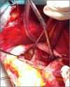
Giant circumflex artery with bilobed saccular aneurysm and right atrial fistula mimicking a bi-atrial hydatid cyst: a case report
- PMID: 36908687
- PMCID: PMC9997557
- DOI: 10.1093/jscr/rjad108
Giant circumflex artery with bilobed saccular aneurysm and right atrial fistula mimicking a bi-atrial hydatid cyst: a case report
Abstract
Coronary artery fistulas (CAF) are rare anomalies that pose a significant diagnostic and therapeutic challenge. Most of them originate from the right coronary artery and are congenital. They are often associated with coronary aneurysms. We report the case of a 38-year-old Black man who presented with exertion dyspnea. Transthoracic echocardiography found what was thought to be a bi-atrial hydatid cyst, alongside a right atrial shunt. Cardiac magnetic resonance imaging showed a cystic lesion hypointense on T1 and T2 sequences, located next to the left atrium as well as an aneurysmal circumflex artery shunting in the right atrium. Coronary angiography and computed tomography angiography confirmed the bilobed circumflex saccular aneurysm and CAF. The patient underwent a successful surgery, which consisted of closure of the fistula using two patches. He was discharged after an uneventful postoperative course. Our case report illustrates the diagnostic difficulty of CAF and the importance of multimodal imaging.
Keywords: Coronary aneurysm; Coronary artery fistula; Coronary cameral fistula; Multimodal imaging.
Published by Oxford University Press and JSCR Publishing Ltd. © The Author(s) 2023.
Figures


References
-
- Loukas M, Germain AS, Gabriel A, John A, Tubbs RS, Spicer D. Coronary artery fistula: a review. Cardiovasc Pathol 2015;24:141–8. - PubMed
-
- Luo L, Kebede S, Wu S, Stouffer GA. Coronary artery fistulae. Am J Med Sci 2006;332:79–84. - PubMed
-
- Chiu CZ, Shyu KG, Cheng JJ, Lin SC, Lee SH, Hung HF, et al. . Angiographic and clinical manifestations of coronary fistulas in Chinese people: 15-year experience. Circ J 2008;72:1242–8. - PubMed
Publication types
LinkOut - more resources
Full Text Sources

