Review
. 2023 Feb 24:17:1047778.
doi: 10.3389/fnins.2023.1047778. eCollection 2023.

Blood-brain barrier endothelial cells in neurodegenerative diseases: Signals from the "barrier"

Affiliations
Review

Blood-brain barrier endothelial cells in neurodegenerative diseases: Signals from the "barrier"

Yiwen Yuan et al. Front Neurosci. .

Abstract

As blood-brain barrier (BBB) disruption emerges as a common problem in the early stages of neurodegenerative diseases, the crucial roles of barrier-type brain endothelial cells (BECs), the primary part of the BBB, have been reported in the pathophysiology of neurodegenerative diseases. The mechanisms of how early vascular dysfunction contributes to the progress of neurodegeneration are still unclear, and understanding BEC functions is a promising start. Our understanding of the BBB has gone through different stages, from a passive diffusion barrier to a mediator of central-peripheral interactions. BECs serve two seemingly paradoxical roles: as a barrier to protect the delicate brain from toxins and as an interface to constantly receive and release signals, thus maintaining and regulating the homeostasis of the brain. Most previous studies about neurodegenerative diseases focus on the loss of barrier functions, and far too little attention has been paid to the active regulations of BECs. In this review, we present the current evidence of BEC dysfunction in neurodegenerative diseases and explore how BEC signals participate in the pathogenesis of neurodegenerative diseases.

Keywords: Alzheimer’s disease; Parkinson’s disease; blood–brain barrier; endothelial cells; neurodegenerative disease; neurovascular unit.


Conflict of interest statement

The authors declare that the research was conducted in the absence of any commercial or financial relationships that could be construed as a potential conflict of interest.

Figures
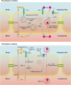
FIGURE 1
FIGURE 1
Brain endothelial cell transport in physiological and pathological conditions. Under physiological conditions, RAGE regulates the influx of Aβ to the brain, while LRP1 mediates the efflux of Aβ. The LRP1-dependent transendothelial Aβ transport is regulated by PICALM. PICALM guides Aβ to Rab5-positive early endosome and Rab11-positive transcytotic vesicle to the luminal side of BECs. The α-syn efflux is also an LRP1-dependent process, but the underlying mechanisms are still unclear. In the luminal-abluminal direction, the α-syn trafficking across BECs is directed by Rab7/VPS35 trafficking pathway. The GLUT1 is densely expressed on the abluminal membrane of the BECs, regulating the glucose transport across BBB. Under pathological conditions, the upregulation of RAGE and downregulation of LRP1 lead to the deposition of Aβ in the brain. The CCR5 antagonist is reported to alleviate the Aβ deposition by increasing the LRP1 expression. The LRP1 deficiency in BECs contributes to BBB breakdown in an Aβ-independent way by activation of cyclophilin A-MMP9 pathway in BECs. Similarly, the loss of GLUT1 in BECs leads to BBB breakdown. A GLP-1R agonist can reverse the reduced GLUT1 expression. The RBC-EVs can carry α-syn across BBB, but whether α-syn in these RBC-EVs can induce α-syn oligomerization in BECs like EV-derived α-syn in other recipient cells is still unclear. BEC, barrier type brain endothelial cell; RAGE, the receptor for advanced glycation end products; Aβ, β-amyloid; LRP1, the low-density lipoprotein receptor-related protein 1; PICALM, the phosphatidylinositol binding clathrin assembly protein; α-syn, α-synuclein; GLUT1, glucose transporter 1; BBB, blood–brain barrier; GLP-1R, glucagon-like peptide-1 receptor; RBC, red blood cell; EVs, extracellular vesicles.
FIGURE 2
FIGURE 2
Immune responses and stem cell activities in physiological and pathological conditions. Under physiological conditions, BECs secret VEGF and TGFβ in the autocrine way to maintain the integrity of BBB. Moreover, IL-25 released from BECs inhibits leukocyte trafficking in physiological conditions. In the brain, BECs secret neurotrophic factors to regulate the proliferation, differentiation, and migration of NSCs, promoting neurogenesis. BEC-derived TGFβ1 regulates the differentiation of OPCs from NPCs. However, the exact role of BECs in the myelination process is still unknown. In disease states, activated BECs secret IL-1, IL-6, IL-8, and GM-CSF to promote the trafficking of leukocytes across BBB. Periparial ApoE4 activates BECs and promotes leukocyte trafficking across BBB. The upregulation of VCAM1 in BECs is regulated by the binding of C3a and C3aR in BECs, activating the microglia and impairing the NSC activities. The chemokine CCL5 released by BECs recruits the resident brain microglia. Considering the crucial roles of BECs in neurogenesis and myelination, the studies of the contents and functions of BECs secretions in the regeneration failure of neurons and oligodendrocytes can provide insights into the pathophysiological process of neurodegenerative diseases. BECs, barrier type brain endothelial cells; NSCs, neural stem cells; TGFβ, transforming growth factor beta; OPCs, oligodendrocyte precursor cells; NPCs, neural progenitor cells; SHH, sonic hedgehog; VEGF, vascular endothelial growth factors; BBB, blood–brain barrier; GM-CSF, granulocyte-macrophage colony-stimulation factor; ApoE4, apolipoprotein E4; VCAM1, vascular cell adhesion molecule 1.

References

    1. Alam P., Holst M. R., Lauritsen L., Nielsen J., Nielsen S. S. E., Jensen P. H., et al. (2022). Polarized alpha-synuclein trafficking and transcytosis across brain endothelial cells via Rab7-decorated carriers. Fluids Barriers CNS 19:37. 10.1186/s12987-022-00334-y - DOI - PMC - PubMed
    1. Ali M., Falkenhain K., Njiru B. N., Murtaza-Ali M., Ruiz-Uribe N. E., Haft-Javaherian M., et al. (2022). VEGF signalling causes stalls in brain capillaries and reduces cerebral blood flow in Alzheimer’s mice. Brain 145 1449–1463. 10.1093/brain/awab387 - DOI - PMC - PubMed
    1. Alvarez J. I., Dodelet-Devillers A., Kebir H., Ifergan I., Fabre P. J., Terouz S., et al. (2011). The Hedgehog pathway promotes blood-brain barrier integrity and CNS immune quiescence. Science 334 1727–1731. 10.1126/science.1206936 - DOI - PubMed
    1. Alvarez-Buylla A., Cebrian-Silla A., Sorrells S. F., Nascimento M. A., Paredes M. F., Garcia-Verdugo J. M., et al. (2022). Comment on “Impact of neurodegenerative diseases on human adult hippocampal neurogenesis”. Science 376:eabn8861. 10.1126/science.abn8861 - DOI - PMC - PubMed
    1. Arai K., Lo E. H. (2009). An oligovascular niche: Cerebral endothelial cells promote the survival and proliferation of oligodendrocyte precursor cells. J. Neurosci. 29 4351–4355. 10.1523/JNEUROSCI.0035-09.2009 - DOI - PMC - PubMed