. 2023 Mar 6:11:e14867.
doi: 10.7717/peerj.14867. eCollection 2023.

Six new species of free-living nematodes (Nematoda: Enoplida) from deep-sea cold seeps on Hikurangi Margin, New Zealand

Affiliations

Six new species of free-living nematodes (Nematoda: Enoplida) from deep-sea cold seeps on Hikurangi Margin, New Zealand

Daniel Leduc. PeerJ. .

Abstract

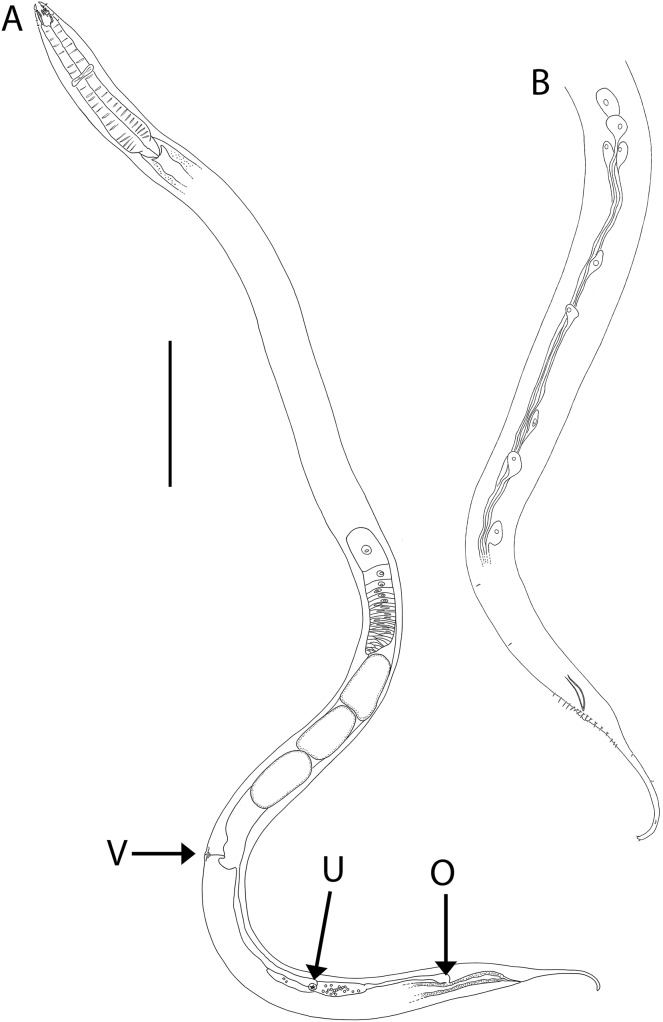
Little is known about the taxonomy of deep-sea nematode species inhabiting cold seep habitats. An opportunity to characterize the nematode species communities of New Zealand cold seeps was provided by a 2019 research voyage to New Zealand's Hikurangi Margin, during which macrofauna cores were obtained at two seeps at approximately 1,250 and 2,000 m water depth. Here, six new species of the orderEnoplida are described. Metacylicolaimus catherinae sp. nov. represents the first record of the genus for the New Zealand Exclusive Economic Zone and for the deep sea globally. Halalaimus talaurinus sp. nov., Thalassoalaimus duoporus sp. nov. and Crenopharynx crassipapilla sp. nov. are only the second species of their respective genera to be described/recorded from New Zealand waters, and Oncholaimus adustus sp. nov. is the eighth species of the genus to be recorded from the region. Rhabdodemania zealandiaensis sp. nov. was among the most abundant and widespread species found at the Hikurangi Margin seep sites. A few specimens had been found in a previous ecological study of meiofaunal nematode communities on Chatham Rise, a submarine ridge south of Hikurangi Margin. It is possible that this species has a preference for seep environments due to elevated food availability, however it does not seem to be exclusively found in seeps. We find no evidence for an affinity between nematode seep communities in New Zealand and elsewhere, which is consistent with the high variability in nematode community observed to date among regions. Ongoing work on the ecology and distribution of nematode communities at the Hikurangi Margin seep sites will help determine spatial patterns in abundance and species distributions in more detail, including the identification of any species/taxa with affinities with seeps.

Keywords: Cold seeps; Deep-sea; Enoplea; Nematoda; Southwest Pacific; Taxonomy.


Conflict of interest statement

The author declares that they have no competing interests.

Figures

Figure 1
Figure 1. Metacylicolaimus catherinae sp. nov.
(A) Male pharyngeal body region; (B and C) female cephalic region; (D) female reproductive system; (E) female posterior body region; (F) male posterior body region; (G) male copulatory apparatus. Scale bar: A = 300 μm, B and C = 70 μm, D = 800 μm, E = 175 μm, F = 215 μm, G = 100 μm.
Figure 2
Figure 2. Metacylicolaimus catherinae sp. nov.
Light micrographs. (A and C) Male cephalic region; (B) lateral view of mid-body region showing type I loxometaneme (lm) and epifermal gland (eg); (D) male copulatory apparatus; (E) sperm; (F) ventral view of vulva. Scale bar: A and C = 20 μm, B = 13 μm, D = 16 μm, E = 30 μm, F = 50 μm.
Figure 3
Figure 3. Oncholaimus adustus sp. nov.
(A) Male pharyngeal body region; (B) female cephalic region; (C) female posterior body region; (D) female cephalic region; (E) male copulatory apparatus. Scale bar: A = 200 μm, B and D = 116 μm, C = 130 μm, E = 70 μm.
Figure 4
Figure 4. Oncholaimus adustus sp. nov.
(A) Entire female showing location of vulva (V), uvette (U) and osmium (O); (B) male posterior body region showing caudal glands. Scale bar: A = 500 μm, B = 250 μm.
Figure 5
Figure 5. Oncholaimus adustus sp. nov.
Light micrographs. (A–C) Male cephalic region showing amphid, anterior sensilla, buccal cavity and armature, and secretory-excretory pore; (D) female anterior body region; (E) intestine wall. Scale bar: A–C = 50 μm, D = 45 μm, E = 55 μm.
Figure 6
Figure 6. Halalaimus talaurinus sp. nov.
(A) Female anterior body region; (B) male anterior body region; (C) male pharyngeal body region; (D) posterior body region; (E) male copulatory apparatus; (F) male posterior body region (tail tip broken). Arrows show position of cuticle pores. Scale bar: A and B = 25 μm, C = 100 μm, D = 62 μm, E and F = 55 μm.
Figure 7
Figure 7. Halalaimus talaurinus sp. nov.
(A) Entire male; (B) female reproductive system. Scale bar = 200 μm.
Figure 8
Figure 8. Halalaimus talaurinus sp. nov.
Light micrographs. (A) Male testis showing sperm; (B) male copulatory apparatus; (C) male caudal ala; (D–F) male cephalic region showing amphidial aperture, anterior sensilla and cuticle thickness. Scale bar: A–C = 20 mm; D–F = 15 μm.
Figure 9
Figure 9. Thalassoalaimus duoporus sp. nov.
(A) Female anterior body region; (B) female cephalic region; (C) male pharyngeal body region; (D) female pharyngeal body region; (E) male posterior body region; (F) male posterior body region; (G) female posterior body region. Scale bar: A = 25 μm, B = 22 μm, C and D = 122 μm, E = 33 μm, F = 75 μm, G = 45 μm.
Figure 10
Figure 10. Thalassoalaimus duoporus sp. nov.
(A) Female anterior body region and reproductive system; (B) entire male. Scale bar: A = 500 μm, B = 610 μm.
Figure 11
Figure 11. Thalassoalaimus duoporus sp. nov.
Light micrographs. (A and B) Male anterior body region showing anterior sensilla, buccal cavity and amphid; (C) junction of intestine and posterior portion of pharynx in female; (D) secretory-excretory pore and ampulla; (E) male posterior body region; (F) female posterior body region. Scale bar: A and B = 10 μm, C, E and F = 22 μm, D = 17 μm.
Figure 12
Figure 12. Crenopharynx crassipapilla sp. nov.
(A) Anterior body region of female; (B) male cephalic region; (C) female posterior body region; (D and E) male copulatory apparatus showing distal portion of spicules and gubernaculum; (F) male posterior body region; (G) sperm; (H) male copulatory apparatus showing entire spicule. Scale bar: A = 50 μm, B = 35 μm, C = 200 μm, D and E = 100 μm, F = 150 μm, G = 15 μm, H = 240 μm.
Figure 13
Figure 13. Crenopharynx crassipapilla sp. nov.
(A) Entire male; (B) female reproductive system. Scale bar: A = 500 μm, B = 330 μm.
Figure 14
Figure 14. Crenopharynx crassipapilla sp. nov.
Light micrographs of male. (A–C) Cephalic region showing outline of cephalic capsule, anterior sensilla and buccal cavity; (D–E) Anterior body region showing body and anterior sensilla, amphid aperture and secretory-excretory pore. Scale bar: A–C = 20 μm, D–G = 28 μm.
Figure 15
Figure 15. Rhabdodemania zealandiaensis sp. nov.
(A) Male pharyngeal region; (B) female cephalic region; (C and D) male cephalic region; male anterior pharyngeal region; (F) male posterior body region; (G) female posterior body region. Scale bar: A = 250 μm, B and D = 36 μm, C = 48 μm, E = 85 μm, F and G = 80 μm.
Figure 16
Figure 16. Rhabdodemania zealandiaensis sp. nov.
(A) Entire female; (B) entire male; (C) posterior portion of female reproductive system. Scale bar: A = 500 μm, B = 550 μm, C = 380 μm.
Figure 17
Figure 17. Rhabdodemania zealandiaensis sp. nov.
Light micrographs. (A) Male anterior body region showing buccal cavity, pharynx and secretory-excretory pore; (B) male anterior body region showing cuticle surface and amphidial nerve; (C) vulva; (D and E) male copulatory apparatus; (F) posterior portion of male intestine, vas deferens and copulatory supplements. Scale bar: A and B = 20 μm, C = 40 μm, D–F = 32 μm.

References

    1. Alekseev VM, Linnik OV. Halalaimus dolgovi sp. n. (Nematoda, Oxystominidae), a new nematode of the marine genus from Lake Khanka. Zoologicheskii Zhurnal. 1994;73:111–115. [In Russian]
    1. Allgén CA. Weitere beiträge zur kenntnis der marinen nematodenfauna der Campbellinsel. Nyt Magazin for Naturvidenskaberne. 1932;70:97–198.
    1. Allgén CA. Kleine mitteilungen über freilebende nematoden I–IV. Zoologischer Anzeiger. 1954;153:88–95.
    1. Baco AR, Rowden AA, Levin LA, Smith CR, Bowden DA. Initial characterization of cold seep faunal communities on the New Zealand Hikurangi margin. Marine Geology. 2010;272(1–4):251–259. doi: 10.1016/j.margeo.2009.06.015. - DOI
    1. Baird W. Catalogue of the entozoan, or intestinal worms, contained in the collection of the British Museum. London: British Museum; 1853. p. 132.

Publication types

LinkOut - more resources