[Preprint]. 2023 Feb 27:2023.02.27.530189.
doi: 10.1101/2023.02.27.530189.

Smaug regulates germ plasm synthesis and primordial germ cell number in Drosophila embryos by repressing the oskar and bruno 1 mRNAs

Affiliations

Smaug regulates germ plasm synthesis and primordial germ cell number in Drosophila embryos by repressing the oskar and bruno 1 mRNAs

Najeeb U Siddiqui et al. bioRxiv. .

Update in

Abstract

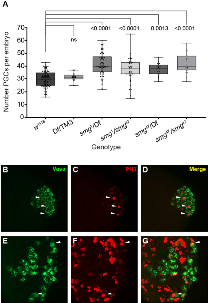
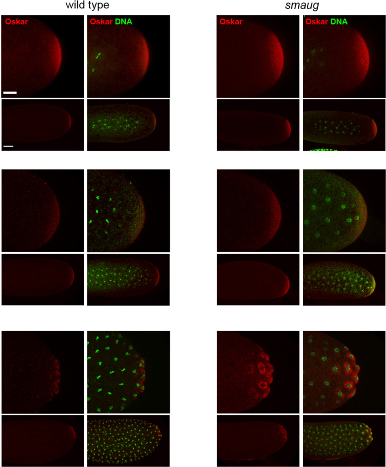
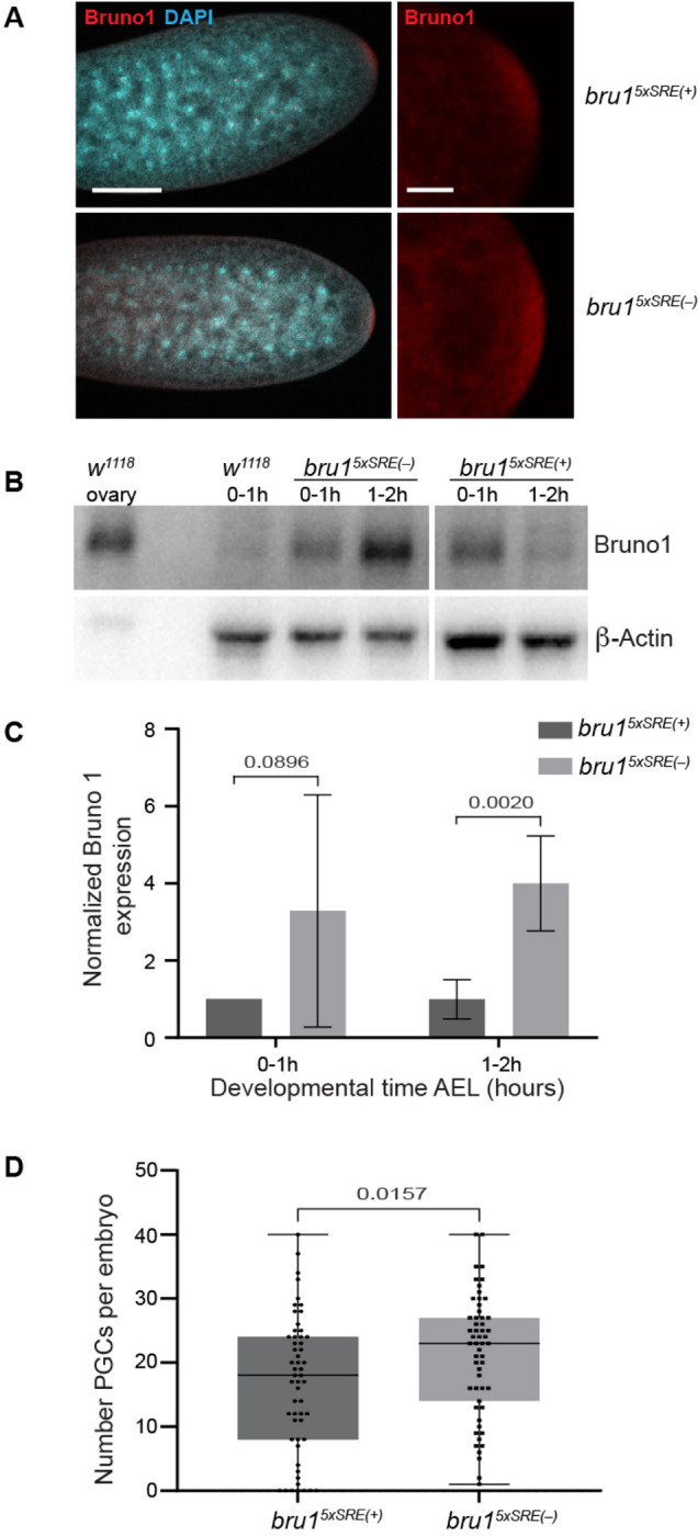
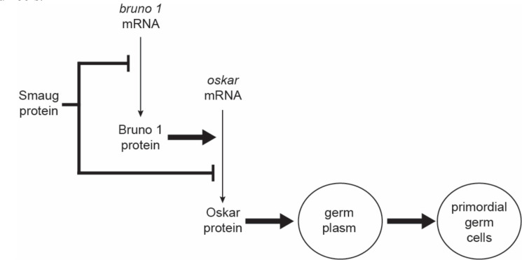
During Drosophila oogenesis, the Oskar (OSK) RNA-binding protein (RBP) determines the amount of germ plasm that assembles at the posterior pole of the oocyte. Here we identify the mechanisms that regulate the osk mRNA in the early embryo. We show that the Smaug (SMG) RBP is transported into the germ plasm of the early embryo where it accumulates in the germ granules. SMG binds to and represses translation of the osk mRNA itself as well as the bruno 1 (bru1) mRNA, which encodes an RBP that we show promotes germ plasm production. Loss of SMG or mutation of SMG's binding sites in the osk or bru1 mRNAs results in ectopic translation of these transcripts in the germ plasm and excess PGCs. SMG therefore triggers a post-transcriptional regulatory pathway that attenuates germ plasm synthesis in embryos, thus modulating the number of PGCs.


Conflict of interest statement

Competing interests: All authors declare they have no competing interests.

Figures

Figure 1.
Figure 1.. SMG protein is enriched in the germ plasm of early embryos.
(A) Live imaging of Venus-SMG shows that SMG protein is enriched in the germ plasm as early as nuclear cycle (NC) 5. Still images were taken from Movie S1. The time after imaging began is indicated at the top right of each still image in minutes and seconds. White arrowheads indicate the boundaries of Venus-SMG in the germ plasm in the first three images; in the fourth image the arrowheads point to Venus-SMG associated with nuclei in budding PGCs. The transgene insertion was on the second chromosome (see Materials and Methods): P{w+[Venus-SMG]:6}. (B) Live imaging of mCherry-SMG (red) showing association of SMG with the spindle poles (green; detected with Jupiter-GFP) upon arrival of nuclei at the posterior (NC8) and incorporation into the PGCs when they bud (NC9). Still images were taken from Movie S2. The mCherry-SMG transgene insertion was on the second chromosome (see Materials and Methods): P{w+[mCherry-SMG]:7}.
Figure 2.
Figure 2.. SMG is a component of the germ granules.
(A) SMG co-localizes with VAS, a known component of germ granules. Panels are still images taken from movies of embryos co-expressing mCherry-SMG (red) and VAS-GFP (green). The boxed areas in the left-hand images are shown at higher magnification on the right. See associated Movie S3. The mCherry-SMG transgene insertion was on the third chromosome (see Materials and Methods): P{w+[mCherry-SMG]:8}. (B) Immuno-electron microscopy shows that, in the pole cells, SMG (10 nm gold: small particles) is found in germ granules along with VAS (15 nm gold: large particles, indicated with white arrowheads). The boxed area in the left-hand image is shown at higher magnification in the center. In the posterior somatic cells (right panel), SMG (10 nm gold) is enriched in electron-dense, non-membrane-bound organelles. Scale bars: 0.2 μm (C) SMG and VAS also co-localize in the apical region of the posterior soma, representing germ granules that are not taken up into the PGCs when they bud. White arrowheads point to co-localization in a PGC; white arrows point to co-localization in the apical somatic cytoplasm. See also associated Figure S1. The mCherry-SMG transgene insertion was on the second chromosome (see Materials and Methods): P{w+[mCherrySMG]:7}.
Figure 3.
Figure 3.. Extra PGCs form in smg mutants.
(A) Box plots showing the number of PGCs in embryos from a variety of wild-type or smg-mutant mothers. P values are from the Kruskal Wallis One-Way ANOVA followed by Dunn’s test for multiple comparisons. (B-G) Confocal images showing VAS (green) and Phospho-histone H3 (PH3; red) in wild type (w1118) (B, C, D) or in smg mutants (E, F, G). The smg mutant genotype of the females from which the embryos shown in (E-G) were obtained was smg1/Df(3L)Scf-R6. White arrowheads point to VAS-positive cells that are also PH3 positive. Note that in smg mutants the somatic nuclei continue to divide at this stage (23), and therefore are PH3-positive but VAS-negative. Three-dimensional reconstructions were used to distinguish the VAS-positive, PH3-positive PGC cells from the VAS-negative, PH3-positive somatic nuclei (see Materials and Methods).
Figure 4.
Figure 4.. Excess OSK protein is produced in the germ plasm of smg mutants.
Anti-OSK immunofluorescence in wild-type (left) or smg-mutant (right) embryos. The latter were from smg-mutant mothers (genotype: smg1/Df(3L)Scf-R6). The top and middle sets of panels show the posterior pole of embryos prior to budding of the PGCs; the bottom set of panels shows the posterior pole of embryos during budding of the PGCs. Images are z-stack-projected confocal images of OSK (red) and DNA as visualized with pico-green (green). Scale bar in the low magnification image is 50 μm; that in the high magnification image is 25 μm. See also associated Figure S2.
Figure 5.
Figure 5.. Mutation of SREs in the osk mRNA results in increased OSK protein and PGCs.
(A) Confocal microscope images of NC 5 embryos expressing osk2xSRE(+) or osk2xSRE(−) in an osk0/Df(osk) background. Red: OSK, blue: DAPI. In both genotypes, OSK is localized to the germ plasm. Scale bar in the low magnification image is 100 μm; that in the high magnification image is 25 μm. (B) A representative western blot of embryos expressing osk2xSRE(+) or osk2xSRE(−) in an osk0/Df(osk) background. (C) Quantification of OSK in the two genotypes described in (B). Values were normalized to the actin loading control and then to osk2xSRE(+) at the 0–1 hr time point. n = 5. (D) Box plots of PGC numbers in embryos expressing osk2xSRE(+) or osk2xSRE(−) in an osk0/Df(osk) background. Each dot or square represents the PGC count in a single embryo. P values in (C) and (D) are from the one-sided Wilcoxon rank sum test.
Figure 6.
Figure 6.. SMG represses bru1 mRNA in the germ plasm.
Anti-BRU1 immunofluorescence in wild-type (left) or smg-mutant (right) embryos. The latter were from smg-mutant mothers (genotype: smg1/Df(3L)Scf-R6). The top and middle sets of panels show the posterior pole of embryos prior to budding of the PGCs and the bottom set of panels shows the posterior pole of embryos during budding of the PGCs. Images are z-stack-projected confocal images of BRU1 (red); DNA as visualized with pico-green (green). Scale bar in the low magnification image is 50 μm; that in the high magnification image is 25 μm.
Figure 7.
Figure 7.. Mutation of SREs in the bru1 mRNA results in increased BRU1 protein and PGCs.
(A) Confocal microscope images of NC 7 embryos expressing bru15xSRE(+) or bru15xSRE(−). Red: BRU1, blue: DAPI. In both genotypes, BRU1 is localized to the germ plasm. Scale bar in the low magnification image is 100 μm; that in the high magnification image is 25 μm. (B) A representative western blot of embryos expressing bru15xSRE(+) or bru15xSRE(−) in a wild-type background. (C) Quantification of BRU1 in the two genotypes described in (B). Values were normalized to the actin loading control and then to bru15xSRE(+) at the 0–1 hr time point. n = 4. (D) Box plots of PGC numbers in embryos expressing bru15xSRE(+) or bru15xSRE(−). Each dot or square represents the PGC count in a single embryo. P values in (C) and (D) are from the one-sided Wilcoxon rank sum test.
Figure 8.
Figure 8.. Pathway for regulation of PGC number in embryos.
SMG protein is synthesized and transported into the germ plasm of the early embryo. There SMG represses translation of the osk and bru1 mRNAs thus attenuating synthesis of OSK protein and modulating PGC number.

References

    1. A. P. Mahowald, Assembly of the Drosophila germ plasm. Int Rev Cytol 203, 187–213 (2001). - PubMed
    1. Houston D. W., King M. L., Germ plasm and molecular determinants of germ cell fate. Curr Top Dev Biol 50, 155–181 (2000). - PubMed
    1. Trcek T., Lehmann R., Germ granules in Drosophila. Traffic 20, 650–660 (2019). - PMC - PubMed
    1. Rongo C. et al., Germ plasm assembly and germ cell migration in Drosophila. Cold Spring Harb Symp Quant Biol 62, 1–11 (1997). - PubMed
    1. Lehmann R., Germ Plasm Biogenesis--An Oskar-Centric Perspective. Curr Top Dev Biol 116, 679–707 (2016). - PMC - PubMed

Publication types