[Preprint]. 2023 Feb 27:2023.02.26.530139.
doi: 10.1101/2023.02.26.530139.

Active maintenance of CD8+ T cell naïvety through regulation of global genome architecture

Affiliations

Active maintenance of CD8+ T cell naïvety through regulation of global genome architecture

Brendan E Russ et al. bioRxiv. .

Update in

Abstract

The differentiation of naïve CD8+ cytotoxic T lymphocytes (CTLs) into effector and memory states results in large scale changes in transcriptional and phenotypic profiles. Little is known about how large-scale changes in genome organisation reflect or underpin these transcriptional programs. We utilised Hi-C to map changes in the spatial organisation of long-range genome contacts within naïve, effector and memory virus-specific CD8+ T cells. We observed that the architecture of the naive CD8+ T cell genome was distinct from effector and memory genome configurations with extensive changes within discrete functional chromatin domains. However, deletion of the BACH2 or SATB1 transcription factors was sufficient to remodel the naïve chromatin architecture and engage transcriptional programs characteristic of differentiated cells. This suggests that the chromatin architecture within naïve CD8+ T cells is preconfigured to undergo autonomous remodelling upon activation, with key transcription factors restraining differentiation by actively enforcing the unique naïve chromatin state.


Conflict of interest statement

DECLARATION OF INTERESTS S.J.T. is a member of the scientific advisory board for Medicago, Inc., QC, Canada. No funding from Medicago was provided for this work. A.W.G. is a member of the scientific advisory board of ArsenalBio.

Figures

Figure 1.
Figure 1.. Conservation of higher order chromatin structures during CTL differentiation.
Sort purified, naïve (CD44loCD62Lhi) CD45.1+ CD8+ OT-I CTLs were adoptively transferred into CD45.2+ congenic C57BL/6J mice prior to recipients being infected with A/HKx31-OVA. Effector (CD44hi CD62Llo) and memory (CD44hi) OT-Is were isolated and sort purified either 10 or 60 days p.i., respectively. Virus-specific CTL were compared with sort purified CD4+CD8+ (double positive) thymocytes from C57BL/6J mice. A) Eigenvectors calculated at 1Mb resolution for chromosome 17 of naïve CTL, with A and B compartments shown in light blue and dark blue, respectively. While minor variation in A/B compartment structure was observed between differentiation states (dashed boxes), compartment structures and proportion of the genome in each compartment was largely conserved with differentiation. B) Changes in compartment from A to B and B to A with differentiation did not, on average, coincide with changes in gene transcription. Changes in A/B compartment upon naïve (blue) to effector (green) differentiation versus average transcript frequency (log counts per million – cpm) are shown as an example. C) Heatmap showing intra-TAD interaction intensities for chromosome 17. D) Heatmaps showing interaction frequency within a 10Mb window of chr17, for naïve, effector and memory CTL, with insulation scores calculated for each at 50Kb resolution.
Figure 2.
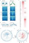
Figure 2.. Distinct higher order chromatin structures within distinct CD8+ T cell populations.
A) MDS plot showing relationship between Hi-C samples derived from double positive (CD4+CD8+) thymocytes and naïve, effector and memory OT-1 CTLs. B) Hi-C data (50Kb bins) normalised using ICED method showing interaction frequency at Sox4, Prickle1 and Satb1 loci in naïve, effector and memory OT-1 CTLs. Track below memory panel shows genes, with purple arrow highlighting genes of interest and their direction of transcription. C) ATAC-Seq data for Naïve, effector and memory OT-1 CTLs. D) Pairwise correlation of binned interaction frequencies (50kb) for naïve and effector (N-E), and naïve and memory (N-M) samples, with dotted line indicating 0 on the y axis. E) Bottom panel shows normalised RNA-Seq counts (Russ et al, 2014) F) Quantification of domain changes identified by pairwise correlation (50Kb) analysis depicted in D.
Figure 3.
Figure 3.. Loss and gain of cis regulatory interactions underscores CTL differentiation state specific gene transcription profiles.
A) Numbers of cis interactions unique to each differentiation state, determined by pairwise comparisons using multiHiCcompare (50Kb resolution, 0.05 FDR). B) GSEA analysis comparing genes connected by loops enriched in one condition over another (Y axis), against RNA-seq data derived from matching samples (Russ et al., 2014). Circle sizes reflect adjusted p values (-log10) and colour represents normalised enrichment score (NES), with red indicating enrichment versus the first RNA-seq condition listed in pairwise comparison, and blue indicating enrichment is the second RNA-seq condition listed. C-E) Examples of loci where loops were lost and gained upon differentiation (blue loops are present in naïve over effector or memory; red loops are gained on differentiation).
Figure 4.
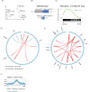
Figure 4.. Hi-C Loops border active regions containing differentiation state specific enhancers.
A) ATAC-seq signal (log2) within and surrounding loops that are present in naïve but not effector CTL, or vice versa. Loops are scaled to occupy 100kb, and ATAC-seq signal is shown for 100kb up and downstream of the loop borders. B) Enrichment of active and poised (H3K4me1+ H3K4me2+) transcriptional enhancers that occur in naive CD8+ T cells but not effector T cells within loops that occur in naive but not effector CD8+ T cells (upper panel) and vice versa (lower panel). C, D) Cis interactions connect gene regulatory elements. Circos plots show the gene neighbourhood of Satb1 (C) and Klrg1 (D) in naïve and effector OT-1 CTLs, respectively. Tracks in order from outside to the centre: are genes, H3K4me1 (blue), H3K4me2 (lite green), H3K4me3 (dark green), ATAC-Seq (pink), Hi-C interactions (naïve over effector CTLs (B) and effector over naïve CTLs (C)) shown as ribbons. E) Enrichment of transcription factor binding at TEs unique to naïve or effector (Russ et al., 2017) was performed using curated transcription factor ChIP-Seq data through the CistromeDB Toolbox (Zheng et al., 2019).
Figure 5.
Figure 5.. BACH2 enforces a naïve chromatin architecture.
(A) MDS plot showing relationship between naïve Bach2−/− and naïve Satb1m1Anu/m1Anu (described in figure 7) Hi-C samples and naïve, effector and memory OT-1 CTLs. (B) Loss and gain of cis interaction in naïve Bach2−/− CD8+ T cells in comparison with WT naïve and virus specific OT-1 CD8+ T cells. C) GSEA analysis comparing genes connected by loops gained in naïve Bach2−/− CD8+ T cells relative to naïve WT CD8+ T cells against RNA-seq data derived from naïve and effector CTLs samples (Russ et al., 2014). P values and normalised enrichment score (NES) are shown. (D, E) Examples of changes in looping architecture in naïve Bach2−/− CD8+ T cells relative to naïve WT CD8+ T cells (blue loops are enriched in WT naïve over naïve Bach2−/− CD8+ T cells and red loops are enriched in naïve Bach2−/− CD8+ T cells over WT). (F) Loops that occur in naïve Bach2−/− CD8+ T cells but not WT naive CD8+ T cells are enriched for active and poised (H3K4me1+ H3K4me2+) transcriptional enhancers that occur in effector CD8+ T cells but not naïve T cells.
Figure 6.
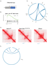
Figure 6.. SATB1 maintains CD8+ T cell naïve chromatin architecture.
(A) Loss and gain of cis interactions in naïve Satb1m1Anu/m1Anu CD8+ T cells in comparison with WT naïve OT-1 T cells. (B) GSEA analysis comparing genes connected by loops gained in naïve Satb1m1Anu/m1Anu CD8+ T cells relative to naïve OT-1 T cells against RNA-seq data derived from matching samples (Nussing et al. under revision). Normalised enrichment score (NES) is shown. (C) Loops lost at the type 1 chemokine locus in naïve Satb1m1Anu/m1Anu CD8+ T cells relative to naïve OT-1 cells. (D) Hi-C contact maps showing the Ccl5 encoding locus in naïve OT-1, Satb1m1Anu/m1Anu naïve, and effector OT-1 CTLs. (E) Loops lost at the Satb1 locus in naïve Bach2−/− CD8+ T cells relative to naïve OT-1 cells.
Figure 7.
Figure 7.. Altered chemokine expression in mice following deletion of cis interacting elements mapped by Hi-C.
(A) Identification of an interactions between the Ccl5 gene promoter and previously identified transcriptional enhancers at −5kb and −20kb from the Ccl5 transcription start site (Russ et al., 2017). Data is presented as a virtual 4C plot, showing naïve and effector Hi-C data, with the arrow indicating a zone of increased interaction in effector CTLs. (B) Chromatin accessibility data (mapped by FAIRE) in effector CTL, showing the positioning of CRISPR deletions made in separate mouse lines to remove the −5 and −20 transcriptional enhancers. (C) Wild-type and enhancer deletion mice were infected intranasally with 104 pfu A/HKx31influenza virus, and lymphocytes were collected from the bronchiolar lavage (BAL) fluid on d10 for analysis by flow cytometry to assay CCL4 and CCL5 expression in CD8+ and CD4+ T cells. (D) Reduced H3K27Ac at the Ccl5 locus in in vitro cultured enhancer deletion effector CTLs. Naïve CTL from WT (blue) and −5 (red) and −20 (green) enhancer deletion mice were stimulated with plate bound αCD3 and αCD28 and cultured for 5 days before ChIP assays were performed to measure histone acetylation at the promoter and enhancers of Ccl5. Data are pooled from 3 independent cultures, and error bars are SEM. Data are expressed relative to a total input.

References

    1. Agarwal S., and Rao A. (1998). Modulation of chromatin structure regulates cytokine gene expression during T cell differentiation. Immunity 9, 765–775. - PubMed
    1. Avni O., Lee D., Macian F., Szabo S.J., Glimcher L.H., and Rao A. (2002). T(H) cell differentiation is accompanied by dynamic changes in histone acetylation of cytokine genes. Nat Immunol 3, 643–651. - PubMed
    1. Barski A., Cuddapah S., Cui K., Roh T.Y., Schones D.E., Wang Z., Wei G., Chepelev I., and Zhao K. (2007). High-resolution profiling of histone methylations in the human genome. Cell 129, 823–837. - PubMed
    1. Battulin N., Fishman V.S., Mazur A.M., Pomaznoy M., Khabarova A.A., Afonnikov D.A., Prokhortchouk E.B., and Serov O.L. (2015). Comparison of the three-dimensional organization of sperm and fibroblast genomes using the Hi-C approach. Genome Biol 16, 77. - PMC - PubMed
    1. Beagan J.A., Duong M.T., Titus K.R., Zhou L., Cao Z., Ma J., Lachanski C.V., Gillis D.R., and Phillips-Cremins J.E. (2017). YY1 and CTCF orchestrate a 3D chromatin looping switch during early neural lineage commitment. Genome Res 27, 1139–1152. - PMC - PubMed

Publication types