. 2023 Feb 27:18:101669.
doi: 10.1016/j.bonr.2023.101669. eCollection 2023 Jun.

Metabolic crosstalk between stromal and malignant cells in the bone marrow niche

Affiliations

Metabolic crosstalk between stromal and malignant cells in the bone marrow niche

Hernán A Tirado et al. Bone Rep. .

Abstract

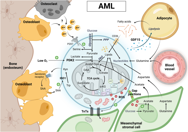
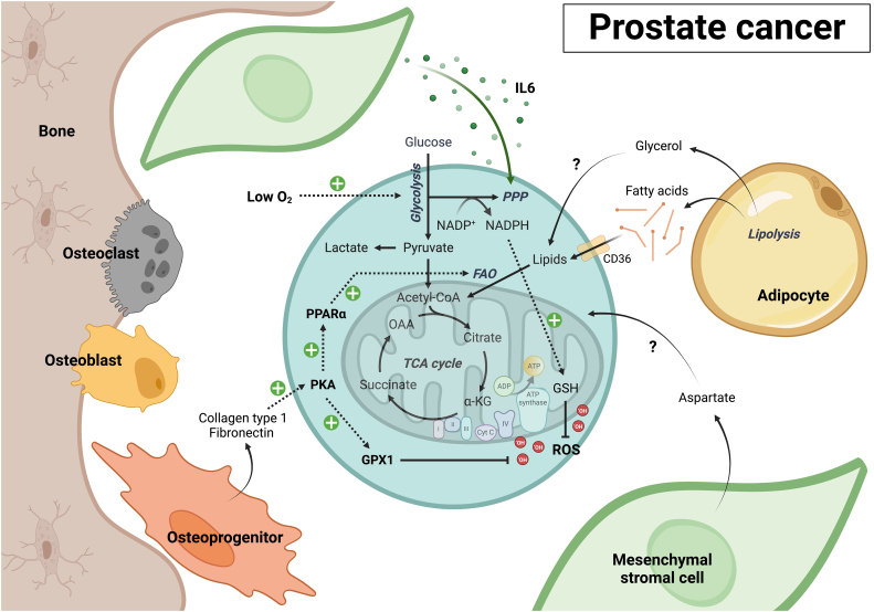
Bone marrow is the primary site of blood cell production in adults and serves as the source of osteoblasts and osteoclasts that maintain bone homeostasis. The medullary microenvironment is also involved in malignancy, providing a fertile soil for the growth of blood cancers or solid tumors metastasizing to bone. The cellular composition of the bone marrow is highly complex, consisting of hematopoietic stem and progenitor cells, maturing blood cells, skeletal stem cells, osteoblasts, mesenchymal stromal cells, adipocytes, endothelial cells, lymphatic endothelial cells, perivascular cells, and nerve cells. Intercellular communication at different levels is essential to ensure proper skeletal and hematopoietic tissue function, but it is altered when malignant cells colonize the bone marrow niche. While communication often involves soluble factors such as cytokines, chemokines, and growth factors, as well as their respective cell-surface receptors, cells can also communicate by exchanging metabolic information. In this review, we discuss the importance of metabolic crosstalk between different cells in the bone marrow microenvironment, particularly concerning the malignant setting.

Keywords: Bone marrow; Bone metastasis; Cell metabolism; Cellular communication; Leukemia; Stromal cells.


Conflict of interest statement

The authors declare that they have no known competing financial interests or personal relationships that could have appeared to influence the work reported in this paper.

Figures

Fig. 1
Fig. 1
The cellular composition of the bone marrow niche. Several stromal cell types are involved in the maintenance of HSCs and the support of hematopoiesis in the bone marrow. In the central bone marrow niche, LepR+ adipo-CAR cells and sinusoidal endothelial cells provide CXCL12 and SCF to support HSC retention and function, while megakaryocytes secrete CXCL4 and TGF-β1, preserving HSC quiescence. Myelopoiesis also takes place in the central bone marrow, in niches distinct from those where HSCs are found. NG2+ periarteriolar cells and non-myelinating Schwann cells secrete respectively CXCL12 and TGF-β, involved in maintaining quiescent HSCs near arterioles in the endosteal and central bone marrow regions. In the endosteal niche, Osteolectin+LepR+ osteo-CAR cells provide CXCL12 and SCF to lymphoid-biased HSCs and early lymphoid progenitors. Osteoprogenitors further support B cell maturation, while mature osteoblasts maintain early T cell progenitors. The role of mature adipocytes in the bone marrow is complex and incompletely understood, but they are thought to support HSCs by providing fatty acids.
Fig. 2
Fig. 2
Metabolic crosstalk between AML cells and bone marrow stromal cells. AML LSCs with a more glycolytic profile home to the hypoxic endosteal bone marrow niche, where high environmental ATP –from a yet unknown cellular source– additionally promotes leukemic serine biosynthesis. Metabolic crosstalk in the endosteal niche also includes a vicious cycle of AML-derived kynurenine and osteoblast-derived acute-phase protein serum amyloid A (SAA) production, leading to niche remodeling in favor of cancer progression. AML LSCs and blasts with a more oxidative metabolism may preferentially localize close to and interact with adipocytes or MSCs. Release of GDF15 by AML cells triggers adipocyte lipolysis, providing fatty acids for oxidation in the AML cells. Transfer of ROS from AML cells to MSCs stimulates acetate production, which is also taken up by AML cells to fuel their TCA cycle and support lipid synthesis. AML mitochondria damaged by excessive ROS can be transferred to MSCs via TnTs, and the cancer cells receive healthy mitochondria via a similar mechanism in return. MSCs also provide glutamine-derived aspartate to AML cells, allowing the latter to synthetize nucleotides necessary for proliferation and DNA damage repair.
Fig. 3
Fig. 3
Metabolic interactions in the ALL bone marrow niche. The ALL bone marrow is characterized by an accumulation of MSCs at the expense of osteoblasts. MSCs convert glucose to lactate, which fuels the mitochondrial metabolism of ALL cells in addition to fatty acids provided by adipocytes. High ROS levels promote the exchange of damaged versus healthy mitochondria between ALL cells and MSCs via TnTs. MSCs also help ALL cells to maintain their redox balance by converting cystine to cysteine, which after transfer to the leukemic cells supports reduced glutathione (GSH) synthesis. MSCs further protect ALL cells from amino acid-depleting therapies such as L-Asparaginase or PEG-Arginase by respectively providing asparagine or arginine on which the ALL cells depend for their survival and growth.
Fig. 4
Fig. 4
Short- and long-distance metabolite exchanges in MM. Avid glutamine consumption by MM cells leads to osteoblast depletion and bone destruction. Secretion of 2-deoxy-d-ribose (2DDR) by MM cells further contributes to this phenotype. Low microenvironmental glutamine levels trigger glutamine synthesis in MSCs, but this process does not appear to be sufficient in osteoblasts, which need large amounts of glutamine to differentiate and function. Urea, released in large amounts due to the hyperactive nitrogen metabolism of MM cells, stimulates the growth of nitrogen-recycling bacteria in the gut that through glutamine synthesis promote MM progression. Besides glutamine catabolism, MM cells exhibit high rates of glycolysis, a phenotype that is reinforced by bone marrow hypoxia. Low oxygen levels in the bone marrow also promote production of the immune-suppressive metabolite adenosine by the cancer cells. MM cells additionally take up lactate produced by MSCs and induce fatty acid release by adipocytes, although it is still unclear whether these metabolites fuel oxidative phosphorylation or have a different metabolic fate in the MM cells. As observed for other blood cancer cells, MM cells exhibit mitochondrial exchange with MSCs, trading their damaged for functional mitochondria.
Fig. 5
Fig. 5
Subtype-dependent metabolic crosstalk in bone-metastatic breast cancer. Most metastatic breast cancer cells in the bone microenvironment exhibit an active mitochondrial metabolism fueled by glucose, glutamine, MSC-derived lactate, and adipocyte-derived fatty acids. Mitochondria transferred from endothelial cells and MSCs further boost the oxidative capacity of the cancer cells. Glutamine metabolism in metastatic breast cancer cells also supports the biosynthesis of serine, a metabolite that is not only essential for cancer cell survival in the metastatic niche, but that additionally promotes osteoclastogenesis and bone destruction. TNBC cells induce bone loss through a different mechanism. These cells exhibit high rates of glycolysis and fermentation, releasing high amounts of lactate that promotes osteoclast activity and bone resorption. In turn, secretion of PUFAs by osteoclasts promotes proliferation, migration, and survival of breast cancer cells. While the osteoblast-osteoclast balance is disturbed in the bone marrow invaded by breast cancer cells, osteoblasts are still present and secrete several metabolites that favor tumor growth, including LPA, PGE2 and deoxycholate. Osteocytes can have pro- or anti-tumorigenic effects on breast cancer cells depending on their relative secretion of ATP and adenosine. This ratio can change in response to oxidative stress, and ectonucleotidase expression by the cancer cells can further shift the ratio between these two metabolites in favor of tumor cell growth and migration.
Fig. 6
Fig. 6
Metabolic communication between stromal and prostate cancer cells in bone. Current insights in the metabolic communication occurring in bone-metastatic prostate cancer are very limited in comparison to other cancers growing in the bone marrow. Co-culture with MSCs stimulates glycolysis, TCA cycle activity and redox metabolism in prostate cancer cells. Unique to prostate cancer cells growing in the bone marrow microenvironment is their high rate of PPP activity, a pathway that is stimulated by IL6 released by MSCs. While relatively limited, metastatic prostate cancer cells do exhibit mitochondrial oxidative metabolism, which may be fueled by fatty acids released by bone marrow adipocytes. Adipocytes also release glycerol, although its uptake by and metabolic fate in bone-metastatic prostate cancer cells remains unknown. While no metabolic interactions with mature osteoblasts have been described yet, osteoprogenitors release fibronectin and collagen type 1, which activate protein kinase A (PKA) signaling in prostate cancer cells. PKA increases expression of several metabolic genes in the cancer cells, including some involved in fatty acid metabolism and ROS detoxification. Aspartate, an amino acid highly enriched in the bone marrow, may also fuel mitochondrial metabolism of metastatic prostate cancer cells like it does in primary tumors, although this remains to be investigated.

References

    1. Acar M., et al. Deep imaging of bone marrow shows non-dividing stem cells are mainly perisinusoidal. Nature. 2015;526(7571):126–130. - PMC - PubMed
    1. Agarwal P., et al. Mesenchymal niche-specific expression of Cxcl12 controls quiescence of treatment-resistant leukemia stem cells. Cell Stem Cell. 2019;24(5):769–784 e6. - PMC - PubMed
    1. Allaman I., et al. Astrocyte-neuron metabolic relationships: for better and for worse. Trends Neurosci. 2011;34(2):76–87. - PubMed
    1. Ambrosi T.H., Chan C.K.F. Skeletal stem cells as the developmental origin of cellular niches for hematopoietic stem and progenitor cells. Curr. Top. Microbiol. Immunol. 2021;434:1–31. - PMC - PubMed
    1. Arber D.A., et al. International consensus classification of myeloid neoplasms and acute leukemias: integrating morphologic, clinical, and genomic data. Blood. 2022;140(11):1200–1228. - PMC - PubMed

LinkOut - more resources