. 2023 Feb 13;2(3):pgad043.
doi: 10.1093/pnasnexus/pgad043. eCollection 2023 Mar.

The first genetic landscape of inherited retinal dystrophies in Portuguese patients identifies recurrent homozygous mutations as a frequent cause of pathogenesis

Affiliations

The first genetic landscape of inherited retinal dystrophies in Portuguese patients identifies recurrent homozygous mutations as a frequent cause of pathogenesis

Virginie G Peter et al. PNAS Nexus. .

Abstract

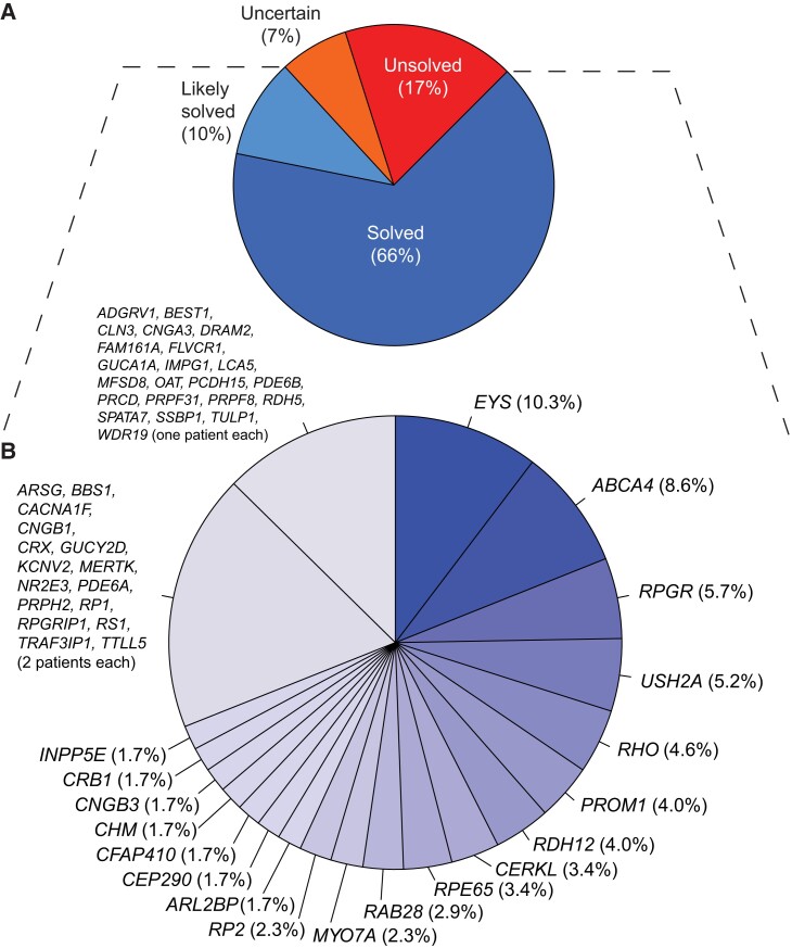
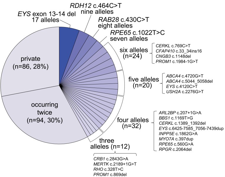
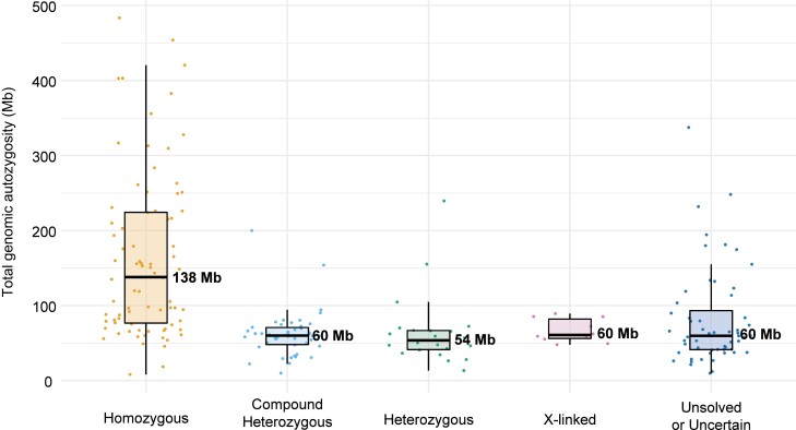
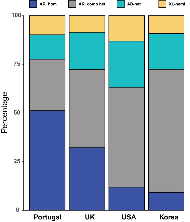
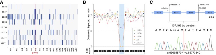
Inherited retinal diseases (IRDs) are a group of ocular conditions characterized by an elevated genetic and clinical heterogeneity. They are transmitted almost invariantly as monogenic traits. However, with more than 280 disease genes identified so far, association of clinical phenotypes with genotypes can be very challenging, and molecular diagnosis is essential for genetic counseling and correct management of the disease. In addition, the prevalence and the assortment of IRD mutations are often population-specific. In this work, we examined 230 families from Portugal, with individuals suffering from a variety of IRD diagnostic classes (270 subjects in total). Overall, we identified 157 unique mutations (34 previously unreported) in 57 distinct genes, with a diagnostic rate of 76%. The IRD mutational landscape was, to some extent, different from those reported in other European populations, including Spanish cohorts. For instance, the EYS gene appeared to be the most frequently mutated, with a prevalence of 10% among all IRD cases. This was, in part, due to the presence of a recurrent and seemingly founder mutation involving the deletion of exons 13 and 14 of this gene. Moreover, our analysis highlighted that as many as 51% of our cases had mutations in a homozygous state. To our knowledge, this is the first study assessing a cross-sectional genotype-phenotype landscape of IRDs in Portugal. Our data reveal a rather unique distribution of mutations, possibly shaped by a small number of rare ancestral events that have now become prevalent alleles in patients.

Keywords: EYS; Portugal; homozygosity; inherited retinal diseases.


Figures

Fig. 1.
Fig. 1.
Genetic classification of the cases analyzed. A) Classification of patients by their diagnostic status at the molecular level. Percentages are computed over the total number of patients in the study. B) Further stratification of patients from the “likely solved” and “solved” classes, by disease gene harboring causative mutations, represented by individual slices of the chart. Genes that were mutated in one or two patients were grouped. Percentages are computed over the total number of patients from the “likely solved” and “solved” classes.
Fig. 2.
Fig. 2.
Prevalence of the mutations detected. Individual slices of the chart refer to specific variants, with the exception of mutations occurring only once (private) or twice, which were grouped. n refers to the total number of alleles identified in a given group. Percentages are computed over the total number of causative alleles detected in patients from the solved and likely solved classes, regardless of the inheritance mode of the disease. The correct HGVS nomenclature for “EYS exon 13-14 del” is EYS c.2024-5718_2260-10064del and for “CFAP410 c.33_34ins16” is CFAP410 c.33_34insAGCTGCACAGCGTGCA; a simplified notation was used here because of space constraints.
Fig. 3.
Fig. 3.
Boxplots of total genomic autozygosity, as a function of the genotypes identified. Levels of autozygosity are expressed as the sum of all homozygous regions detected in each genome, for patients analyzed by NGS procedures. Values relative to individual patients are represented by dots, while median values are indicated by horizontal thick bars, with numbers (Mb: megabases). Standard notation for boxplots applies to other components of the graph. Sixteen index subjects were not analyzed, due to lack of appropriate quality data.
Fig. 4.
Fig. 4.
Relative number of patients, by genotype and mode of inheritance of the disease, in our cohort vs. similar studies in other populations. Data from this work (Portugal) are compared with those from three other large studies from the United Kingdom (12), the United States (7), and Korea (10). Our genotypes are enriched in homozygous and reduced in compound heterozygous recessive mutations. AR-hom, autosomal recessive inheritance, mutation in homozygosis; AR-comp het, autosomal recessive inheritance, mutations in compound heterozygosity; AD-het, autosomal dominant inheritance, mutation in heterozygosis; XL-hemi, X-linked inheritance, mutation in hemizygosis (in males).
Fig. 5.
Fig. 5.
Effects of the DRAM2 intronic variant c.517+5C>A on pre-mRNA splicing. A) Co-segregation analysis, within LL20's family, of genotypes and phenotypes. B) Schematic representation of exons 7 and 8 of DRAM2, including the position of the variant and the main splicing events detected. The canonical mRNA transcript is indicated by “wt”. Isoforms 1 and 2 result from the preferential activation of alternative donor sites, located 16 and 129 nucleotides upstream of the canonical one, respectively. Isoform 1 contains a premature stop triplet at codon 169, whereas isoform 2 bears an in-frame deletion of 43 codons. C) Electrophoresis of RT-PCR products from leukocytes of a control individual (ctrl) and of patients LL20 and LL253. The amplification product of the canonical transcript (wt) is 250 bp in length, while isoform 1 and isoform 2 have sizes of 234 and 121 bp, respectively. water: negative control of amplification. D) Electropherograms of the PCR products shown in (C). The control sample contains a mixture of the canonical mRNA and isoform 1, indicating that the latter transcript is produced, although in minimal quantities, even in the absence of the mutation.
Fig. 6.
Fig. 6.
Features of the prevalent deletion of exons 13 and 14 in EYS. A) Autozygosity plots of eight patients, showing the common homozygous haplotype on chromosome 6 that eventually led to the identification of the most common mutation from our cohort. Homozygous regions are indicated by solid blue bars, while the red vertical, dotted line shows the minimal critical region spanning EYS. B) Coverage plot of four selected homozygotes for this mutation, as well as another patient (LL323, affected sister of index patient LL175), who carries this deletion in compound heterozygosity with another mutation in EYS. The area shaded in blue indicates the approximate location of the deletion, as inferred from the reduction of sequence coverage. C) Schematic representation detailing the deletion and the resulting novel junction. Genomic coordinates are given with respect to build GRCh37-hg19 of the human genome sequence. PCR primers allowing the detection of this junction are also indicated.

References

    1. Ayuso C, Millan JM. 2010. Retinitis pigmentosa and allied conditions today: a paradigm of translational research. Genome Med. 2:34. - PMC - PubMed
    1. Sahel JA, Marazova K, Audo I. 2014. Clinical characteristics and current therapies for inherited retinal degenerations. Cold Spring Harb Perspect Med. 5:a017111. - PMC - PubMed
    1. Retinal Information Network (RetNet). https://web.sph.uth.edu/RetNet/.
    1. Duncan JL, et al. . 2018. Inherited retinal degenerations: current landscape and knowledge gaps. Transl Vis Sci Technol. 7:6. - PMC - PubMed
    1. Ku CA, Pennesi ME. 2020. The new landscape of retinal gene therapy. Am J Med Genet C Semin Med Genet. 184:846–859. - PubMed