Review
. 2023 Feb 28;15(2):759-779.
doi: 10.21037/jtd-22-861. Epub 2023 Jan 31.

Esophageal diverticula: from diagnosis to therapeutic management-narrative review

Affiliations
Review

Esophageal diverticula: from diagnosis to therapeutic management-narrative review

Adrian Constantin et al. J Thorac Dis. .

Abstract

Background and objective: Esophageal diverticulum (ED) is a relatively rare condition, characterized by high etio- and pathophysiological versatility, with an uncommon clinical impact, consequently requiring a complete and complex diagnostic evaluation, so that the therapeutic decision is "appropriate" to a specific case. The aim of the paper is, therefore, a reassessment of the diagnostic possibilities underlying the establishment of the therapeutic protocol and the available therapeutic resources, making a review of the literature, and a non-statistical retrospective analysis of cases hospitalized and operated in a tertiary center.

Methods: Thus, classical investigations (upper digestive endoscopy, barium swallow) need to be correlated with complex, manometric, and imaging evaluations with direct implications in therapeutic management. Moreover, in the absence of a precise etiology, the operative indication needs to be established sparingly, with the imposition of the identification and interception of the pathophysiological mechanisms through the therapeutic gesture.

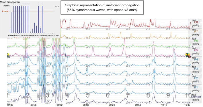
Key content and findings: The identification of the pathophysiological mechanisms is mandatory for the management of diverticular disease, the result obtained-restoring swallowing and comfort/good quality of life in the postoperative period-is directly related to the chosen therapeutic procedure. In addition, management appears to be a difficult goal in the context of the low incidence of ED but also of the results that emphasize important differences in the reports in the medical literature. Although ED is a benign condition, surgical techniques are demanding, impacted by significant morbidity and mortality. The causes of these results are multiple: possible localizations anywhere in the esophagus, diverticulum size/volume from a few millimeters to an impressive one, over 10-12 cm, metabolic impact in direct relation to the alteration swallowing, numerous diverticular complications but, perhaps most importantly, alteration of the quality of the diverticular wall by inflammatory phenomena, with an impact on the quality of the suture.

Conclusions: The accumulation of cases in a tertiary profile center, with volume/hospital, respectively volume/surgeon + gastroenterologist could be a solution in improving the results. One consequence would be the identification of alternative solutions to open surgical techniques, a series of minimally invasive or endoscopic variants can refine these results.

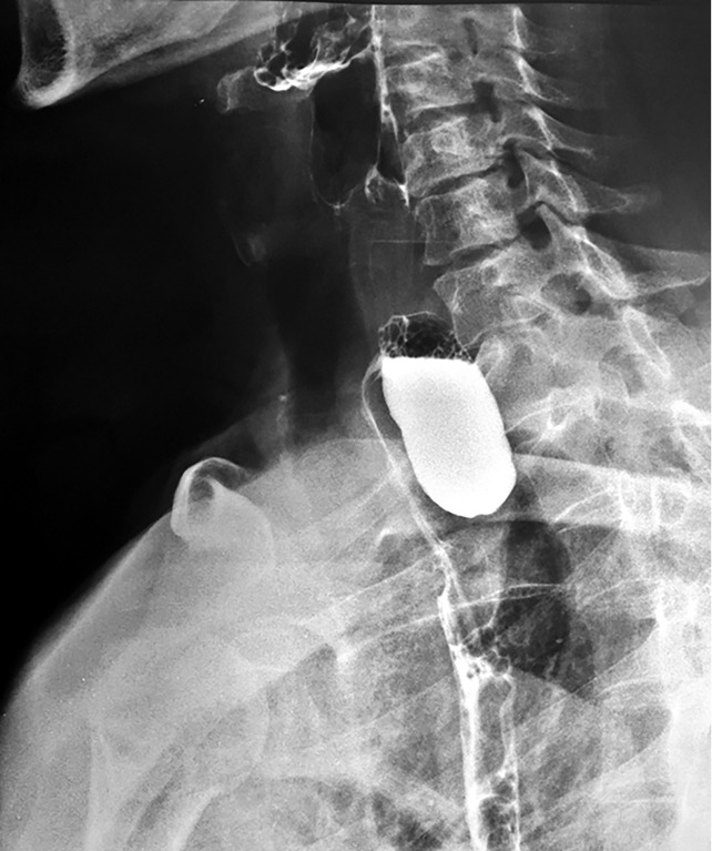
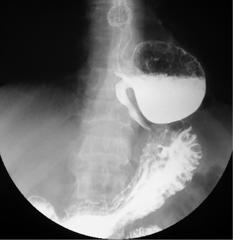
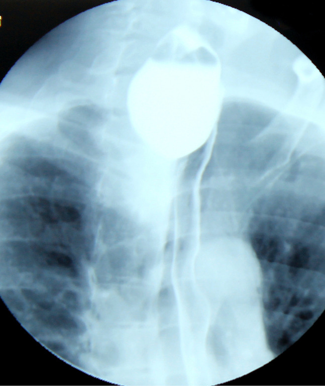
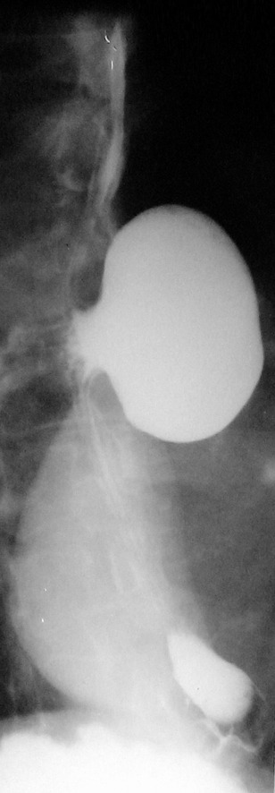
Keywords: Esophageal diverticula (Zenker, Rokitansky, Killian-Jamieson); diagnostic; management approaches.


Conflict of interest statement

Conflicts of Interest: All authors have completed the ICMJE uniform disclosure form (available at https://jtd.amegroups.com/article/view/10.21037/jtd-22-861/coif). The authors have no conflicts of interest to declare.

Figures

Figure 1
Figure 1
Barium swallow. Voluminous Zenker diverticulum (10 cm length, 4 cm width) with significant retention, plunging through the upper opening, with a mass effect on the esophagus that appears displaced laterally, with secondary dysphagia.
Figure 2
Figure 2
Barium swallow. Retention of the barium tracer in a giant midthoracic diverticulum approximately 15 cm in diameter, with lateral displacement of the esophagus.
Figure 3
Figure 3
Barium swallow. The barium tracer fixed at the level of a giant midthoracic 15 cm in diameter diverticulum (A), with the association of an esophageal motility disorder [secondary (B) and tertiary (C) contraction waves are highlighted].
Figure 4
Figure 4
Contrast radiology that highlights a retentive, voluminous epiphrenic diverticulum (8–10 cm in diameter). The diverticulum is located above the diaphragm, with pulmonary and right atrium compression.
Figure 5
Figure 5
Voluminous Zenker diverticulum, plunging through the upper thoracic opening in the thorax, with retentive character and with a mass effect on the esophagus (barium swallow).
Figure 6
Figure 6
Midthoracic diverticulum-large communication with the esophageal lumen (barium swallow), with good passage of contrast substance, slightly retentive.
Figure 7
Figure 7
Association between a midthoracic diverticulum and esophageal motility disorder-segmental hyperperistalsis with irregular and non-propulsive contractions (barium swallow).
Figure 8
Figure 8
Pathological association of an epiphrenic ED with a large hiatal rolling hernia, approx. 1/2 of the stomach fixed in the chest and with a degree of organo-axial gastric volvulus. ED, esophageal diverticula.
Figure 9
Figure 9
Endoscopic examination-the presence of an important mediothoracic esophageal diverticulum (“double barrel shotgun” aspect).
Figure 10
Figure 10
Endoscopic examination-retentive mid-thoracic diverticulum.
Figure 11
Figure 11
CT image of the cervical sagittal plane. Large Zenker diverticulum, located above the thoracic opening, with a thickened wall (blue arrows), with the retention of the contrast substance administered orally. CT, computed tomography.
Figure 12
Figure 12
CT scan cross section. Significant thickening of the diverticular wall, with the appearance of an extraesophageal infiltration (blue arrows), with the suspicion of a neoplastic degeneration (confirmed malignancy by endoscopic biopsy). CT, computed tomography.
Figure 13
Figure 13
3D tomographic reconstruction of the thoracic esophagus. A midthoracic diverticulum (blue arrows) and its direct relationship with aortic artery.
Figure 14
Figure 14
Conventional esophageal manometry-motility disorder type diffuse esophageal spasm (>30% synchronous waves, waves over 180 mmHg).

References

    1. Hussain T, Maurer JT, Lang S, et al. Pathophysiology, diagnosis and treatment of Zenker’s diverticulum. HNO 2017;65:167-76. 10.1007/s00106-016-0302-z - DOI - PubMed
    1. Wang ZM, Zhang SC, Teng X. Esophageal diverticulum serves as a unique cause of bronchoesophageal fistula in children: A case report. Medicine (Baltimore) 2017;96:e9492. 10.1097/MD.0000000000009492 - DOI - PMC - PubMed
    1. Sonbare DJ. Pulsion Diverticulum of the Oesophagus: More than just an Out Pouch. Indian J Surg 2015;77:44-8. 10.1007/s12262-013-0955-8 - DOI - PMC - PubMed
    1. Little RE, Bock JM. Pharyngoesophageal diverticuli: diagnosis, incidence and management. Curr Opin Otolaryngol Head Neck Surg 2016;24:500-4. 10.1097/MOO.0000000000000309 - DOI - PubMed
    1. Thomas ML, Anthony AA, Fosh BG, et al. Oesophageal diverticula. Br J Surg 2001;88:629-42. 10.1046/j.1365-2168.2001.01733.x - DOI - PubMed