. 2023 Feb 18:31:674-688.
doi: 10.1016/j.omtn.2023.02.020. eCollection 2023 Mar 14.

Antisense oligonucleotide therapy corrects splicing in the common Stargardt disease type 1-causing variant ABCA4 c.5461-10T>C

Affiliations

Antisense oligonucleotide therapy corrects splicing in the common Stargardt disease type 1-causing variant ABCA4 c.5461-10T>C

Melita Kaltak et al. Mol Ther Nucleic Acids. .

Abstract

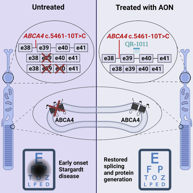
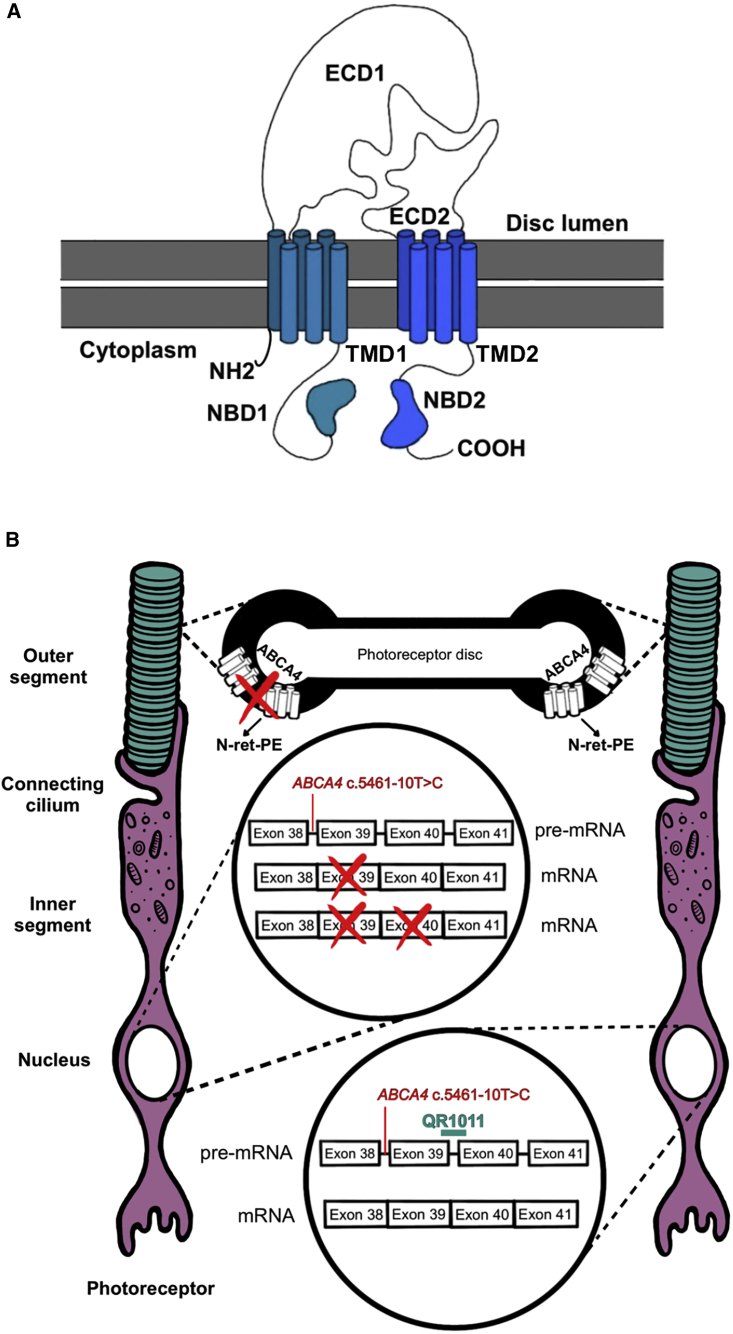
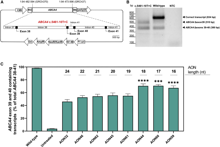
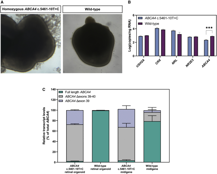
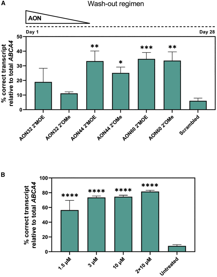
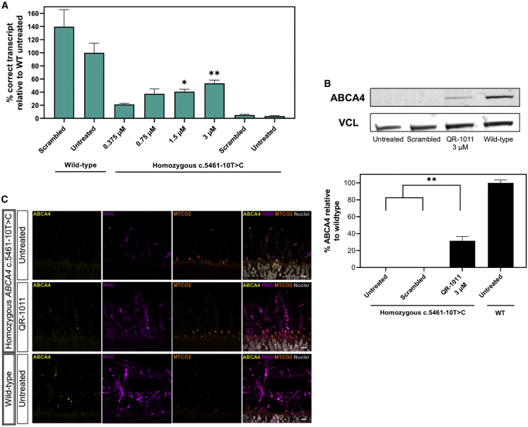
Stargardt disease type 1 (STGD1) is the most common hereditary form of maculopathy and remains untreatable. STGD1 is caused by biallelic variants in the ABCA4 gene, which encodes the ATP-binding cassette (type 4) protein (ABCA4) that clears toxic byproducts of the visual cycle. The c.5461-10T>C p.[Thr1821Aspfs∗6,Thr1821Valfs∗13] variant is the most common severe disease-associated variant, and leads to exon skipping and out-of-frame ABCA4 transcripts that prevent translation of functional ABCA4 protein. Homozygous individuals typically display early onset STGD1 and are legally blind by early adulthood. Here, we applied antisense oligonucleotides (AONs) to promote exon inclusion and restore wild-type RNA splicing of ABCA4 c.5461-10T>C. The effect of AONs was first investigated in vitro using an ABCA4 midigene model. Subsequently, the best performing AONs were administered to homozygous c.5461-10T>C 3D human retinal organoids. Isoform-specific digital polymerase chain reaction revealed a significant increase in correctly spliced transcripts after treatment with the lead AON, QR-1011, up to 53% correct transcripts at a 3 μM dose. Furthermore, western blot and immunohistochemistry analyses identified restoration of ABCA4 protein after treatment. Collectively, we identified QR-1011 as a potent splice-correcting AON and a possible therapeutic intervention for patients harboring the severe ABCA4 c.5461-10T>C variant.

Keywords: ABCA4; MT: Oligonucleotides: Therapies and Applications; RNA therapy; Stargardt disease; antisense oligonucleotides; retinal organoids; splicing correction.


Conflict of interest statement

M.K., P.d.B., K.D., T.H., W.B., G.P., and J.S. were employed by ProQR Therapeutics during this project. K.D. is inventor on the international patent application (WO2018189376) that has been filed by ProQR Therapeutics describing methods and means for AON therapy for Stargardt disease type 1. The rest of the co-authors declare that they have no competing interests.

Figures

None
Graphical abstract
Figure 1
Figure 1
Structure of ABCA4 and the splicing modulating effect of QR-1011 (A) The complex structure of the ABCA4 protein involves two transmembrane domains (TMD1 and TMD2), each with six transmembrane helices. In addition, the protein structure displays two glycosylated extra cytoplasmatic domains (ECD1 and ECD2) and two nucleotide-binding domains (NBD1 and NBD2) where ATP hydrolysis takes place. (B) In presence of the frameshift variant ABCA4 c.5461-10T>C, generated transcripts lack either the exon 39 or exons 39 and 40; this splicing defect hampers the production of functional ABCA4 protein and toxic retinoid products (N-retinylidene-phosphatidylethanolamine [PE]) cannot be removed from the photoreceptor’s outer segments, leading to the accumulation of A2E and lipofuscin granules, key pathogenic features for STGD1. The splicing modulating activity of QR-1011 is designed to restore wild-type splicing and include exons 39 and 40 in ABCA4.
Figure 2
Figure 2
Splice-predictive midigene detected AON-induced ABCA4 exon inclusion (A) The midigene incorporates the ABCA4 genomic region between intron 37 and 41, together with the ABCA4 c.5461-10T>C variant. The construct is flanked by RHO 3 and RHO 5 exons that contain strong splicing donor and acceptor sites, while the expression is initiated by the CMV promotor. (B) Expression of the ABCA4 c.5461-10T>C midigene in HEK293 cells showed two truncated ABCA4 isoforms: the ABCA4 Δexon39 and ABCA4 Δexons 39–40, but no full length ABCA4. The wild-type midigene displayed mostly expression of the correct transcript with a low level of the single and double skip isoforms. (C) The final AON screening with lead candidate AONs applied on midigene transfected cells demonstrated that all versions of AON32 resulted in splice correction. Data are shown as mean ± standard error of the mean, n = 6, ∗∗∗p ≤ 0.001, ∗∗∗∗p ≤ 0.0001.
Figure 3
Figure 3
Morphological and transcript comparison of wild-type and gene-edited ROs (A) Morphology assessed at day 120 of organoid differentiation suggests ROs derived from homozygous c.5461-10T>C and the control isogenic parent cell line displayed neural retina (thin light rim at the margin of the ROs) and the brush border with photoreceptor cells. (B) Analyses of the total transcript expression of CRX, NRL, NR2E3, USH2A, and ABCA4 (see Table S3). CRX and the photoreceptor markers were expressed similarly in both groups. ABCA4 was expressed at higher levels in wild-type organoids. Data are shown as mean ± standard error of the mean, n = 4. (C) Comparison of ABCA4 transcript expression in c.5461-10T>C and wild-type ROs and midigenes showed that the relative levels of the three transcripts are comparable between RO and midigene for both WT and the c.5461-10T>C variant. Data are shown as mean ± standard error of the mean, ∗∗∗p ≤ 0.0001.
Figure 4
Figure 4
Gene-edited and patient-derived homozygous c.5461-10T>C ROs show high levels of rescued ABCA4 transcript upon AON treatment (A) Transcript analysis of gene edited ROs treated with a 1.5 μM 2′MOE-modified and 2′OMe-modified AONs for 28 days. (Top) The 4-week-long treatment used a wash-out regimen in which the AON was added only at day 0 of treatment and its concentration was halved by each medium change every other day. (B) The percentage of splicing correction in patient-derived ROs treated with QR-1011. For the 2 × 10 μM group, QR-1011 was administered at treatment day 0 and 14. A dose-dependent increase in splice correction was observed from 21.2% to 53.4%. Data are shown as mean ± standard error of the mean, n = 6 per condition. Asterisks display the significant differences vs. scrambled oligo or untreated (∗p ≤ 0.05, ∗∗p ≤ 0.01, ∗∗∗p ≤ 0.001, ∗∗∗∗p ≤ 0.0001).
Figure 5
Figure 5
Low concentrations of QR-1011 can restore RNA splicing and rescue of the wild-type protein in patient-derived c.5461-10T>C ROs (A) Splice adjustment efficacy with clinically relevant dosages of QR-1011 in ROs deriving from a biallelic c.5461-10T>C patient cell line. After 56 days of treatment, all patient-derived RO samples showed splice restoring activity of QR-1011, while the scrambled AON showed no effect on ABCA4 splicing. Data are shown as mean ± standard error of the mean, n = 6. (B) Western blot analysis (n = 3) of treated patient-derived ROs and wild-type ROs revealed restoration of ABCA4 protein after QR-1011 treatment. Untreated c.5461-10T>C ROs and those subjected to treatment with scrambled AON contained no detectable protein. All samples were normalized to the average signal obtained from wild-type ROs. Vinculin (VCL) was used as a loading control. Data are shown as mean ± standard error of the mean, n = 3. ∗p ≤ 0.05, ∗∗p ≤ 0.01. (C) ABCA4 protein immunoreactivity (yellow) in treated patient-derived organoids colocalized within the outer segments (OS, rhodopsin, magenta) of photoreceptor cells and resembled the localization found in wild-type ROs. The inner segments (IS) were visualized with the mitochondrial-targeting antibody MTCO2 (orange) and DAPI nuclear staining is shown in gray. Scale, 10 μm.

References

    1. Allikmets R., Singh N., Sun H., Shroyer N.F., Hutchinson A., Chidambaram A., Gerrard B., Baird L., Stauffer D., Peiffer A., et al. A photoreceptor cell-specific ATP-binding transporter gene (ABCR) is mutated in recessive Stargardt macular dystrophy. Nat. Nat. Genet. 1997;15:236–246. doi: 10.1038/ng0397-236. - DOI - PubMed
    1. Mata N.L., Weng J., Travis G.H. Biosynthesis of a major lipofuscin fluorophore in mice and humans with ABCR-mediated retinal and macular degeneration. Proc. Natl. Acad. Sci. USA. 2000;97:7154–7159. doi: 10.1073/pnas.130110497. - DOI - PMC - PubMed
    1. Quazi F., Lenevich S., Molday R.S. ABCA4 is an N-retinylidene-phosphatidylethanolamine and phosphatidylethanolamine importer. Nat. Commun. 2012;3:925. doi: 10.1038/ncomms1927. - DOI - PMC - PubMed
    1. Weng J., Mata N.L., Azarian S.M., Tzekov R.T., Birch D.G., Travis G.H. Insights into the function of Rim protein in photoreceptors and etiology of Stargardt's disease from the phenotype in abcr knockout mice. Cell. 1999;98:13–23. doi: 10.1016/S0092-8674(00)80602-9. - DOI - PubMed
    1. Cremers F.P., van de Pol D.J., van Driel M., den Hollander A.I., van Haren F.J., Knoers N.V., Tijmes N., Bergen A.A., Rohrschneider K., Blankenagel A., et al. Autosomal recessive retinitis pigmentosa and cone-rod dystrophy caused by splice site mutations in the Stargardt's disease gene ABCR. Hum. Mol. Genet. 1998;7:355–362. doi: 10.1093/hmg/7.3.355. - DOI - PubMed

LinkOut - more resources