

The association of left atrial mechanics with left ventricular morphology in patients with hypertrophic cardiomyopathy: a cardiac magnetic resonance study
- PMID: 36910882
- PMCID: PMC9995245
- DOI: 10.5114/pjr.2023.125213
The association of left atrial mechanics with left ventricular morphology in patients with hypertrophic cardiomyopathy: a cardiac magnetic resonance study
Abstract
Purpose: Hypertrophic cardiomyopathy (HCM) is related with structural and pathologic changes in the left atrium (LA) and left ventricle (LV). The aim of this study was to explore the association between LA mechanics and LV characteristics in patients with HCM using cardiac magnetic resonance feature tracking (CMR-FT).
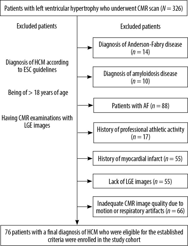
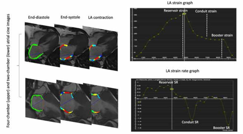
Material and methods: A total of 76 patients with HCM and 26 healthy controls were included in the study. The parameters including the extent of LV late gadolinium enhancement (LGE-%) and the LV early diastolic longitudinal strain rate (edLSR) were assessed for LV. LA conduit, booster, and reservoir functions were assessed by LA fractional volumes and strain analyses using CMR-FT. HCM patients were classified as HCM patients without LGE, with mild LGE-% (0% < LGE-% ł 10%), and prominent LGE-% (10% < LGE-%).
Results: HCM patients had worse LA functions compared with the controls (p < 0.05). The majority of LA functional indices were more impaired in HCM patients with regard to LGE. LA volumes were higher in HCM patients with prominent LGE-% compared with HCM patients with mild LGE-% (p < 0.05). However, only a minority of LA functional parameters differed between the 2 groups. LA strain parameters showed weak to modest correlations with LV LGE-% and LV edLSR.
Conclusions: LV characteristics, to some extent, influence LA mechanics, but they might not be the only factor inducing LA dysfunction in patients with HCM.
Keywords: cardiac magnetic resonance; hypertrophic cardiomyopathy; late gadolinium enhancement; left atrium.
© Pol J Radiol 2023.
Conflict of interest statement
The authors report no conflict of interest.
Figures


References
-
- Maron BJ. Hypertrophic cardiomyopathy: a systematic review. JAMA 2002; 287: 1308-1320. - PubMed
-
- Authors/Task Force members, Elliott PM, et al. . 2014 ESC Guidelines on diagnosis and management of hy-pertrophic cardiomyopathy: the Task Force for the Diagnosis and Management of Hypertrophic Cardio-myopathy of the European Society of Cardiology (ESC). Eur Heart J 2014; 35: 2733-2779. - PubMed
-
- Geske JB, Sorajja P, Nishimura RA, et al. . Evaluation of left ventricular filling pressures by Doppler echo-cardiography in patients with hypertrophic cardiomyopathy: correlation with direct left atrial pressure measurement at cardiac catheterization. Circulation 2007; 116: 2702-2708. - PubMed
-
- Tülüce K, Yakar Tülüce S, Yavuzgil O, et al. . The left atrial phasic functions and the relationship with plas-ma N-terminal pro-B-type natriuretic peptide levels and symptomatic states in patients with hypertrophic cardiomyopathy. Anadolu Kardiyol Derg 2014; 14: 719-727. - PubMed
-
- Yang H, Woo A, Monakier D,et al. . Enlarged left atrial volume in hyper-trophic cardiomyopathy: a marker for disease severity. J Am Soc Echocardiogr 2005; 18: 1074-1082. - PubMed
LinkOut - more resources
Full Text Sources