

Current diagnostic and therapeutic strategies for the management of lymphatic insufficiency in patients with hypoplastic left heart syndrome
- PMID: 36911024
- PMCID: PMC9999027
- DOI: 10.3389/fped.2023.1058567
Current diagnostic and therapeutic strategies for the management of lymphatic insufficiency in patients with hypoplastic left heart syndrome
Abstract
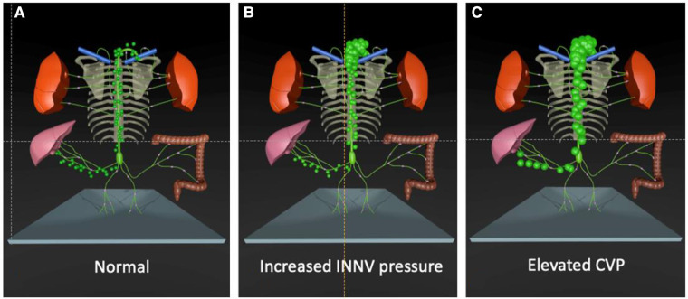
Children with hypoplastic left heart syndrome share unique hemodynamic features that alter lymphatic integrity at all stages of palliation. Lymphatic congestion is almost universal in this patient group to some extent. It may lead to reversal of lymphatic flow, the development of abnormal lymphatic channels and ultimately decompression and loss of protein rich lymphatic fluid into extra lymphatic compartments in prone individuals. Some of the most devastating complications that are associated with single ventricle physiology, notably plastic bronchitis and protein losing enteropathy, have now been proven to be lymphatic in origin. Based on the new pathophysiologic concept new diagnostic and therapeutic strategies have recently been developed. Dynamic contrast magnetic resonance lymphangiography is now mainstay in diagnosis of lymphatic insufficiency and allows a thorough assessment of anatomy and function of the main lymphatic compartments through intranodal, intrahepatic and intramesenteric lymphatic imaging. Contrast enhanced ultrasound can evaluate thoracic duct patency and conventional fluoroscopic lymphangiography has been refined for evaluation of patients where magnetic resonance imaging cannot be performed. Novel lymphatic interventional techniques, such as thoracic duct embolization, selective lymphatic duct embolization and liver lymphatic embolization allow to seal abnormal lymphatic networks minimally invasive and have shown to resolve symptoms. Innominate vein turn-down procedures, whether surgical or interventional, have been designed to reduce lymphatic afterload and increase systemic preload effectively in the failing Fontan circulation. Outflow obstruction can now be managed with new microsurgical techniques that create lympho-venous anastomosis. Short term results for all of these new approaches are overall promising but evidence is sparse and long-term outcome still has to be defined. This review article aims to summarize current concepts of lymphatic flow disorders in single ventricle patients, discuss new emerging diagnostic and therapeutic strategies and point out lacks in evidence and needs for further research on this rapidly growing topic.
Keywords: dynamic contrast magnetic resonance lymphangiography; fontanoperation; hypoplastic left heart syndrome; innominate vein turn-down procedures; lymphatic insufficiency; lymphatic interventional techniques; plastic bronchitis; protein-losingenteropathy.
© 2023 Bauer, Dori, Scala, Tulzer and Tulzer.
Conflict of interest statement
The authors declare that the research was conducted in the absence of any commercial or financial relationships that could be construed as a potential conflict of interest.
Figures


Similar articles
-
Lymphatic Imaging and Intervention in Congenital Heart Disease.J Soc Cardiovasc Angiogr Interv. 2023 Sep 27;3(1):101174. doi: 10.1016/j.jscai.2023.101174. eCollection 2024 Jan. J Soc Cardiovasc Angiogr Interv. 2023. PMID: 39131972 Free PMC article. Review.
-
Lymphatic Management in Single-Ventricle Patients.Semin Thorac Cardiovasc Surg Pediatr Card Surg Annu. 2020;23:41-47. doi: 10.1053/j.pcsu.2020.03.001. Semin Thorac Cardiovasc Surg Pediatr Card Surg Annu. 2020. PMID: 32354546 Review.
-
The Lymphatic System in the Fontan Patient-Pathophysiology, Imaging, and Interventions: What the Anesthesiologist Should Know.J Cardiothorac Vasc Anesth. 2022 Aug;36(8 Pt A):2669-2678. doi: 10.1053/j.jvca.2021.07.049. Epub 2021 Jul 31. J Cardiothorac Vasc Anesth. 2022. PMID: 34446325 Review.
-
Innominate Vein Turn-down Procedure for Failing Fontan Circulation.Semin Thorac Cardiovasc Surg Pediatr Card Surg Annu. 2020;23:34-40. doi: 10.1053/j.pcsu.2020.01.002. Semin Thorac Cardiovasc Surg Pediatr Card Surg Annu. 2020. PMID: 32354545
-
Case Report: Transcatheter interventional procedure to innominate vein turn-down procedure for failing fontan circulation.Front Pediatr. 2024 Feb 6;12:1341443. doi: 10.3389/fped.2024.1341443. eCollection 2024. Front Pediatr. 2024. PMID: 38379912 Free PMC article.
Cited by
-
[The lymphatic system-an interdisciplinary challenge].Radiologie (Heidelb). 2025 May;65(5):325-331. doi: 10.1007/s00117-025-01438-w. Epub 2025 Mar 27. Radiologie (Heidelb). 2025. PMID: 40146254 Review. German.
-
Lymphatic failure and lymphatic interventions: Knowledge gaps and future directions for a new frontier in congenital heart disease.Semin Pediatr Surg. 2024 Jun;33(3):151426. doi: 10.1016/j.sempedsurg.2024.151426. Epub 2024 May 23. Semin Pediatr Surg. 2024. PMID: 38820801 Free PMC article. Review.
-
The Nutmeg Lung Pattern in a Fetus with Hypoplastic Left Heart Syndrome and Turner Syndrome.Pediatr Cardiol. 2025 Apr 27. doi: 10.1007/s00246-025-03873-x. Online ahead of print. Pediatr Cardiol. 2025. PMID: 40287864
-
Lymphatic Disorder Management in Pediatric Patients With Congenital Heart Disease in European Pediatric Cardiology Centers: Current Status, Disparities, and Future Considerations.J Am Heart Assoc. 2024 Nov 19;13(22):e036597. doi: 10.1161/JAHA.124.036597. Epub 2024 Nov 7. J Am Heart Assoc. 2024. PMID: 39508150 Free PMC article.
References
Publication types
LinkOut - more resources
Full Text Sources