. 2023 Apr 14;9(4):1033-1045.
doi: 10.1021/acsinfecdis.3c00034. Epub 2023 Mar 13.

Evaluation of Antiviral Activity of Gemcitabine Derivatives against Influenza Virus and Severe Acute Respiratory Syndrome Coronavirus 2

Affiliations

Evaluation of Antiviral Activity of Gemcitabine Derivatives against Influenza Virus and Severe Acute Respiratory Syndrome Coronavirus 2

Hyeon-Min Cha et al. ACS Infect Dis. .

Abstract

Gemcitabine is a nucleoside analogue of deoxycytidine and has been reported to be a broad-spectrum antiviral agent against both DNA and RNA viruses. Screening of a nucleos(t)ide analogue-focused library identified gemcitabine and its derivatives (compounds 1, 2a, and 3a) blocking influenza virus infection. To improve their antiviral selectivity by reducing cytotoxicity, 14 additional derivatives were synthesized in which the pyridine rings of 2a and 3a were chemically modified. Structure-and-activity and structure-and-toxicity relationship studies demonstrated that compounds 2e and 2h were most potent against influenza A and B viruses but minimally cytotoxic. It is noteworthy that in contrast to cytotoxic gemcitabine, they inhibited viral infection with 90% effective concentrations of 14.5-34.3 and 11.4-15.9 μM, respectively, maintaining viability of mock-infected cells over 90% at 300 μM. Resulting antiviral selectivity was comparable to that of a clinically approved nucleoside analogue, favipiravir. The cell-based viral polymerase assay proved the mode-of-action of 2e and 2h targeting viral RNA replication and/or transcription. In a murine influenza A virus-infection model, intraperitoneal administration of 2h not only reduced viral RNA level in the lungs but also alleviated infection-mediated pulmonary infiltrates. In addition, it inhibited replication of severe acute respiratory syndrome virus 2 infection in human lung cells at subtoxic concentrations. The present study could provide a medicinal chemistry framework for the synthesis of a new class of viral polymerase inhibitors.

Keywords: SARS-CoV-2; antiviral agent; gemcitabine derivatives; influenza virus; polymerase inhibitor.


Conflict of interest statement

The authors declare no competing financial interest.

Figures

Figure 1
Figure 1
Screening of a chemical library for identification of hit compounds against influenza virus. (A) Cell-based antiviral assay against influenza A viruses. MDCK cells infected with influenza A virus, either A/Puerto Rico/8/34 (H1N1; PR8) or A/Hong Kong/8/68 (H3N2; HK), at a multiplicity of infection (MOI) of 0.001, were treated with 10 μM of each compound from a chemical library composed of 488 nucleoside analogues. On day 3 postinfection, cell viability was measured using 3-(4, 5-dimethylthiazolyl-2)-2, 5-diphenyltetrazolium bromide (MTT). Values from mock-infected and virus-infected, mock-treated cells were defined as 100 and 0%, respectively. Ten micromolar concentration of RBV was used as an internal control. The scatter plot shows relative antiviral activity (%) of each compound against the two viruses. Active compounds of interest are labeled. (B) Chemical structure of gemcitabine and its derivatives, 1, 2a, and 3a, identified from the cell culture-based screening against influenza A viruses, as shown in (A). (C) Dose–response curve of antiviral activity and cytotoxicity of gemcitabine and the hit compounds. MDCK cells were mock-infected (black) or infected with three viruses, PR8 (red), HK (blue), and B/Lee/40 (Lee; green), individually at an MOI of 0.001 for 1 h at 35 °C. After removal of unabsorbed virus, they were treated with threefold serial dilutions (from 300 to 0.05 μM) of each compound among gemcitabine (upper left), 1 (upper right), 2a (lower left), and 3a (lower right). On day 3, relative antiviral activity (left y-axis) and cell viability (right y-axis) were determined using an MTT assay in which values from 0.6% DMSO-treated cells and 0.6% DMSO-treated, virus-infected cells were defined 100 and 0%, respectively. Values are expressed as means ± SEM from three different samples.
Figure 2
Figure 2
Antiviral activity and cytotoxicity of chemically optimized gemcitabine derivatives, 2e, 2f, 2h, and 3c. (A) Dose–response curve showing antiviral activity (left y-axis) and cytotoxicity (right y-axis) of chemically modified compounds from 2a and 3a. MDCK cells, mock-infected (black) or infected with three different viruses, PR8 (red), HK (blue), and B/Lee/40 (Lee; green), were treated with increasing concentrations of compounds 2e (left upper), 2f (right upper), 2h (left lower), and 3c (right lower). On day 3 postinfection, the percentage of viable cells was determined by an MTT assay. Chemical structures are inserted within the graphs, in which modified parts are highlighted in red. Values are expressed as the mean ± SEM from three samples. (B) Inhibition of influenza viral NP expression in the presence of 2e and 2h. PR8 virus was used to infect MDCK cells at an MOI of 0.01 for 1 h at 35 °C. The virus-infected cells were treated with 0.1, 1, and 10 μM gemcitabine (GEM), 2e or 2h. The following day, cell lysates were harvested for immunoblotting of viral NP by using β-actin as a loading control. The proteins are labeled on the right of the blots. (C) Reduction of viral RNA titers by 2e and 2h. Cells were infected and treated with compounds, as shown in (B). Culture supernatants were harvested 1 day postinfection for viral RNA preparation. Two-step quantitative reverse transcription-polymerase chain reaction (qRT-PCR) was performed using an influenza A virus-specific universal primer and an NS gene-specific primer set. Viral genome copies were calculated from Ct value changes relative to those from PR8-infected, DMSO-treated cells. Data are expressed as means ± SEM from three independent experiments. P-values below 0.05 were considered statistically significant following a two-way analysis of variance (ANOVA), with Dunnett’s multiple comparison test.
Scheme 1
Scheme 1. Synthesis of Gemcitabine Derivatives, Compounds 1, 2a–2i, and 3a–3g
INT; intermediate. Different substituents (R) are displayed below the synthesis scheme.
Figure 3
Figure 3
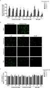
Inhibition of influenza A viral polymerase activity by 2e and 2h. (A) Time course dose–response graph of viral polymerase activity in the presence of 2e and 2h. HeLa cells were cotransfected with plasmids comprising an influenza viral replicon system amplifying both strands of EGFP transcripts. At 4 h post-transfection, cells were treated with increasing concentrations (11, 33, and 100 μM) of 2e or 2h or RBV as a positive control. Polymerase activity was measured by quantifying the number of fluorescent spots per well for 72 h at 6 h intervals. (B) Representative fluorescent microscopy images showing inhibition of EGFP-expressing influenza viral polymerase activity by compounds 2e and 2h. Negative control samples were untransfected, while positive control samples were transfected with the minigenome replicon plasmids and 0.2% DMSO-treated. Gemcitabine derivatives, 2e and 2h, and RBV were used to treat the transfected HeLa cells at concentrations of 11, 33, and 100 μM. The images were obtained at 24 h post-treatment. Original magnification, ×200. (C) Cytotoxicity of 2e and 2h. Samples were prepared as described in (A). Confluence was analyzed by measuring object counts per well on the bright-field microscopy images at different time points. In (A) and (C), values are expressed as means ± SEM from experiments in triplicate. Statistical significance was determined by two-way ANOVA, with Dunnett’s multiple comparison test compared to the mock-treated samples. *, P < 0.05; **, P < 0.01; ***, P < 0.001; ****, P < 0.0001.
Figure 4
Figure 4
In vivo antiviral activity of 2h in an influenza virus-infected mouse model. (A) Schematic illustration of in vivo studies using compound 2h to treat influenza A virus infection. Four hours before intranasal infection with mouse-adapted PR8 strain (maPR8) at a dose of 5 MLD50, BALB/c mice were intraperitoneally administered with 2h (5 mg/kg) or orally with oseltamivir phosphate (OSV-P; 5 mg/kg). At 4 h after virus infection, mice were additionally given the same dose of OSV-P. Mice were subsequently treated daily with 2h once a day (QD) or with OSV-P twice a day (BID) for additional 4 days. Mock-infected mice and maPR8-infected mice were used as controls. Eight mice were assigned to each group. On day 5, five mice were sacrificed for viral RNA titration in the lungs and the rest three mice for lung histopathology analysis. (B) Reduction of viral RNA copies in the lungs after treatment of mice with 2h. As described in (A), five mice in each group were sacrificed to prepare total RNA from the lung tissues. Viral mRNA was quantified by qRT-PCR using an oligo(dT) and influenza A virus NS genome-specific primers. GAPDH mRNA level was calculated to normalize the viral mRNA expression. Each symbol represents an individual mouse. Statistical analysis was performed using an ordinary one-way ANOVA, with Dunnett’s multiple comparison test compared to the virus-infected, mock-treated group (virus only). ***, P < 0.001; ****, P < 0.0001. (C) Histopathological analysis of maPR8-infected lung tissues after treatment with compound 2h. Three mice in each group were sacrificed at day 5 postinfection, and lung tissues were fixed with formalin for hematoxylin and eosin (H&E) staining. Tissues from mock-infected mice (Mock) and A/H1N1 maPR8 virus-infected mice (maPR8) were used as controls. Images from 2h-treated mice (maPR8 + Compd 2h) are displayed for comparison with OSV-P-treated samples (maPR8 + OSV-P). Original magnification, ×100.
Figure 5
Figure 5
Anti-SARS-CoV-2 activity of compound 2h in human lung cells. (A) Immunofluorescence microscopy. Calu-3 cells infected with SARS-CoV-2 at an MOI of 0.05 were treated with various concentrations of compound 2h, 1.2, 3.7, 11.1, and 33.3 μM, for 2 days at 37 °C, where 0.2% DMSO was used as a delivery vehicle control. After fixing and permeabilization, cells were stained with an anti-S antibody and Alexa Fluor 488-conjugated goat anti-mouse IgG (green; upper panels). Cell nuclei were counter stained with DAPI (blue). Merged images are presented in the lower panels. (B) Dose–response curves of antiviral activity and cytotoxicity. SARS-CoV-2-infected Calu-3 cells were treated with increasing concentration of compound 2h or RDV as depicted in (A). Antiviral activity was determined by normalizing reciprocal fluorescence intensity from SARS-CoV-2-infected cells (red line), while cell viability was estimated by measuring viability of mock-infected cells with MTT (black line). EC50, CC50, and SI values are recorded below each graph.

References

    1. Bouvier N. M.; Palese P. The biology of influenza viruses. Vaccine 2008, 26, D49–D53. 10.1016/j.vaccine.2008.07.039. - DOI - PMC - PubMed
    1. Matrosovich M. N.; Matrosovich T. Y.; Gray T.; Roberts N. A.; Klenk H. D. Human and avian influenza viruses target different cell types in cultures of human airway epithelium. Proc. Natl. Acad. Sci. U.S.A. 2004, 101, 4620–4624. 10.1073/pnas.0308001101. - DOI - PMC - PubMed
    1. Kawaguchi A.; Naito T.; Nagata K. Involvement of influenza virus PA subunit in assembly of functional RNA polymerase complexes. J. Virol. 2005, 79, 732–744. 10.1128/jvi.79.2.732-744.2005. - DOI - PMC - PubMed
    1. Martin K.; Heleniust A. Nuclear transport of influenza virus ribonucleoproteins: the viral matrix protein (M1) promotes export and inhibits import. Cell 1991, 67, 117–130. 10.1016/0092-8674(91)90576-k. - DOI - PubMed
    1. Hu B.; Guo H.; Zhou P.; Shi Z. L. Characteristics of SARS-CoV-2 and COVID-19. Nat. Rev. Microbiol. 2021, 19, 141–154. 10.1038/s41579-020-00459-7. - DOI - PMC - PubMed

Publication types