. 2023 Apr 15;207(8):1055-1069.
doi: 10.1164/rccm.202203-0450OC.

SOX17 Deficiency Mediates Pulmonary Hypertension: At the Crossroads of Sex, Metabolism, and Genetics

Affiliations

SOX17 Deficiency Mediates Pulmonary Hypertension: At the Crossroads of Sex, Metabolism, and Genetics

Shreya Sangam et al. Am J Respir Crit Care Med. .

Abstract

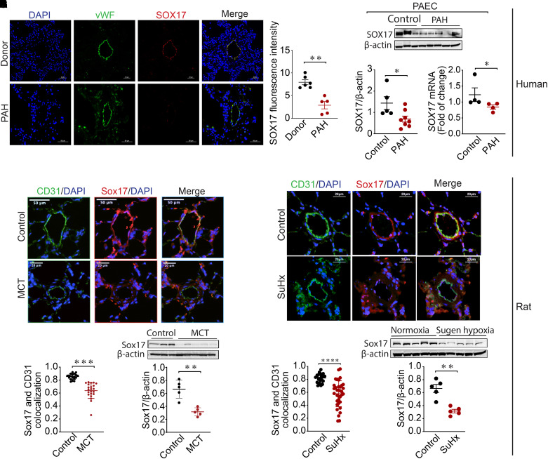
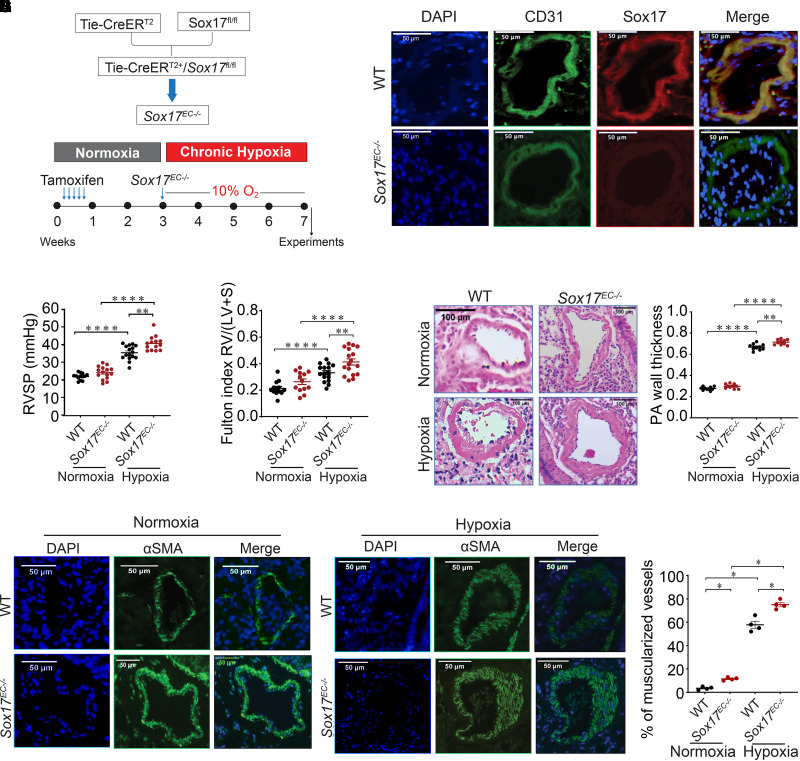
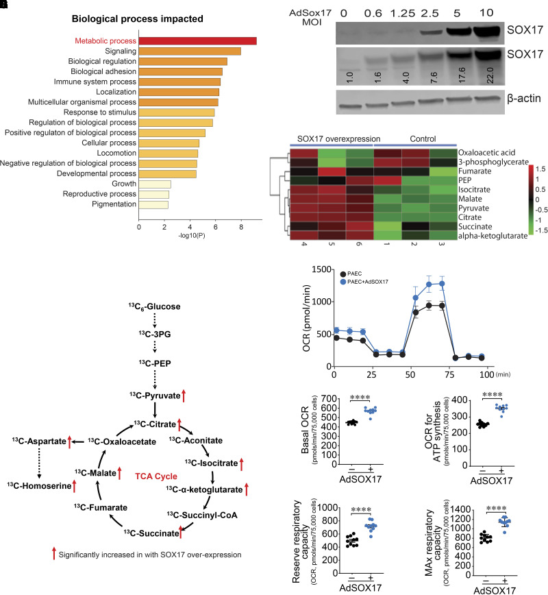
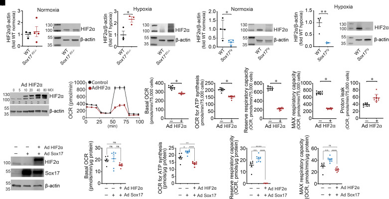
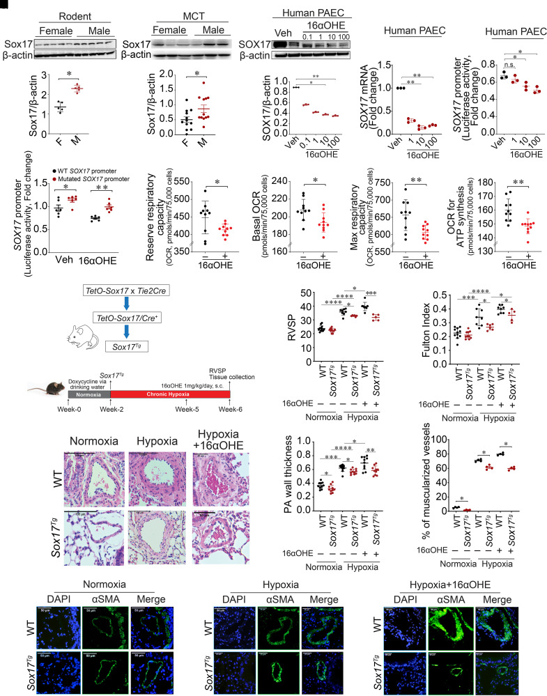
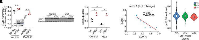
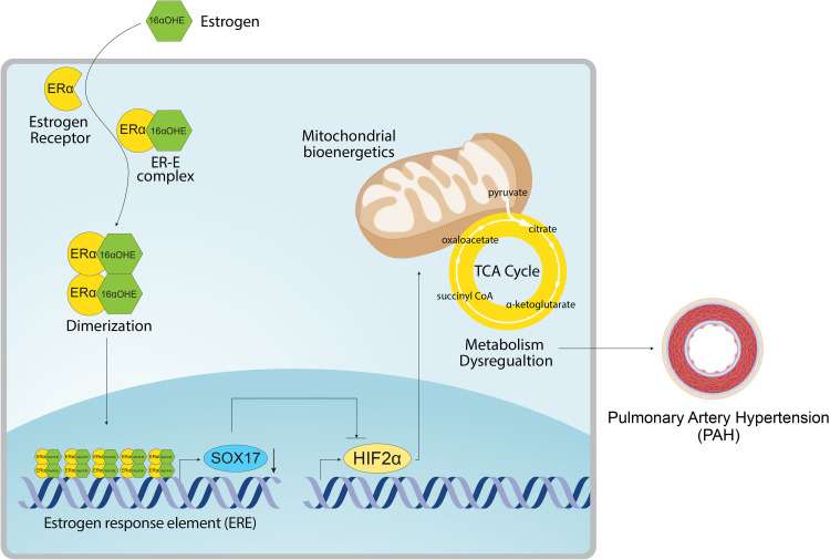
Rationale: Genetic studies suggest that SOX17 (SRY-related HMG-box 17) deficiency increases pulmonary arterial hypertension (PAH) risk. Objectives: On the basis of pathological roles of estrogen and HIF2α (hypoxia-inducible factor 2α) signaling in pulmonary artery endothelial cells (PAECs), we hypothesized that SOX17 is a target of estrogen signaling that promotes mitochondrial function and attenuates PAH development via HIF2α inhibition. Methods: We used metabolic (Seahorse) and promoter luciferase assays in PAECs together with the chronic hypoxia murine model to test the hypothesis. Measurements and Main Results: Sox17 expression was reduced in PAH tissues (rodent models and from patients). Chronic hypoxic pulmonary hypertension was exacerbated by mice with conditional Tie2-Sox17 (Sox17EC-/-) deletion and attenuated by transgenic Tie2-Sox17 overexpression (Sox17Tg). On the basis of untargeted proteomics, metabolism was the top pathway altered by SOX17 deficiency in PAECs. Mechanistically, we found that HIF2α concentrations were increased in the lungs of Sox17EC-/- and reduced in those from Sox17Tg mice. Increased SOX17 promoted oxidative phosphorylation and mitochondrial function in PAECs, which were partly attenuated by HIF2α overexpression. Rat lungs in males displayed higher Sox17 expression versus females, suggesting repression by estrogen signaling. Supporting 16α-hydroxyestrone (16αOHE; a pathologic estrogen metabolite)-mediated repression of SOX17 promoter activity, Sox17Tg mice attenuated 16αOHE-mediated exacerbations of chronic hypoxic pulmonary hypertension. Finally, in adjusted analyses in patients with PAH, we report novel associations between a SOX17 risk variant, rs10103692, and reduced plasma citrate concentrations (n = 1,326). Conclusions: Cumulatively, SOX17 promotes mitochondrial bioenergetics and attenuates PAH, in part, via inhibition of HIF2α. 16αOHE mediates PAH development via downregulation of SOX17, linking sexual dimorphism and SOX17 genetics in PAH.

Keywords: 16α-hydroxyestrone; SRY-related HMG-box 17; hypoxia-inducible factor 2α; metabolism; pulmonary arterial hypertension.


Figures

Figure 1.
Figure 1.
SOX17 (SRY-related HMG-box 17) is downregulated in pulmonary arterial hypertension (PAH). (A and B) Representative (A) and quantitative (B) immunofluorescence staining for DAPI (blue), vWF (green), and SOX17 (red) in human lungs from a non-PAH donor (n = 6) and patients with PAH (n = 5). (C) Western blot demonstrating SOX17 protein concentrations in pulmonary artery endothelial cells (PAECs) isolated from non-PAH control and subjects with PAH. (D) Densitometric analysis of SOX17 normalized to β-actin in PAECs from control subjects (n = 5) and patients with PAH (n = 8). (E) Real-time PCR analysis showing mRNA fold change of SOX17 normalized to actin in PAECs from control (n = 4) subjects and subjects with PAH (n = 4). (F) Representative immunofluorescence images showing DAPI (blue), CD31 (cluster of differentiation 31) (green), and Sox17 (red) staining in lung sections from control and monocrotaline (MCT)–exposed rats. (G) Quantitative immunofluorescence of SOX17 in control and MCT-exposed rat samples. (H) Western blot showing protein concentrations of Sox17 in MCT versus control rat lungs and densitometric analysis of Sox17 normalized to β-actin in MCT (n = 5) and control (n = 5) rats. (I) Representative immunofluorescence images showing DAPI (blue), CD31 (green), and Sox17 (red) staining in lung sections from control and rats exposed to Sugen–hypoxia (SuHx). (J) Quantitative immunofluorescence of SOX17 in control and SuHx-exposed rat samples. (K) Western blot showing the protein concentrations of Sox17 in lungs from SuHx versus normoxic control rats and densitometric analysis of Sox17 normalized to β-actin in SuHx (n = 5) and normoxic control (n = 5) rats. Data are represented as mean ± SEM. *P < 0.05, **P < 0.01, ***P < 0.001, and ****P < 0.0001 versus controls. vWF = von Willebrand factor.
Figure 2.
Figure 2.
Endothelial deficiency of Sox17 (SRY-related HMG-box 17) (Sox17EC−/−) augments chronic hypoxia-induced murine pulmonary hypertension (PH). (A) Schema that reflects modeling of murine hypoxic PH. (B) Representative immunostaining against Sox17 (red) and CD31 (green) in mouse lung section from control and Sox17EC−/− mice (scale bar, 50 μm). (C) RVSP measurements in wild-type (WT) and Sox17EC−/− mice under normoxia and hypoxia (n = 10–16 per group). (D) Fulton index (RV/[LV + S]) measurements in WT control and Sox17EC−/− mice exposed to normoxia or hypoxia (n = 10–16 per group). (E) Representative hematoxylin and eosin images of PAs (categorized into diameter ⩽ 50 μm and diameter > 50 μm) showing wall thickness in WT and Sox17EC−/− mice under normoxia or hypoxia. (F) Summarized data showing PA wall thickness (wall area/total area) in WT and Sox17EC−/− mice. (G) Representative αSMA (α-smooth muscle actin) staining in WT and Sox17EC−/− mice under normoxic conditions. (H) Representative αSMA staining in WT and Sox17EC−/− mice under hypoxic conditions. (I) Percentage of muscularized vessels in control and Sox17EC−/− mice under normoxia and hypoxia (n = 4 per group) and quantitative immunofluorescence of αSMA in mouse lung samples. Data are represented as mean ± SEM. *P < 0.05, **P < 0.01, and ****P < 0.0001. CD31 = cluster of differentiation 31; PA = pulmonary artery; RV/(LV + S) = ratio of right ventricular weight to the sum of left ventricular and septal weight; RVSP = right ventricular systolic pressure.
Figure 3.
Figure 3.
SOX17 (SRY-related HMG-box 17) augments oxidative phosphorylation and mitochondrial function in endothelial cells. (A) Gene Ontology analysis highlights metabolic processes as the top targets of SOX17 deficiency. (B) PAECs were transduced with increasing multiplicities of infection (MOIs) of AdSOX17. Western blot analysis was used to confirm the overexpression of SOX17. An MOI of 20 was used. (C) A heatmap illustrating alterations in metabolite concentrations in SOX17-overexpressed endothelial cells (ECs) compared with control ECs. (D) Targeted gas chromatography–mass spectrometry analysis identifies increases in TCA cycle metabolites in SOX17 overexpressing ECs. (E–I) Increasing SOX17 expression enhances mitochondrial bioenergetics (E), as determined by increases in basal oxygen consumption rate (OCR) (F), OCR for ATP generation (G), and reserve (H) and MAX (I) respiratory capacity. Data are represented as mean ± SEM. ****P < 0.0001. 3PG = 3-phosphoglyceric acid; AdSOX17 = adenovirus containing human SOX17; CoA = coenzyme A; MAX = maximal; PAEC = pulmonary artery endothelial cell; PEP = phosphoenolpyruvate; TCA = tricarboxylic acid.
Figure 4.
Figure 4.
SOX17 (SRY-related HMG-box 17) attenuates HIF2α (hypoxia-inducible factor 2α)–mediated inhibition of oxidative phosphorylation and mitochondrial function in endothelial cells. (A and B) HIF2α protein concentrations are unchanged in Sox17EC−/− mice under normoxic conditions (A) but significantly increased by hypoxia (B). (C and D) HIF2α protein concentrations are significantly decreased in Sox17Tg mice under both normoxic (C) and hypoxic (D) conditions. (E) Western blot analysis was used to confirm overexpression of HIF2α. An MOI of 40 was used. (F–K) Increasing HIF2α expression attenuates mitochondrial bioenergetics (F), as determined by decreases in basal oxygen consumption rate (OCR) (G), OCR for ATP generation (H), reserve (I) and maximal (J) respiratory capacity, and increases in the proton leak (K). (L–P) Coexpressing HIF2α reverses the positive effect of SOX17 overexpression on mitochondrial bioenergetics; (L) Western blot indicating overexpression of SOX17 and HIF2α. HIF2α coexpression with SOX17 decreases basal respiration (M), amount of oxygen consumed for ATP generation (N), reserve respiratory capacity (O), and maximal respiratory capacity (P) compared with SOX17 overexpression alone. Data are represented as mean ± SEM. *P < 0.05, **P < 0.01, and ****P < 0.0001. Ad = adenovirus; MAX = maximal; MOI = multiplicity of infection; ns = not significant; WT = wild-type.
Figure 5.
Figure 5.
Endothelial cell–derived SOX17 (SRY-related HMG-box 17) is a transcriptional target of 16α-hydroxyestrone (16αOHE) and attenuates hypoxic pulmonary hypertension (PH) in mice. (A) Sox17 expression in female (n = 5) and male (n = 5) rat lungs at baseline. (B) Densitometric graph of Sox17 expression normalized to β-actin in female and male rat lungs. (C) Representative western blot showing Sox17 expression in female and male rat lungs exposed to monocrotaline (MCT). (D) Densitometric graph of Sox17 expression normalized to β-actin in female (n = 12) and male (n = 12) rat lungs exposed to MCT. (E) Representative western blot showing protein expression of SOX17 in pulmonary artery endothelial cells (PAECs) treated with 16αOHE (0.1–100 nM). (F) Densitometric analysis of SOX17 normalized to β-actin in vehicle (Veh)– and 16αOHE-treated PAECs. (G) mRNA expression of SOX17 in PAECs treated with 16αOHE (1–100 nM). (H) A dual-luciferase assay was used to measure relative promoter luciferase activities. 16αOHE exposure reduced SOX17 promoter activity compared with Veh in PAECs transfected with pGL3-SOX17 promoter plasmid. (I) Dual-luciferase assay showing luciferase activities of wild-type (WT) and mutated SOX17 promoter in Veh- and 16αOHE-treated PAECs. (J–M) We exposed human PAECs to 16αOHE (10 nM) and measured mitochondrial bioenergetics. Real-time measurements of mitochondrial OCRs on the mitochondrial biogenetic profile, including basal respiration (K) and the amount of oxygen consumed for ATP generation (M), were reduced, together with reserve respiratory capacity (J) and Max respiratory capacity (L). (N) Schema that reflects modeling of murine hypoxic PH. (O and P) RVSP (O) and Fulton index (ratio of right ventricular weight to the sum of left ventricular and septal weight) (P) measurements in WT and Sox17Tg mice under normoxia, hypoxia, or hypoxia with or without 16αOHE. (Q) Representative hematoxylin and eosin images of lung sections showing pulmonary artery wall thickness (PAWT) in WT and Sox17Tg mice. (R) Summarized data showing PAWT measurements (wall area/total area) in WT and Sox17Tg mice. (S) Percentage of muscularized vessels in WT and Sox17Tg mice under normoxia, hypoxia, or hypoxia with or without 16αOHE (n = 4 per group). (T) Representative αSMA staining in WT and Sox17Tg mice under normoxic or hypoxic conditions with or without 16αOHE. Data are represented as mean ± SEM. *P < 0.05, **P < 0.01, ***P < 0.001, and ****P < 0.0001. αSMA = α-smooth muscle actin; F = female; M = male; Max = maximal; OCRs = oxygen consumption rates; PA = pulmonary arterial; RVSP = right ventricular systolic pressure; s.c. = subcutaneous.
Figure 6.
Figure 6.
16α-Hydroxyestrone (16αOHE) represses SOX17 (SRY-related HMG-box 17) promoter through ERα (estrogen receptor α). (A) pGL3-SOX17 promoter plasmid was cotransfected with scrambled RNA (scRNA) or siRNA against ERα (siERα) in pulmonary artery endothelial cells (PAECs) and later exposed to 16αOHE versus vehicle. Dual-luciferase assay shows the relative luciferase activity of SOX17 promoter; representative western blot ERα protein concentrations in scRNA- and siERα-transfected human PAECs to confirm silencing. (B) Western blot showing Sox17 protein concentrations in wild-type (WT) (n = 5) and ERα−/− (n = 3) control rats and WT (n = 4) and ERα−/− (n = 3) monocrotaline (MCT) rats. (C) Densitometric analysis of Sox17 normalized to β-actin in ERα−/− control and MCT rats compared with WT. (D) Correlation analysis between ESR1 and SOX17 mRNA (fold change) showing a negative correlation in PAECs from patients with pulmonary arterial hypertension (PAH). Pearson correlation coefficient (r) and P value are shown. (E) rs10103692 was significantly associated with reduced citrate concentrations (metabolite quantitative trait locus, n = 1,326, β = −2.04 × 106 for each copy of G allele, SD = 0.75 × 105, P = 0.007, for non–log10-transformed citrate concentrations, n = 1,165 for AA, n = 150 for AG, and n = 10 for GG) in subjects of European ancestry after adjusting for age, sex, time between diagnosis and blood draw (PAH vintage), and population stratification. Data are represented as mean ± SEM. *P < 0.05 and **P < 0.01. ESR1 = estrogen receptor 1.
Figure 7.
Figure 7.
Summary schema. The schema reveals the overall proposed mechanism demonstrated in this work. Specifically, we report a novel signaling cascade involving 16αOHE-mediated repression of SOX17 (SRY-related HMG-box 17) expression and promoter activity via ERα. To understand the protective effects of SOX17 in vivo, we further connect SOX17-mediated inhibition of HIF2α with enhanced oxidative phosphorylation and mitochondrial function in endothelial cells. 16αOHE = 16α-hydroxyestrone; CoA = coenzyme A; ERα = estrogen receptor α; HIF2α = hypoxia-inducible factor 2α; TCA = tricarboxylic acid.

Comment in

References

    1. Karnes JH, Wiener HW, Schwantes-An TH, Natarajan B, Sweatt AJ, Chaturvedi A, et al. Genetic admixture and survival in diverse populations with pulmonary arterial hypertension. Am J Respir Crit Care Med . 2020;201:1407–1415. - PMC - PubMed
    1. Rhodes CJ, Batai K, Bleda M, Haimel M, Southgate L, Germain M, et al. UK NIHR BioResource Rare Diseases Consortium; UK PAH Cohort Study Consortium; US PAH Biobank Consortium Genetic determinants of risk in pulmonary arterial hypertension: international genome-wide association studies and meta-analysis. Lancet Respir Med . 2019;7:227–238. - PMC - PubMed
    1. Gräf S, Haimel M, Bleda M, Hadinnapola C, Southgate L, Li W, et al. Identification of rare sequence variation underlying heritable pulmonary arterial hypertension. Nat Commun . 2018;9:1416. - PMC - PubMed
    1. Zhu N, Welch CL, Wang J, Allen PM, Gonzaga-Jauregui C, Ma L, et al. Rare variants in SOX17 are associated with pulmonary arterial hypertension with congenital heart disease. Genome Med . 2018;10:56. - PMC - PubMed
    1. Wu Y, Wharton J, Walters R, Vasilaki E, Aman J, Zhao L, et al. The pathophysiological role of novel pulmonary arterial hypertension gene SOX17. Eur Respir J . 2021;58:2004172. - PubMed

Publication types