Review
. 2023 Aug;44(8):1521-1535.
doi: 10.1038/s41401-023-01068-9. Epub 2023 Mar 13.

Regulated cell death pathways in cardiomyopathy

Affiliations
Review

Regulated cell death pathways in cardiomyopathy

Shu-Yuan Sheng et al. Acta Pharmacol Sin. 2023 Aug.

Abstract

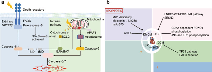
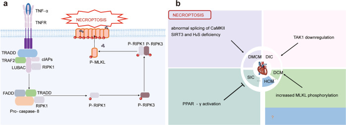
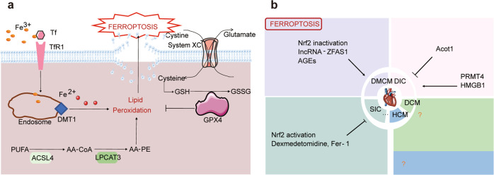
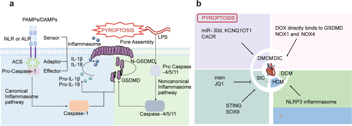
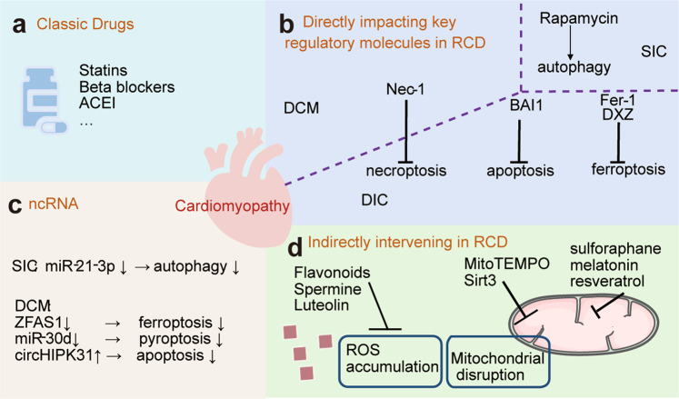
Heart disease is a worldwide health menace. Both intractable primary and secondary cardiomyopathies contribute to malignant cardiac dysfunction and mortality. One of the key cellular processes associated with cardiomyopathy is cardiomyocyte death. Cardiomyocytes are terminally differentiated cells with very limited regenerative capacity. Various insults can lead to irreversible damage of cardiomyocytes, contributing to progression of cardiac dysfunction. Accumulating evidence indicates that majority of cardiomyocyte death is executed by regulating molecular pathways, including apoptosis, ferroptosis, autophagy, pyroptosis, and necroptosis. Importantly, these forms of regulated cell death (RCD) are cardinal features in the pathogenesis of various cardiomyopathies, including dilated cardiomyopathy, diabetic cardiomyopathy, sepsis-induced cardiomyopathy, and drug-induced cardiomyopathy. The relevance between abnormity of RCD with adverse outcome of cardiomyopathy has been unequivocally evident. Therefore, there is an urgent need to uncover the molecular and cellular mechanisms for RCD in order to better understand the pathogenesis of cardiomyopathies. In this review, we summarize the latest progress from studies on RCD pathways in cardiomyocytes in context of the pathogenesis of cardiomyopathies, with particular emphasis on apoptosis, necroptosis, ferroptosis, autophagy, and pyroptosis. We also elaborate the crosstalk among various forms of RCD in pathologically stressed myocardium and the prospects of therapeutic applications targeted to various cell death pathways.

Keywords: cardiomyopathy; cell death; cell signaling.


Conflict of interest statement

The authors declare no competing interests.

Figures

Fig. 1
Fig. 1. Schematic illustration of apoptosis pathway and apoptosis in cardiomyopathies.
a Apoptosis pathway. The extrinsic pathway is initiated by the binding of death ligands to their canonical death receptors and subsequent formation of DISC, which then activates caspase 8 and the death effector caspases 3 and 7, leading to apoptosis. In the intrinsic pathway, numerous stimuli lead to permeabilization of the mitochondrial outer membrane (MOMP) and further leakage of proapoptotic proteins such as Cytochrome c. Afterward, released Cytochrome c binds Apaf-1 to form apoptosome which then activates caspase 9 and subsequently triggers the activation of the downstream effector caspase 3 and caspase 7, leading to apoptosis. BAK/BAX which can be activated by tBID promotes to the MOMP while BCL-2 inhibits its function. b Apoptosis in cardiomyopathy. In DMCM, accumulated AGEs promote apoptosis while Melatonin, Lin28a, miR675, or deficiency of Mst1 inhibit apoptosis. In DIC, CDK2-dependent FOXO1 phosphorylation, as well as, JNK and ERK phosphorylation promote apoptosis while activation of FNDC5 Wnt/PCP-JNK pathway or SESN2 inhibits apoptosis. In DCM, activation of TP53 pathway and BAG3 mutation promote apoptosis.
Fig. 2
Fig. 2. Schematic illustration of necroptosis pathway and necroptosis in cardiomyopathies.
a Necroptosis pathway. TNF-α activates TNFR1 and then recruits regulatory proteins such as TRADD and RIPK1 to form complex I as well as subsequent Complex II. When caspase 8 is inhibited, phosphorylated RIPK1 and phosphorylated RIPK3 recruit as well as phosphorylate its substrate MLKL. Activated MLKL oligomerizes and inserts into plasma membrane to execute necroptosis, finally causing plasma membrane rupture and release of DAMPs and PAMPs. b Necroptosis in cardiomyopathy. In DMCM, abnormal splicing of CaMKII and SIRT3 as well as H2S deficiency promotes necroptosis. In DIC, TAK1 downregulation promotes necroptosis. In DCM, increased MLKL phosphorylation promotes necroptosis. In SIC, activation of PPAR-γ inhibits necroptosis.
Fig. 3
Fig. 3. Schematic illustration of ferroptosis pathway and ferroptosis in cardiomyopathies.
a Ferroptosis pathway. Both Iron-dependent lipid peroxidation and decompensated anti-oxidation system can launch the course of ferroptosis. Iron in circulation can be taken up in the form of Fe3+ and converted to Fe2+ inside of endosome. GPX4 is the major antioxidant defense and GSH is an indispensable cofactor for its activity. Cysteine utilization, after its conversion from cystine, is a major limiting factor for the GSH biosynthesis while cystine uptake mainly relies on System Xc- which imports extracellular cystine by exchanging intracellular glutamate. AA-PE is the main substrates of peroxidation, its synthesis needs the catalyzation by ACSL4 and LPCAT3. b Ferroptosis in cardiomyopathy. In DMCM, Nrf2 inactivation, IncRNA-ZFAS1 and AGEs accumulation promote ferroptosis. In DIC, PRMT4 and HMGB1 promote ferroptosis, while Acot1 inhibits ferroptosis. In SIC, Nrf2 activation and administrations of dexmedetomidine or Fer-1 inhibit ferroptosis.
Fig. 4
Fig. 4. Schematic illustration of autophagy pathway and autophagy in cardiomyopathies.
a Autophagy pathway. The canonical autophagy is initiated when the mTORC1 activity is suppressed and ULK1 complex is activated, which subsequently activate Class III PI3K Complex as well as lead to the formation of phagophores. Two distinct ubiquitin-like conjugation systems participate in the elongation of the phagophores: one involves ATG5-ATG12-ATG16L and another involves the LC3-PE. Afterwards, autophagosomes fuse with lysosomes to form autophagolysosomes where cargoes are degraded. b Autophagy in cardiomyopathy. In DMCM, AMPK, DCRF and Heme oxygenase-1 promote autophagy. In DIC, TFEB and prior starvation promote autophagy while GATA4 inhibits autophagy. In DCM, mutation of LMNA or PLEKHM2 inhibits autophagy. In HCM, TSC1 promotes autophagy while Mybpc3-targeted knock-in or loss of Vps34 inhibits autophagy. In SIC, Beclin-1 and miR-22 knock-out promote autophagy while ALDH2 inhibits autophagy.
Fig. 5
Fig. 5. Schematic illustration of pyroptosis pathway and pyroptosis in cardiomyopathies.
a Pyroptosis pathway. In the canonical form, various insults lead to the assembly of the inflammasome complex consisting of sensors, adaptors, and effector, which then activates caspase 1. Activated caspase 1 mediates the pro IL-1β/IL-18 maturation and GSDMD cleavage. The truncated N-GSDMD is the final executioner of pyroptosis by forming pores in plasma membrane and releasing intracellular contents. In noncanonical inflammasome pathway, LPS directly actives pro-caspase 4/5/11, which further cleaves GSDMD to generate N-GSDMD. b Pyroptosis in cardiomyopathy. In DMCM, miR-30d, KCNQ1OT1 and CACR promote pyroptosis. In DIC, Dox directly binds to GSDMD or upregulated NOX1 and NOX4 promote pyroptosis. In DCM, activated NLRP3 inflammasome promotes pyroptosis. In SIC, STING and SOX9 promote pyroptosis while irisin or JQ1 inhibit pyroptosis.
Fig. 6
Fig. 6. Schematic illustration of therapeutic potential targeted to intervene regulated cell death.
a Standard therapy including classic drugs is far from satisfaction. b ncRNAs have emerged as potential therapeutic targets in broad range of cardiomyopathy hearts: miR-21-3p antisense inhibitor alleviates inappropriate autophagy in SIC. In DCM, cardiac protection was achieved by inhibition of the ncRNA ZFAS1, miR-30d, or overexpression of circHIPK3, which respectively attenuates ferroptosis, pyroptosis and apoptosis. c Directly impact the key regulatory molecules in cell death pathway may serve as a potential effective therapy for the disease: Nec-1 blocks the process of necroptosis against DCM and DIC; BAI1 alleviates cardiomyocyte necrosis as well as apoptosis while Fer-1 and DXZ suppress ferroptosis in the setting of DIC; Rapamycin restores autophagy in SIC to remit cardiac dysfunction. d Indirectly intervening RCD by aiming at upstream signaling may also prove to be effective: Natural products such as Flavonoids, Spermine, and Luteolin eliminate excess ROS accumulation; Sirt3 and MitoTEMPO protect mitochondrial homeostasis.

References

    1. McKenna WJ, Maron BJ, Thiene G. Classification, epidemiology, and global burden of cardiomyopathies. Circ Res. 2017;121:722–30. doi: 10.1161/CIRCRESAHA.117.309711. - DOI - PubMed
    1. Lannou S, Mansencal N, Couchoud C, Lassalle M, Dubourg O, Stengel B, et al. The Public Health Burden of Cardiomyopathies: insights from a Nationwide Inpatient Study. J Clin Med. 2020;9:920–33. doi: 10.3390/jcm9040920. - DOI - PMC - PubMed
    1. Yamada T, Nomura S. Recent findings related to cardiomyopathy and genetics. Int J Mol Sci. 2021;22:15222–34. doi: 10.3390/ijms222212522. - DOI - PMC - PubMed
    1. Elliott PM, Anastasakis A, Borger MA, Borggrefe M, Cecchi F, Charron P, et al. 2014 ESC Guidelines on diagnosis and management of hypertrophic cardiomyopathy: the Task Force for the Diagnosis and Management of Hypertrophic Cardiomyopathy of the European Society of Cardiology (ESC) Eur Heart J. 2014;35:2733–79. doi: 10.1093/eurheartj/ehu284. - DOI - PubMed
    1. Sorajja P, Pedersen WA, Bae R, Lesser JR, Jay D, Lin D, et al. First experience with percutaneous mitral valve plication as primary therapy for symptomatic obstructive hypertrophic cardiomyopathy. J Am Coll Cardiol. 2016;67:2811–8. doi: 10.1016/j.jacc.2016.03.587. - DOI - PubMed