. 2023 Apr;24(4):676-689.
doi: 10.1038/s41590-023-01444-x. Epub 2023 Mar 13.

A single-amino acid substitution in the adaptor LAT accelerates TCR proofreading kinetics and alters T-cell selection, maintenance and function

Affiliations

A single-amino acid substitution in the adaptor LAT accelerates TCR proofreading kinetics and alters T-cell selection, maintenance and function

Wan-Lin Lo et al. Nat Immunol. 2023 Apr.

Abstract

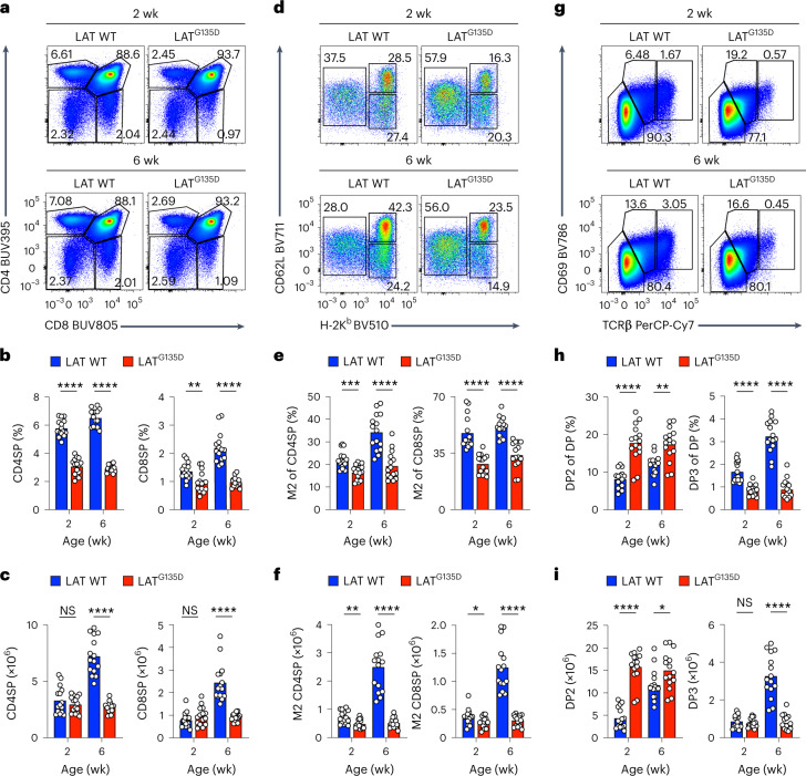
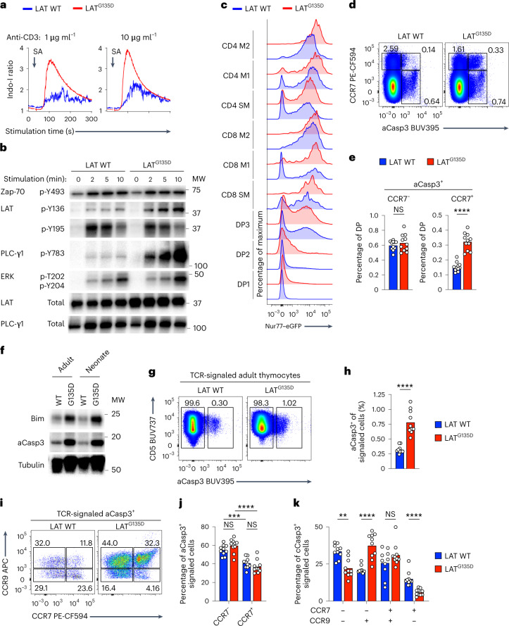
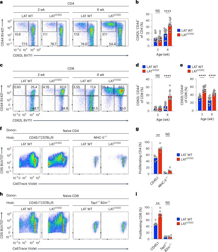
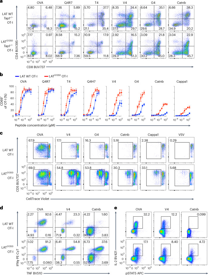
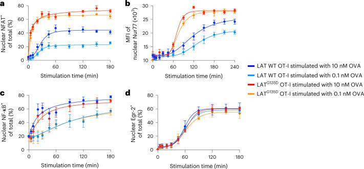
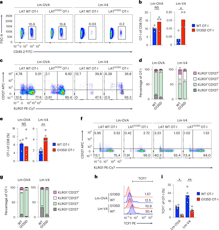
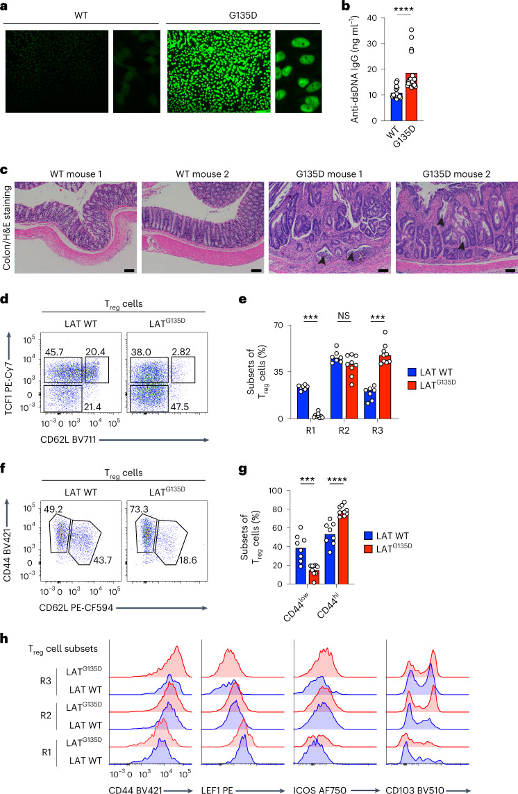
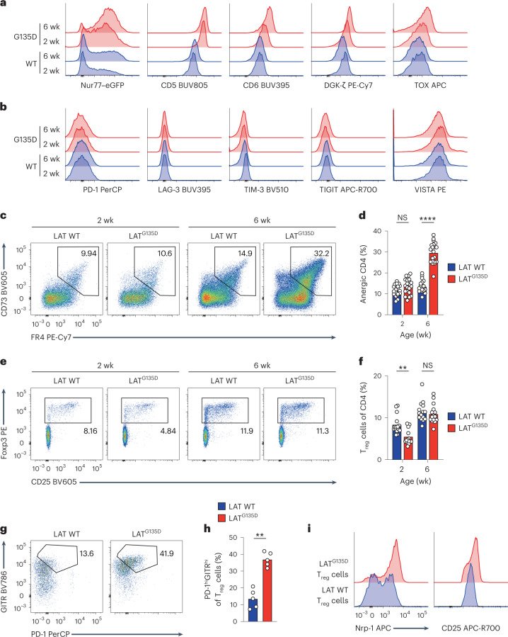
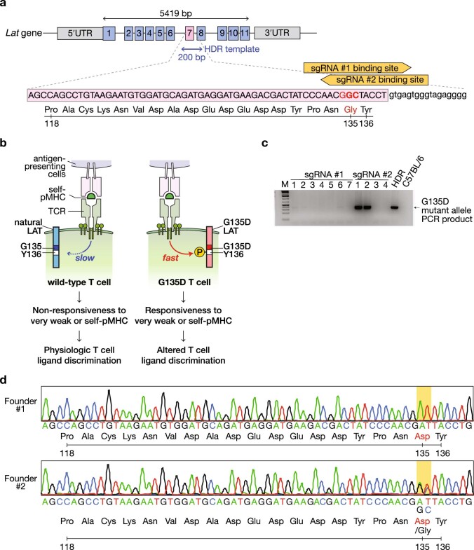
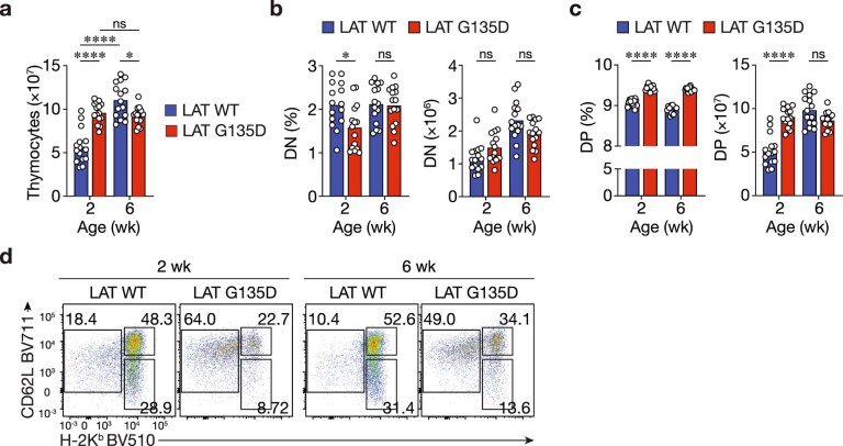
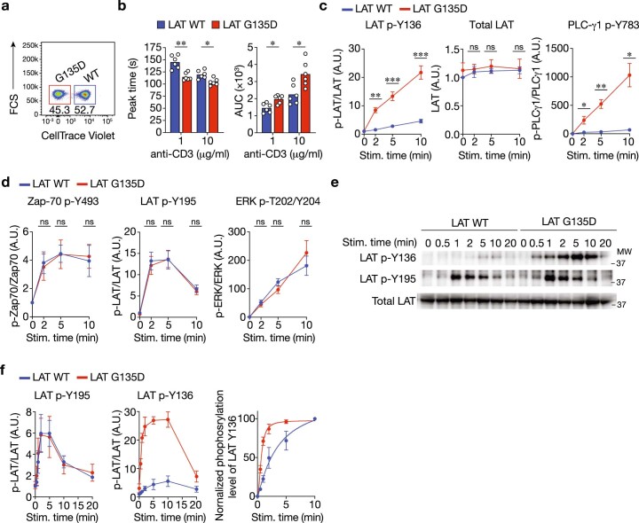
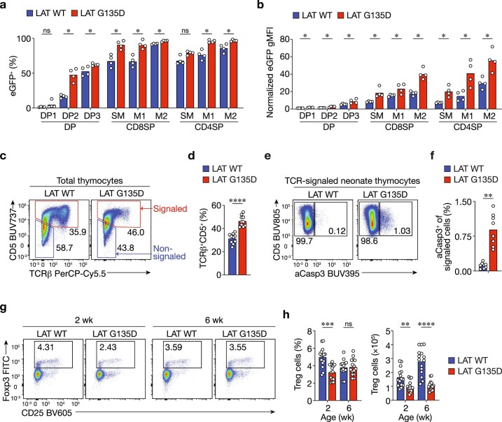
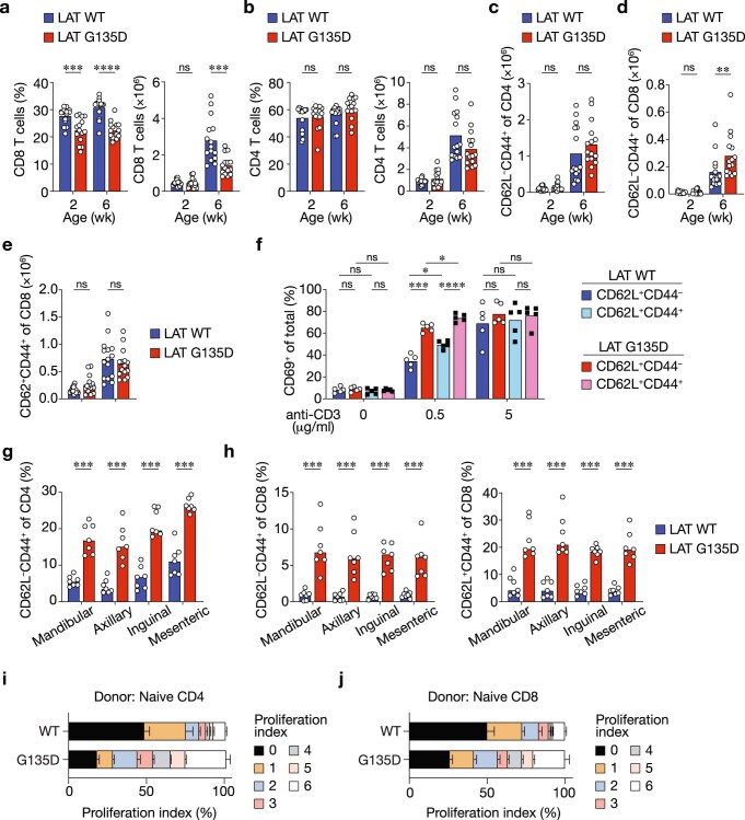
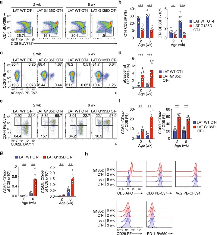
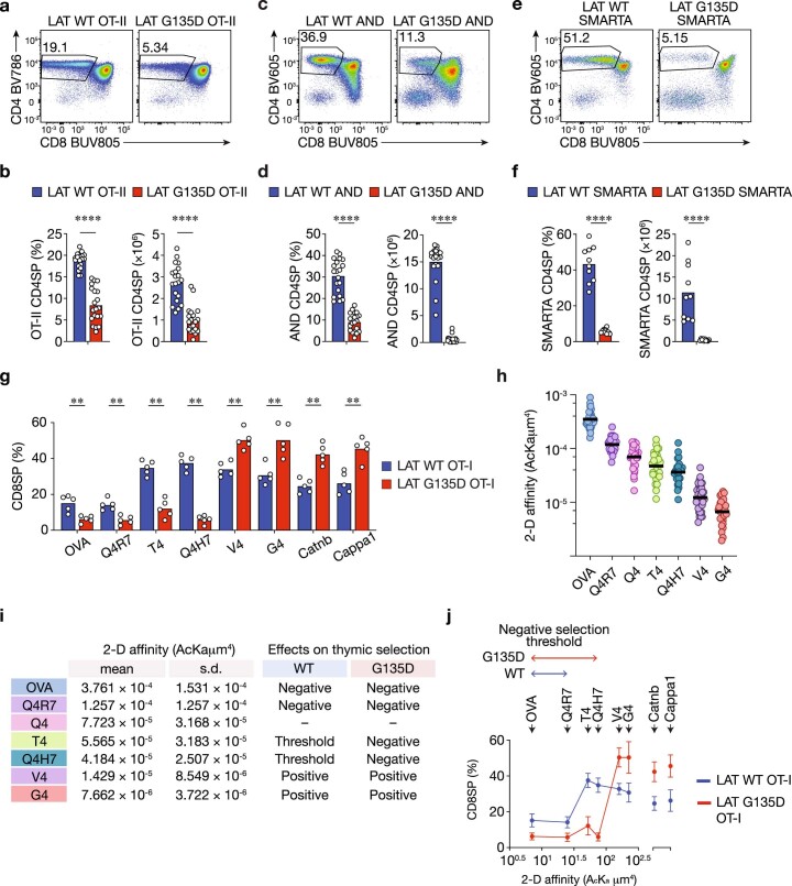
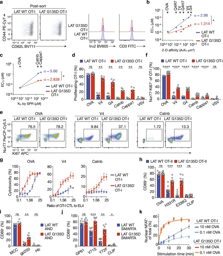
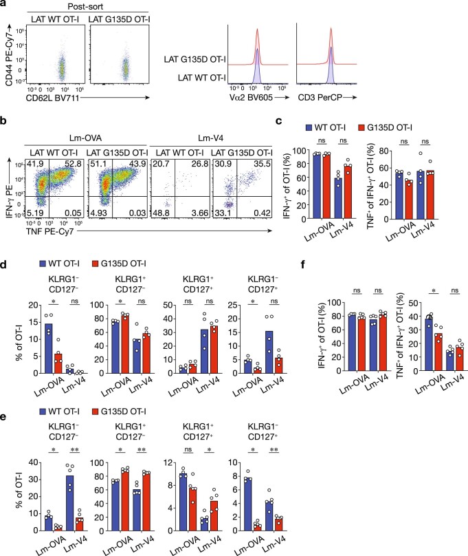
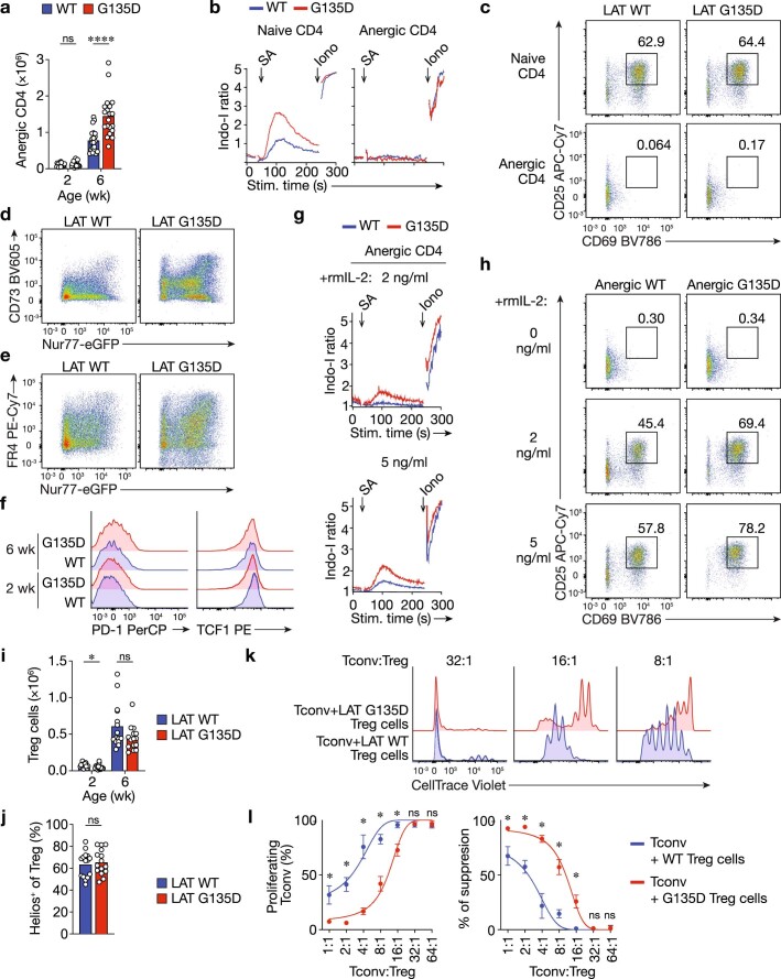
Mature T cells must discriminate between brief interactions with self-peptides and prolonged binding to agonists. The kinetic proofreading model posits that certain T-cell antigen receptor signaling nodes serve as molecular timers to facilitate such discrimination. However, the physiological significance of this regulatory mechanism and the pathological consequences of disrupting it are unknown. Here we report that accelerating the normally slow phosphorylation of the linker for activation of T cells (LAT) residue Y136 by introducing an adjacent Gly135Asp alteration (LATG135D) disrupts ligand discrimination in vivo. The enhanced self-reactivity of LATG135D T cells triggers excessive thymic negative selection and promotes T-cell anergy. During Listeria infection, LATG135D T cells expand more than wild-type counterparts in response to very weak stimuli but display an imbalance between effector and memory responses. Moreover, despite their enhanced engagement of central and peripheral tolerance mechanisms, mice bearing LATG135D show features associated with autoimmunity and immunopathology. Our data reveal the importance of kinetic proofreading in balancing tolerance and immunity.


Conflict of interest statement

A.M. is a cofounder of Arsenal Biosciences, Spotlight Therapeutics and Survey Genomics. A.M. serves on the board of directors at Spotlight Therapeutics and Survey Genomics and is board observer at Arsenal Biosciences. A.M. is a member of the scientific advisory boards of Arsenal Biosciences, Spotlight Therapeutics, Survey Genomics and NewLimit. A.M. owns stock in Arsenal Biosciences, Spotlight Therapeutics, NewLimit, Survey Genomics, PACT Pharma and Merck. A.M. has received fees from Arsenal Biosciences, Spotlight Therapeutics, NewLimit, 23andMe, PACT Pharma, Juno Therapeutics, Trizell, Vertex, Merck, Amgen, Genentech, AlphaSights, Rupert Case Management, Bernstein and Analytical, Life Science & Diagnostics Association (ALDA). A.M. is an investor in and informal adviser to Offline Ventures and a client of EPIQ Capital Group. The A.M. laboratory has received research support from Juno Therapeutics, Epinomics, Sanofi, GlaxoSmithKline, Gilead and Anthem. D.Z. has a consulting agreement and research collaboration agreement with Pieris Pharmaceuticals. A.W. is a cofounder and consultant of Nurix Therapeutics. A.W. is on the scientific advisory boards at BlueSphere Bio, BridGene Biosciences, Genentech, Jasper Therapeutics and Soteria Biotherapeutics. He receives consulting fees and has received stock options or owns stock in these companies. The other authors declare no competing interests.

Figures

Fig. 1
Fig. 1. LATG135D affects thymopoiesis and decreases the production of single-positive thymocytes.
ai, Cellularity of thymi harvested from wild-type (WT) or LATG135D C57BL/6 mice as neonates (2 weeks old) or as adults (6 weeks old). The data are representative of at least four independent experiments. wk, weeks. a, Representative pseudocolor plots depicting the expression of CD4 and CD8. b,c, Bar graphs summarizing the percentages (b) and absolute numbers (c) of CD4SP (left) and CD8SP cells (right) among live thymocytes. d, Expression of the thymocyte maturation markers CD62L and MHC-I H-2Kb on CD4SP cells, including semi-mature (CD62LH-2Kb–; SM), mature stage 1 (CD62LH-2Kb+; M1) and mature stage 2 cells (CD62L+H-2Kb+; M2). e,f, Bar graphs summarizing the percentages (e) and absolute numbers (f) of CD62L+H-2Kb+ M2 CD4SP (left) and M2 CD8SP thymocytes (right). g, Representative pseudocolor plots showing CD69 and TCRβ expression profiles of DP thymocytes, including preselection DP1 (CD69TCRβ), midselection DP2 (CD69medTCRβmed) and postselection DP3 cells (CD69hiTCRβhi). h,i, Bar graphs summarizing the percentages (h) and absolute numbers (i) of DP2 (left) and DP3 cells (right) among DP thymocytes. In b, c, e, f, h and i, each dot represents an individual mouse (n = 15). In b, **P = 0.0041 and ****P < 0.0001. In c, ****P < 0.0001, NS (not significant) = 0.5393 (left) and NS = 0.2169 (right). In e, ***P = 0.0001 and ****P < 0.0001. In f, ****P < 0.0001, **P = 0.0057 and *P = 0.0295. In h, **P = 0.0012 and ****P < 0.0001. In i, ****P < 0.0001, *P = 0.0209 and NS = 0.6236. Statistical significance was determined by two-tailed Mann–Whitney U-test. Source data
Fig. 2
Fig. 2. LATG135D promotes negative selection in the medulla.
a, Representative calcium traces of wild-type and LATG135D CD53 preselection DP thymocytes were analyzed before and after the addition of streptavidin (SA) to crosslink anti-CD3ε. b, Immunoblot analysis of specific proximal signaling proteins of wild-type or LATG135D CD53 preselection DP thymocytes after crosslinking with anti-CD3ε antibody. MW, molecular weight of protein ladders (kDa). The two columns of labels on the left represent the protein name and the amino acid residue, respectively. The ‘p-’ indicates phosphorylation. c, Histogram of the expression of eGFP in various thymocyte developmental subsets from wild-type and LATG135D Nur77–eGFP reporter mice. d, Pseudocolor plots of the expression of CCR7 and cleaved caspase-3 (aCasp3) in DP thymocytes. e, Bar graphs summarizing the percentages of DP thymocytes undergoing apoptosis in the cortex (CCR7aCasp3+) or ready to migrate to the medulla (CCR7+aCasp3+). Each dot represents a single mouse (n = 11). ****P < 0.0001 and NS = 0.5726. f, Immunoblot analysis of the total protein expression of Bim and aCasp3 of sorted wild-type and LATG135D DP thymocytes. g,h, Analysis of the expression of CD5 and aCasp3 on total thymocytes (CD5+TCRβ+; an example of the gating strategy is shown in Extended Data Fig. 3e). Representative pseudocolor plots (g) and summarized bar graphs (h) are shown. The numbers in g show the percentages of apoptotic (aCasp3+) and nonapoptotic (aCasp3) cells. Each symbol in h represents a single mouse (n = 10). ****P < 0.0001. ik, Representative pseudocolor plots (i) showing the expression of CCR9 and CCR7 on CD5+TCRβ+aCasp3+ thymocytes. The bar graphs show the percentages of CCR7 and CCR7+ CD5+TCRβ+aCasp3+ thymocytes, indicative of clonal deletion in the cortex (CCR7) versus cells ready to migrate to the medulla (CCR7+) (j) and stage of development (CCR9+CCR7, CCR9+CCR7+ or CCR9CCR7; k). Each dot represents an individual mouse (n = 10). **P = 0.0011, ***P = 0.0001, ****P < 0.0001, NS = 0.0892 (j, left), NS = 0.1230 (j, right) and NS = 0.3930 (k). In ak, the data are representative of two (f), three (a,c,ik) or four (b,d,e,g,h) independent experiments. Statistical significance was determined by two-tailed Mann–Whitney U-test. Source data
Fig. 3
Fig. 3. LATG135D augments self-peptide-driven homeostatic proliferation of peripheral T cells.
a,c, Representative pseudocolor plots of the expression of CD62L and CD44 on peripheral spleen CD4 (a) and CD8 T cells (c) from wild-type versus LATG135D neonatal (2 week) and adult (6 week) mice. The numbers associated with the gates show the percentages of naive (CD62L+CD44), central memory (CD62L+CD44+) and effector memory (CD62LCD44+) cells. b,d,e, Bar graphs depicting the percentages of effector memory (CD62LCD44+) cells among peripheral CD4 (b) and CD8 (d) T cells and the percentages of central memory (CD62L+CD44+) cells among peripheral CD8 T cells (e). Each dot represents a single mouse (n = 15). The data are representative of at least five independent experiments. ****P < 0.0001, NS = 0.4302 (b) and NS = 0.9588 (d). fi, Naive CD4 (f,g) or CD8 (h,i) T cells were sorted from 4- to 5-week-old wild-type or LATG135D mice, labeled with CellTrace Violet and adoptively transferred intravenously into congenic hosts (CD45.1+), MHC-II–/– hosts or Tap1–/–B2m–/– hosts (as indicated) that had been sublethally irradiated (300 rads) the day before. The dilution of CellTrace Violet was assessed by flow cytometry 4 d post-transfer. f,h, Representative flow plots of CellTrace Violet dilution and the expression of CD5. g,i, Bar graphs summarizing the percentages of adoptively transferred CD4 (g) and CD8 cells (i) that underwent proliferation. Each dot represents an individual mouse (n = 6 for the CD45.1+ C57BL/6 host and n = 4 for the MHC-II–/– and Tap1–/–B2m–/– hosts. The data were compiled from three independent experiments. **P = 0.0043 (g), **P = 0.0022 (i), NS = 0.3429 (g) and NS = 0.9429 (i). Statistical significance in b, d, e, g and i was determined by two-tailed Mann–Whitney U-test. Source data
Fig. 4
Fig. 4. LATG135D promotes OT-I CD8 T-cell effector function and augments sensitivity to weak ligand stimuli.
a, Fetal thymi from wild-type or LATG135D.OT-I.Rag1–/–.Tap1–/– mice were cultured with OVA peptide, OVA APLs or self-peptides, as indicated. The percentages of CD8SP cells were analyzed on day 4. The data are representative of two independent experiments. Representative flow plots show the development of CD8SP cells in FTOC. b, Naive wild-type or LATG135D.OT-I.Rag1–/– TCR transgenic CD8 T cells were sorted from 4- to 5-week-old mice and stimulated overnight with TCRα–/– antigen-presenting cells pulsed with OVA peptide, OVA APLs or self-peptide Catnb or Cappa1 over a wide range of peptide concentrations (as indicated on the x axis). The upregulation of CD69 was analyzed the next day by flow cytometry. CD69+ cells were plotted against peptide concentrations. The data represent means ± s.d. (n = 3 independent experiments). c, Naive wild-type or LATG135D.OT-I.Rag1–/– TCR transgenic CD8 T cells were sorted from 4- to 5-week-old mice, labeled with CellTrace Violet and cocultured with TCRα–/– antigen-presenting cells pulsed with OVA, APLs (V4 or G4), self-peptide (Catnb or Cappa1) or unrelated peptide (VSV). The fluorescence profile of CellTrace Violet and expression of CD5 were assessed on day 4. The data are representative of at least three independent experiments. d, Representative flow plots depicting the production of the cytokines TNF and IFNγ by naive cells from wild-type or LATG135D.OT-I.Rag1–/– mice stimulated with OVA-, V4- or Catnb-pulsed TCRα–/– antigen-presenting cells overnight. The data are representative of three independent experiments. e, Naive cells from wild-type or LATG135D.OT-I.Rag1–/– mice were sorted from 4- to 5-week-old mice and stimulated with OVA-, V4- or Catnb-pulsed TCRα–/– antigen-presenting cells overnight. The production of IL-2 and expression of pSTAT5 were measured by intracellular staining and flow cytometry. The data are representative of four independent experiments. Source data
Fig. 5
Fig. 5. LATG135D-mediated signaling promotes NFAT1 and Nur77 translocation into the nucleus.
ad, Naive wild-type or LATG135D.OT-I.Rag1–/– CD8 T cells from 4- to 5-week-old mice were sorted and subjected to nuclear staining with CellTrace Blue dyes, then stimulated in vitro with 10 or 0.1 nM OVA peptide-pulsed TCRα–/– splenocytes over a time course of 180 or 240 min (as indicated on x axis). Cell nuclei were isolated according to a published protocol, fixed and permeabilized, then subjected to antibody staining for NFAT1, Nur77, NF-κB or Egr-2. Nuclear NFAT1 (a), Nur77 (b), NF-κB (c) and Egr-2 (d) expression was analyzed by flow cytometry. The percentage of positive nuclei (for NFAT1, NF-κB and Egr-2) or mean fluorescence intensity (MFI; Nur77) for individual conditions was plotted against the stimulation time to depict the nuclear translocation kinetics of transcription factors, as indicated. The data represent means ± s.d. (n = 4 independent experiments). Source data
Fig. 6
Fig. 6. LATG135D augments the CD8 T-cell response in vivo.
a, Representative pseudocolor plots showing the frequency of CD45.2 OT-I T cells among total CD8 T cells on day 7 postinfection of L. monocytogenes expressing OVA (Lm-OVA) or V4 (Lm-V4). b, Bar graphs depicting the frequency of OT-I T cells among total CD8 T cells on day 7 postinfection (n = 4). *P = 0.0286 and NS = 0.2. c, Representative pseudocolor plots of the expression of KLRG1 and CD127 on wild-type or LATG135D.OT-I.Rag1–/– CD8 T cells on day 7 postinfection. d, Bar graphs summarizing the relative distribution of each subset based on the expression of KLRG1 and CD127 (as in c) of wild-type or LATG135D.OT-I.Rag1–/– CD8 T cells on day 7 postinfection (n = 4). e, Bar graphs summarizing the percentage of OT-I T cells among total spleen CD8 T cells 4 days after rechallenge with VSV-OVA (n = 4 for wild-type donor and Lm-OVA first infection, n = 5 for LATG135D donor and Lm-OVA infection, as well as wild-type or LATG135D donor and Lm-V4 first infection). **P = 0.0079 and NS = 0.6863. f, Representative pseudocolor plots of the expression of KLRG1 and CD127 on wild-type or LATG135D.OT-I.Rag1–/– CD8 T cells on day 7 after VSV-OVA rechallenge. g, Bar graphs depicting the frequency of each subset based on the expression of KLRG1 and CD127 on OT-I T cells in the spleen 4 days after rechallenge with VSV-OVA (n = 4 for wild-type donor and Lm-OVA first infection, n = 5 for LATG135D donor and Lm-OVA infection, as well as wild-type or LATG135D donor and Lm-V4 first infection). h, Representative histograms of the expression of TCF1 in wild-type or LATG135D.OT-I.Rag1–/– CD8 T cells analyzed on day 4 after VSV-OVA rechallenge. i, Bar graph quantifying the percentage of TCF1+ cells (the positive horizontal bar gate shown in h) (n = 4 for wild-type donor and Lm-OVA first infection and n = 5 for LATG135D donor and Lm-OVA infection, as well as wild-type or LATG135D donor and Lm-V4 first infection). *P = 0.0159 and **P = 0.0079. In ai, the data are representative of at least three independent experiments. The dots in b, e and i represent individual mice. Statistical significance was determined by two-tailed Mann–Whitney U-test. In d and g, the data represent means ± s.d. Source data
Fig. 7
Fig. 7. Aged female LATG135D mice develop higher titers of anti-dsDNA IgG than wild-type counterparts, along with signs of colitis.
a,b, Sera from aged wild-type or LATG135D female mice (1 year old) were collected and subjected to antinuclear antibody staining (a) and anti-dsDNA IgG titers were measured by ELISA (b) (n = 15 for the wild type and n = 24 for LATG135D). ****P < 0.0001. The data are representative of two independent experiments. c, Histopathological analysis and hematoxylin and eosin (H&E) staining revealed signs of acute and chronic colitis, including abnormal neutrophil infiltration and crypt destruction/distortion (arrowheads), in aged LATG135D female mice that were absent from wild-type littermates. The data are representative of two independent experiments. Scale bars, 100 μm. d, Representative flow cytometry plots of the expression of CD62L and TCF1 on wild-type and LATG135D CD25+Foxp3+ regulatory T cells from 1-year-old mice. e, Bar graph summarizing the frequency of each subset as a proportion of total regulatory T cells. The regulatory T-cell subsets R1, R2 and R3 represent CD62L+TCF1+, CD62LTCF1+ and CD62LTCF1 cells, respectively. ***P = 0.0006 (left), ***P = 0.0002 (right) and NS = 0.1142. The data are representative of three independent experiments. f, Representative flow cytometry plots of the expression of CD62L and CD44 on wild-type and LATG135D CD25+Foxp3+ regulatory T cells from 1-year-old mice. g, Bar graph summarizing the frequency of CD44hi and CD44low regulatory T-cell populations as a proportion of total regulatory T cells. ***P = 0.0010 and ****P < 0.0001. The data are representative of three independent experiments. h, Expression of CD44, LEF1, ICOS and CD103 of wild-type and LATG135D CD25+Foxp3+ regulatory T cells from 1-year-old mice. In b, e and g, statistical significance was determined by two-tailed Mann–Whitney U-test. Source data
Fig. 8
Fig. 8. LATG135D peripheral T cells adapt in an age-dependent manner to maintain tolerance.
a, Representative histograms of the expression of Nur77–eGFP, CD5, CD6, DGK-ζ and TOX in peripheral CD4 T cells from wild-type and LATG135D mice (ages as indicated). The data are representative of at least three independent experiments. b, Representative histograms of the expression of coinhibitory receptors, including PD-1, LAG-3, TIM-3, TIGIT and VISTA on naive peripheral CD4 T cells from wild-type and LATG135D mice (ages as indicated). The data are representative of at least three independent experiments. c, Representative pseudocolor plots showing the expression of CD73 and FR4 on peripheral Foxp3 CD4 T cells from wild-type and LATG135D mice (ages as indicated). d, Bar graph summarizing the percentages of CD73hiFR4hi cells. Each dot represents one mouse (n = 20). The data are representative of and compiled from at least five independent experiments. ****P < 0.0001 and NS = 0.0950. e, Representative pseudocolor plots showing the expression of Foxp3 and CD25 in peripheral CD4 T cells from wild-type and LATG135D mice (ages as indicated). f, Bar graph summarizing the percentages of Foxp3+CD25+ cells. Each dot represents one mouse (n = 15). The data are representative of and compiled from at least five independent experiments. **P = 0.0037 and NS = 0.4423. g, Representative pseudocolor plot showing the expression of PD-1 and GITR in peripheral Foxp3+CD25+ regulatory T cells isolated from the spleens of 6-week-old wild-type and LATG135D mice. h, Bar graph summarizing the percentage of PD-1hiGITRhi as a proportion of total Treg cells. The data are representative of at least five independent experiments. i, Histogram showing the expression of Nrp-1 and CD25. The data are representative of at least five independent experiments. In d, f and h, statistical significance was determined by two-tailed Mann–Whitney U-test. Source data
Extended Data Fig. 1
Extended Data Fig. 1. Generation of LAT G135D knock-in mouse.
a. The cartoon illustrates the genomic locus of mouse Lat. The numbers represent individual exons. The nucleotide and amino acid sequences of exon 7 are depicted. The coding regions are listed in capital letters and the intron immediately following exon 7 is shown in lowercase letters. The orange arrows represent the two sgRNAs used to generate the G135D knock-in mice. b. Illustration of the molecular mechanisms underlying the engineering strategy. In brief, mammalian T cells express natural (wild-type) LAT, which exhibits slow phosphorylation kinetics upon TCR recognition of ligand; this serves as a proofreading bottleneck to create the molecular time delay required for proper TCR ligand discrimination. Only bona fide activating ligands that interact with the TCR with a sufficiently long bond lifetime pass this slow signaling bottleneck to activate T cells (left). On the other hand, the bond lifetime of an interaction between the TCR and self-pMHC is too short to activate T cells (left). Importantly, the phosphorylation of LAT Y136 is regulated by the amino acid preceding Y136. Natural LAT has a small glycine at the −1 position, leading to slow phosphorylation (right); sequence-modifying LAT with a negatively charged aspartate substantially facilitates the rate and magnitude of Y136 phosphorylation (right). G135D mutant LAT therefore bestows on T cells the ability to respond to very weak ligands or self-peptides. c. The PCR genotyping results of the seven pups born to the founder generated with CRISPR/Cas9 using sgRNA #1 and the four pups born to the founder generated with sgRNA #2. Homology-directed repair (HDR) initiated by electroporation provided the repair template for CRISPR/Cas9 and was used as a positive control for PCR screening. gDNA from C57BL/6 parents was used as a negative control. The numbers along the top of the gel represent the individual pups. Pups #1 and #2 from the sgRNA #2 experiments are founders #1 and #2, respectively. The genotyping result was performed once but confirmed with Sanger sequencing analysis. d. Four-color chromatograms of the exon 7 gDNA sequence analyses of founders #1 and #2.
Extended Data Fig. 2
Extended Data Fig. 2. The thymic cellularity of G135D knock-in mice.
a. Bar graphs show the total thymocyte numbers of mice at the indicated ages. wk: weeks. Bar graphs show the frequency (left) or absolute number (right) of double-negative (DN) thymocytes. c. Bar graphs show the frequency (left) or absolute number (right) of double-positive (DP) thymocytes. d. Representative flow plots of the expression of thymocyte maturation markers CD62L and MHC-I H-2Kb on CD8SP. a–c. Each dot represents an individual mouse. n = 15. Two-tailed Mann-Whitney test. *P = 0.0204, ****P < 0.0001, ns = 0.2901 (a); *P = 0.0111, ns = 0.8702 (left), ns = 0.0555 (middle), and ns = 0.0502 (right) (b); ****P < 0.0001 and ns = 0.0502 (c). Two-tailed Mann-Whitney test. Source data
Extended Data Fig. 3
Extended Data Fig. 3. The expression of G135D augments LAT Y136–PLC-γ1 signaling.
a. Representative flow cytometry plot of wild-type or G135D pre-selection CD53 thymocytes labeled with titrated amounts of CellTrace Violet dye. FSC, forward scatter. b. Bar graphs depict the statistical analysis of calcium responses (Fig. 2a) of wild-type or G135D pre-selection CD53 thymocytes in response to anti-CD3ε crosslinking (concentration as indicated). The bar graph at left shows the response time to reach the peak (unit: seconds, s). The bar graphs at the right show the area under curve (AUC) of the calcium responses; the larger the AUC, the stronger the calcium responses. **P = 0.0022 (left), *P = 0.0108 (middle left), *P = 0.0173 (middle right), *P = 0.0152 (right).. Two-tailed Mann-Whitney test. c,d. The band densities of phosphorylated LAT Y136 (c), PLC-γ1 (c), Zap-70 Y493 (d), LAT Y195 (d), ERK T202/Y204 (d) are shown. Data were standardized to the band density of the wild-type no-stimulatory control. Quantified data are summarized from six independent experiments. Error bars represent SEM. LAT p-Y136: ***P = 0.000778 (left), ***P = 0.000329, **P = 0.00127; Total LAT: ns = 0.781024 (middle left), ns = 0.514455 (middle right); ns = 0.852218 (right); PLC-γ1 Y783: **P = 0.001166 (right); *P = 0.005454 (right), *P = 0.023421 (left); Zap-70 p-Y783: ns = 0.663221 (middle left); ns = 0.966048 (middle right); ns = 0.708332 (right); LAT p-Y195: ns = 0.055783 (middle left), ns = 0.898515 (middle right); ns = 0.054248 (right); ERK p-T202/Y204: ns = 0.614684 (middle left), ns = 0.165792 (middle right), ns = 0.091339 (right). Paired student t test (Two sided).. A.U., arbitrary unit. e,f. Immunoblot analysis of wild-type or G135D LAT naive CD4 T cells after crosslinking with 5 μg/ml biotinylated anti-CD3ε antibody at 37 °C (time as indicated above the blots). Phosphorylation of total LAT or phopsho-LAT p-Y136 and p-Y195, was analyzed as indicated. The band densities of phosphorylated LAT Y136 and LAT Y195 are shown in the graphs in (f, left and middle). Normalized percentage of LAT Y136 phosphorylation level in relative to the maximum (f, right). Data are representative of four independent experiments. MW: molecular weight of protein ladders (unit: kDa). Data are presented as mean values ± SEM. Source data
Extended Data Fig. 4
Extended Data Fig. 4. The expression of G135D alters thymic selection.
a,b. Bar graphs summarize the percentage of Nur77-eGFP+ wild-type or G135D thymocytes among the indicated developmental subsets. Each symbol represents one individual mouse; n = 4; *P = 0.0286; ns = 0.571. Two-tailed Mann-Whitney test. c,d. Representative flow plots show the gating strategy for “signaled” or “non-signaled” wild-type or G135D LAT thymocytes (c). The signaled thymocytes are defined as the CD5+TCR+ population. The bar graph represents the frequency of “signaled” thymocytes (d). Each symbol represents one individual mouse; n = 10; ****P < 0.0001. Two-tailed Mann-Whitney test. e,f. Expression of CD5 and cleaved caspase-3 (aCasp3) in signaled thymocytes harvested from wild-type or G135D neonates. Each symbol represents one individual mouse; n = 8; **P = 0.0002. Two-tailed Mann-Whitney test. g,h. Representative flow plots (g) and bar graphs (h) show the expression of Foxp3 and CD25. Each symbol represents one individual mouse; n = 15. **P = 0.0020; ***P = 0.0001; ****P < 0.0001; ns = 0.8943. Two-tailed Mann-Whitney test. wk: weeks. Source data
Extended Data Fig. 5
Extended Data Fig. 5. The expression of G135D in peripheral T cells promotes the emergence of “memory-phenotype” T cells.
a,b. Bar graphs show the percent (left) or absolute number (right) of CD8 (a) or CD4 (b) T cells in the periphery at the indicated ages. wk: weeks. Data are representative of 4 independent results. c–e. Bar graphs show the absolute numbers of CD62LCD44+ CD4 T cells (c), CD62LCD44+ CD8 T cells (d), or CD62L+CD44+ CD8 T cells (e). Data are representative of 4 independent results. Two-tailed Mann-Whitney test. f. CD62L+CD44+ CD8 T cells were sorted from wild-type or LAT G135D mice (8-week old). Sorted CD62L+CD44+ CD8 T cells were then stimulated with anti-CD3 antibody (1 μg/ml) overnight at 37oC. The expression of CD69 was examined on the next day. Bar graphs summarize the percentage of CD69+ T cells. Data are representative of 3 independent experiments. At 0 μg/ml: ns = 0.9807 (bottom left), ns = 0.8325 (bottom right), ns = 0.8784 (middle), ns = 0.9060 (top). At 0.5 μg/ml: P*** = 0.002, P**** < 0.0001, P* = 0.0107 (middle), P* = 0.0221 (top). At 5 μg/ml: ns = 0.7696 (bottom left), ns = 0.9485 (bottom right), ns = 0.9899 (middle), ns = 0.9939 (top). 2-way ANOVA. Data are representative of 4 independent results. g.h. Bar graphs summarize the frequency of CD62LCD44+ CD4 T cells (g), CD62LCD44+ CD8 T cells (h), or CD62L+CD44+ CD8 T cells (h) from various lymph nodes (as indicated) from 6-week old wild-type or G135D mice. n = 7; P*** = 0.0006. Two-tailed Mann-Whitney test. Data are representative of 3 independent results. i,j. Bar graphs summarize the proliferation index as shown in Fig. 3f (in c) and 3 h (in d). n = 4. Data are representative of 3 experiments. Data are presented as mean values ± SD. a-d. Each dot represents an individual mouse. ***P = 0.0005; ****P < 0.0001 (a, left); ***P = 0.0003 (a, right); ns = 0.5949 (a); ns = 0.8381 (b, left); ns = 0.0627 (b, middle left); ns = 8063 (b, middle right); ns = 0.1160 (b, right). Two-tailed Mann-Whitney test. Source data
Extended Data Fig. 6
Extended Data Fig. 6. Thymic and peripheral T cell phenotypes in G135D LAT.OT-I.Rag1–/– mice recapitulate those in G135D mice on the polyclonal C57BL/6 background.
a. Pseudocolor flow plots show the expression of CD4 and CD8 on live thymocytes harvested from neonatal or adult wild-type or G135D LAT.OT-I.Rag1–/– mice. b. Bar graphs show the percentage of CD8 OT-I.Rag1–/– T cells in wild-type or G135D LAT mice at the indicated ages. c,d. Representative flow plots or bar graphs show the expression of CCR7 and cleaved caspase 3 (aCasp3) in DP thymocytes. e,f. Representative pseudocolor plots depict the expression of CD62L and CD44 (e). Bar graphs summarize the frequencies of CD62LCD44+ (left) and CD62L+CD44+ (right) CD8 OT-I.Rag1–/– T cells. g. Bar graphs show the absolute numbers of CD62LCD44+ or CD62L+CD44+ CD8 OT-I.Rag1–/– T cells in wild-type or G135D LAT mice at the indicated ages. h. Representative histograms display the expression of CD5, CD3, Vα2, CD28 and PD-1 on wild-type or G135D LAT.OT-I.Rag1–/– CD8 T cells. a-g. Each dot represents an individual mouse; n = 8 (b); n = 8 (d); n = 5 (f); n = 6 (g); ***P = 0.0002 (b); ***P = 0.0002 (d); **P = 0.0022 (f); **P = 0.0022 (g). Two-tailed Mann-Whitney test. Source data
Extended Data Fig. 7
Extended Data Fig. 7. G135D LAT lowers negative selection threshold and enhances productive positive selection.
a–f. Pseudocolor flow plots show the expression of CD4 and CD8 on live thymocytes harvested from adult wild-type or G135D LAT.OT-II.Rag1–/– mice (a), LAT.AND.Rag1–/– mice (c), or LAT.SMARTA.Rag1–/– mice (e). Bar graphs show the percentage (left) or absolute numbers (right) of CD4SP OT-II.Rag1–/– (b), LAT.AND.Rag1–/– (d), or LAT.SMARTA.Rag1–/– (f) thymocytes in wild-type or G135D LAT mice. P**** = < 0.0001; Two-tailed Mann-Whitney test. Data are representative of 5 independent results. g. Bar graphs show the frequency of developed wild-type or G135D LAT.OT-I.Rag1–/–Tap1–/– CD8SP cells in fetal thymic organ culture (FTOC) on day 4. Dots represent individual FTOC culture. Data are compiled from two experiments. n = 5; P** = 0.0079; Two-tailed Mann-Whitney test. h,i. Dot plots in (b) show the 2-D affinity of OT-I CD8 T cells binding to OVA- or variant-loaded H-2Kb (with murine β2 m) coated red blood cells. The mean ± standard deviation are summarized in the table (c). Each symbol represents one measurement. n = 33 (OVA, Q4, V4); n = 30 (T4, G4); n = 24 (Q4R7; Q4H7). j. Percentage of CD8SP cells developed in the FTOC (as in g) versus the 2-D affinity of OT-I TCR for OVA or APLs (as in h,i). The threshold for negative selection is marked by the horizontal lines on top of the plots. Source data
Extended Data Fig. 8
Extended Data Fig. 8. G135D LAT.OT-I.Rag1–/– CD8 T cells are ultra-sensitive to very weak ligands and self-peptides.
a. Representative flow plots of post-sorted naive CD44CD62L+ wild-type or G135D CD8 T cells. Histogram plots show the expression level of TCR Vα2 or CD3. b. The plot shows the potency (EC15 from Fig. 4b) over the 2-D affinity. The 2-D affinities of individual ligands are included in b,c. Linear regression analysis was performed to obtain the slope (α) as the measure of TCR discriminatory power. The computed α values for wild-type and G135D LAT-expressing T cells are indicated. *P = 0.0200; F = 8.387; DFn = 1; DFd = 8; the two linear regression lines are significantly different. Two-tailed ANCOVA analysis. c. The plot depicts the potency (concentration producing 15% maximal effect, or EC15, from Fig. 4b) over the published 3-D affinity. Linear regression analysis was performed to obtain the slope (α) as the measure of TCR discriminatory power. The computed values for wild-type or G135D LAT-expressing T cells are indicated. *P = 0.0257; F = 37.38; DFn = 1; DFd = 2; the two linear regression lines are significantly different. Two-tailed ANCOVA analysis. d. Bar graphs show the percent of proliferating wild-type LAT.OT-I.Rag1–/– or G135D LAT.OT-I.Rag1–/– CD8 T cells in response to agonist (OVA), very weak OVA APL (V4 &G4), or self-peptide (Catnb & Cappa1) stimuli. Data are representative of five experiments. e,f. Wild-type LAT.OT-I.Rag1–/– or G135D LAT.OT-I.Rag1–/– CD8 T cells were isolated and stimulated with peptide-pulsed TCRα–/– antigen-presenting cells overnight. The expression of Ki67 and endogenous Nur77 (not the eGFP reporter) was analyzed by intracellular staining the next day. Representative pseudocolor plots (b) and bar graphs (c) are shown. Data are representative of three independent experiments. g. Cytotoxicity assays demonstrating wild-type or G135D LAT cytotoxic T lymphocyte (CTL)-mediated killing of EL4 cells pulsed with 1 μM OVA, V4 peptide, or self-peptide Catnb. EL4 cells were mixed with CTL at the indicated ratios. Killing capacities were assessed after 4 hours. Data are representative of four independent experiments. h–j. CD69 upregulation of sorted naive LAT.OT-II.Rag1–/– (h), LAT.AND.Rag1–/– mice (i), or LAT.SMARTA.Rag1–/– (j) CD4 T cells in responded to 1 μM of peptide-pulsed antigen-presenting cells (as indicated). OVA is an agonist for OT-II TCR, whereas H331R and E336Q are two weaker its altered peptide ligands. MCC is an agonist for AND TCR, whereas gp250 is an identified positively selecting self-peptide for AND TCR. GP61 is an agonist for SMARTA TCR, whereas V71S and Y72F are weaker altered peptide ligands for SMARTA TCR. CLIP or Hb peptides are used as negative controls. Data are representative of 5 independent experiments for h, 3 independent experiments for i, and 3 independent experiments for j. k. The first 30 min of NFAT nuclear translocation kinetics in wild-type LAT.OT-I.Rag1–/– or G135D LAT.OT-I.Rag1–/– CD8 T cells in response to various doses of OVA-pulsed TCR Cα–/– antigen-presenting cells, as shown in Fig. 5a. d,f,h,i,j. Each dot represents a sample from an individual mouse. **P = 0.0079; ****P < 0.0001; ns = 0.0222 (d); ns = 0.5021 (f). ***P = 0.0005 (left); ***P = 0.0002 (right); ns = 0.5737 (left); ns = 0.2345 (right) (h); **P = 0.0022; ns = 0.1320 (left); ns = 0.8857 (right) (i); ns = 0.3423; ***P = 0.0002; **P = 0.0011; *P = 0.0049 (j); Two-tailed Mann-Whitney test. Source data
Extended Data Fig. 9
Extended Data Fig. 9. G135D LAT.OT-I.Rag1–/– CD8 T cells exhibit proliferation fitness and skewing toward terminal differentiation during recombinant Listeria infection.
a. Representative flow plots of post-sorted naive CD44CD62L+ wild-type or G135D OT-I CD8 T cells. b. Splenocytes collected from day 7 post-infection were restimulated with corresponding antigens OVA or V4, and the production of cytokines was assessed by intracellular staining and analyzed by flow cytometry. The representative pseudocolor plots depict the production of IFN-γ and TNF after restimulation. The number in each quadrant represents the percentage of OT-I CD8 T cells producing the indicated cytokine. c. Bar graphs summarize the percentage of wild-type or G135D LAT.OT-I.Rag1-/- CD8 T cells producing IFN-γ (left) or TNF+ of IFN-γ + OT-I cells (right). Dots represents single mice; n = 4. ns = 0.9714 (left); ns = 0.0571 (middle left); ns = 0.0571 (middle right); ns = 0.8857 (right). Two-tailed Mann-Whitney test. d,e. Bar graphs show the frequency of the KLRG1CD127,KLRG1+CD127, KLRG1+CD127+, and KLRG1CD127+ subsets of OT-I.Rag1–/– or G135D LAT.OT-I.Rag1–/– CD8 T cells on day 7 post-infection (d) or on day 4 after rechallenge with VSV-OVA (e). Each dot represents one individual mouse; n = 4 (c); n = 4 (WT donor cells primed with Lm-OVA, e) or n = 5 (all other conditions, e). *P = 0.0286 (d); *P = 0.0159 (e); ns = 0.2286 (far left; d); ns = 0.3429 (middle left; d); ns = 0.1143 (middle; d); ns: P > 0.9999 (middle right; d); ns = 0.0571 (far right; d); ns = 0.0635 (e). Two-tailed Mann-Whitney test. f. Splenocytes collected from day 4 post-infection were restimulated with OVA. The production of cytokines was assessed by intracellular staining and analyzed by flow cytometry. Bar graphs summarize the percentage of wild-type or G135D LAT.OT-I.Rag1–/– CD8 T cells producing IFN-γ (left) or TNF+ of IFN-γ+ OT-I cells (right). Dots represent single mice; n = 4 (WT donor, Lm-OVA first infection); n = 5 (G135D donor, Lm-OVA infection; WT or G135D donor, Lm-V4 first infection). *P = 0.0317; ns = 0.2540 (left); ns = 0.0952 (middle left); ns = 0.3095 (right). Two-tailed Mann-Whitney test. Source data
Extended Data Fig. 10
Extended Data Fig. 10. Enlarged G135D anergic T cell populations are functionally hyporesponsive, but IL-2 treatment can restore their function.
a. Bar graphs show the absolute number of CD73+FR4+ CD4 T cells in the periphery at different ages. Data are representative of at least five experiments. Each dot represents one mouse. n = 20. ****P < 0.0001; ns = 0.1894. Two-tailed Mann-Whitney test. wk: weeks. b. Wild-type or G135D LAT CD4 T cells were isolated and stained with antibodies against CD62L, CD44, CD73, and FR4, and then loaded with calcium dye Indo-I and labeled with biotinylated anti-CD3. Wild-type and G135D CD4 T cells were barcoded with different titrations of CellTrace Violet and pooled together, allowing simultaneous analysis of the cells’ calcium responses upon anti-CD3 crosslinking. Ionomycin treatment served as a positive control. Calcium traces recorded over 5 min are shown. Data are representative of three independent experiments. c. Naive or anergic wild-type or G135D LAT CD4 T cells were sorted, and stimulated with plate-bound anti-CD3 and soluble anti-CD28 monoclonal antibodies overnight. The upregulation of CD69 and CD25 was analyzed the next day. Representative pseudocolor contour plots are shown. Data are representative of three independent experiments. d,e. Representative flow cytometry plots show the expression of Nur77-eGFP and CD73 (d) or FR4 (e). Data are representative of at least five independent experiments. f. Representative histograms depict the expression levels of PD-1 and TCF1 in CD73+FR4+Foxp3 anergic CD4 T cells isolated from wild-type or G135D LAT knock-in mice at 2 or 6 weeks of age. Data are representative of two independent experiments. g. CD73+FR4+Foxp3 anergic wild-type or G135D LAT CD4 T cells were sorted and treated with 2 ng/ml or 5 ng/ml recombinant murine IL-2 (rmIL-2; concentration as indicated) overnight and then analyzed for calcium responses. Anergic T cells were labeled with biotinylated anti-CD3 and loaded with calcium dye Indo-I. Calcium responses to anti-CD3 crosslinking were analyzed by flow cytometry for 5 min. Representative calcium traces are shown. Data are representative of three independent experiments. h. CD73+FR4+Foxp3 anergic wild-type or G135D LAT CD4 T cells were sorted and stimulated with plate-bound anti-CD3 and soluble anti-CD28 overnight along with the addition of 2 ng/ml or 5 ng/ml rmIL-2 (concentration as indicated). The upregulation of CD69 and CD25 were analyzed the next day. Representative flow pseudocolor plots are shown. Data are representative of three independent experiments. i. Bar graphs show the absolute number of CD25+Foxp3+ CD4 T cells in the periphery at different ages. Data are representative of at least five experiments. Each dot represents one mouse. n = 15. *P = 0.0164; ns = 0.1261. Two-tailed Mann-Whitney test. j. Bar graphs show the percentages of Helios+ cells among CD25+Foxp3+ CD4 T cells in adult wild-type and G135D mice. Data are representative of at least five experiments. Each dot represents one mouse. n = 15. ns = 0.6312. Two-tailed Mann-Whitney test. k,l. Wild-type or G135D regulatory T (Treg) cells were sorted from wild-type (WT) or G135D Foxp3-RFP+ mice. Polyclonal naive CD8 T cells (Tconv) from CD45.1+ C57BL/J mice were purified and labeled with CellTrace Violet dyes and used as responsive cells. CellTrace Violet-labled CD45.1+ Tconv cells were co-cultured with titrated ratios of regulatory T cells as indicated. Inhibition of Tconv cell proliferation was used as a readout for Treg cell suppressive function. Representative histograms of Tconv cell proliferation are shown in k. Bar graphs in l summarize the proliferation of Tconv cells and the suppressive activity of Treg cells. *P = 0.0286; ns = 0.8857. Two-tailed Mann-Whitney test. Data are representative of five independent experiments. Data are presented as mean values ± SD. Source data

References

    1. Huseby ES, Teixeiro E. The perception and response of T cells to a changing environment are based on the law of initial value. Sci. Signal. 2022;15:eabj9842. doi: 10.1126/scisignal.abj9842. - DOI - PMC - PubMed
    1. Hogquist KA, Jameson SC. The self-obsession of T cells: how TCR signaling thresholds affect fate ‘decisions’ and effector function. Nat. Immunol. 2014;15:815–823. doi: 10.1038/ni.2938. - DOI - PMC - PubMed
    1. Juang J, et al. Peptide–MHC heterodimers show that thymic positive selection requires a more restricted set of self-peptides than negative selection. J. Exp. Med. 2010;207:1223–1234. doi: 10.1084/jem.20092170. - DOI - PMC - PubMed
    1. Pettmann J, et al. The discriminatory power of the T cell receptor. eLife. 2021;10:e67092. doi: 10.7554/eLife.67092. - DOI - PMC - PubMed
    1. Stepanek O, et al. Coreceptor scanning by the T cell receptor provides a mechanism for T cell tolerance. Cell. 2014;159:333–345. doi: 10.1016/j.cell.2014.08.042. - DOI - PMC - PubMed

Publication types

Substances