Review
. 2023 Mar 13;18(1):53.
doi: 10.1186/s13023-023-02649-x.

Multisystem ALK-positive histiocytosis: a multi-case study and literature review

Affiliations
Review

Multisystem ALK-positive histiocytosis: a multi-case study and literature review

Wei Liu et al. Orphanet J Rare Dis. .

Abstract

Background: Anaplastic lymphoma kinase (ALK)-positive histiocytosis, a novel rare histiocytic proliferation, was first described in 2008; it occurs in early infancy with liver and hematopoietic involvement. The spectrum was subsequently broadened to include localized diseases in older children and young adults. However, its full clinicopathological features and molecular lineage have not been fully elucidated.

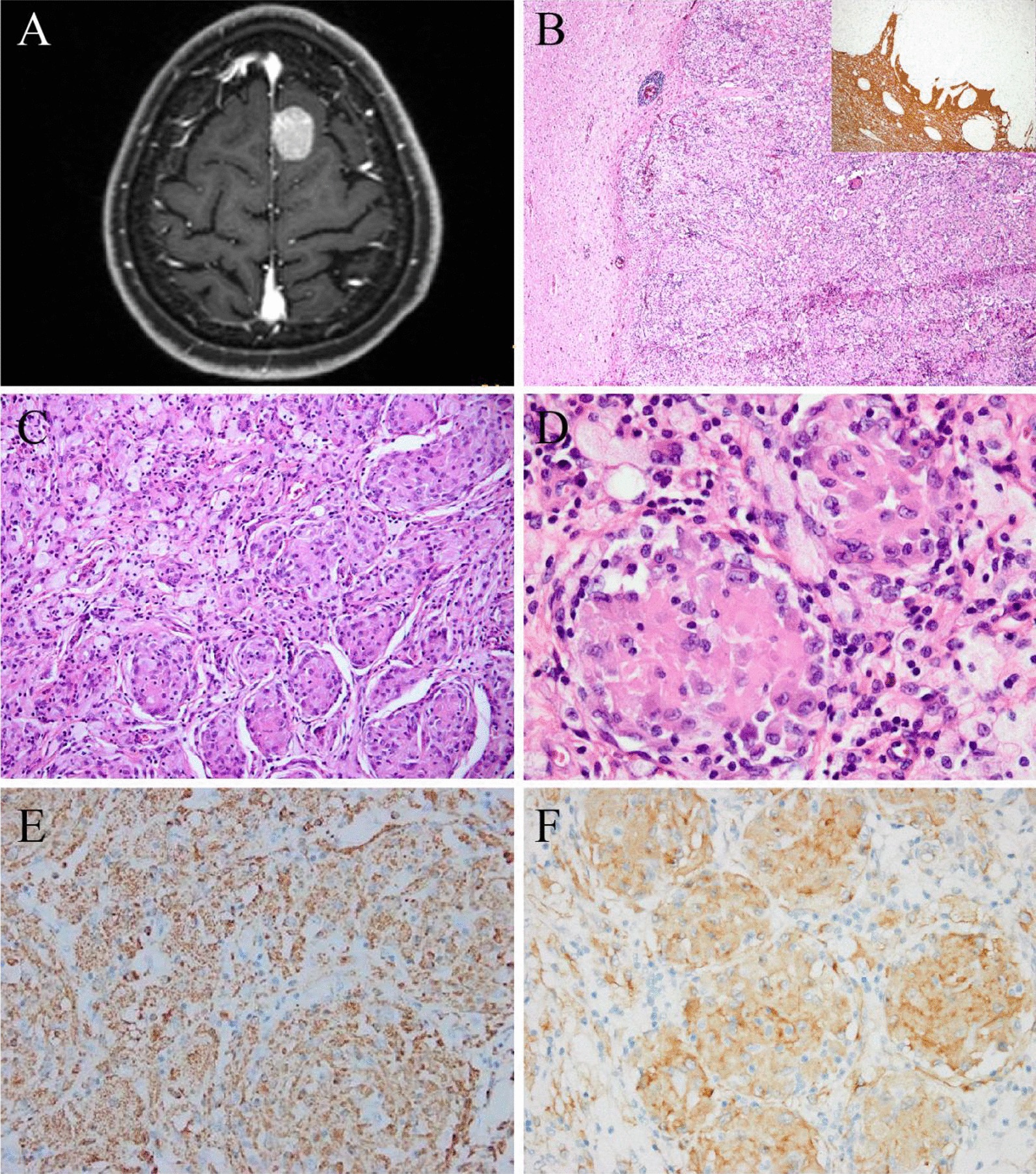
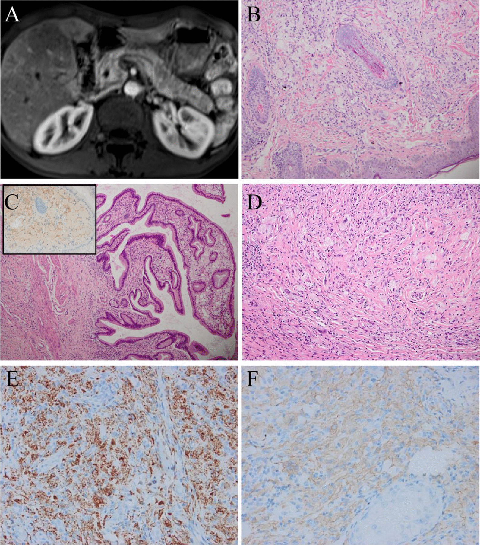
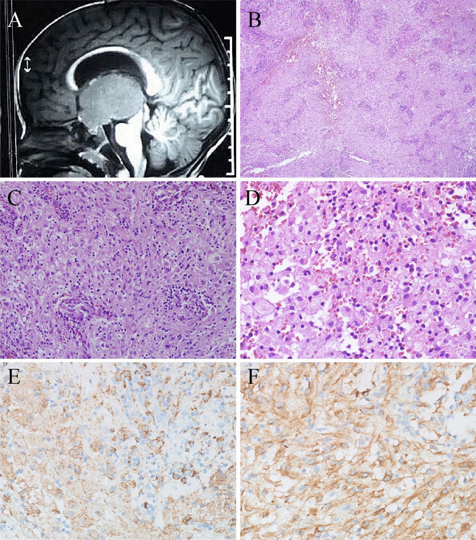
Results: Here, we report four cases of multisystem ALK-positive histiocytosis without hematopoietic involvement. Clinically, three patients were adults aged between 32 and 51 years. Two patients', whose main manifestations were intracranial mass and numerous micronodules in the thoracoabdominal cavity organs and skin papules respectively, had a partial response to ALK inhibitors after surgery. One patient presented with mediastinal neoplasm without surgical treatment, and progressive disease occurred after two years of ALK inhibitor therapy. The fourth patient was a 17-month-old male with a large intracranial mass and presented with a poor response to ALK inhibitor and chemoradiotherapy; he died eight months after surgery. Pathologically, the histiocytes were large, with abundant eosinophilic cytoplasm, and mixed with variable numbers of foamy cells and Touton giant cells. Interstitial fibrosis was also observed. Histiocytes were positive for macrophage markers (CD68 and CD163) and ALK. KIF5B-ALK fusions were detected in two cases, EML4-ALK in one, and both DCTN1-ALK and VRK2-ALK fusions were detected in one case.

Conclusions: We observed that ALK inhibitors present robust and durable responses in adult patients but a poor response in young children with central nervous system involvement. There is no consensus on the optimal treatment regimen and long-term prognosis requires further observation. Moreover, every unusual histiocytic proliferative lesion, especially unresectable and multisystem involvement, should be routinely tested for ALK immunohistochemical staining to identify this rare disease.

Keywords: ALK-positive histiocytosis; EML4-ALK; KIF5B-ALK; Multisystem involvement.


Conflict of interest statement

The authors declare that they have no competing interests.

Figures

Fig. 1
Fig. 1
Radiological and pathological features of case 1. A Chest CT showed a large mass in the upper lobe of the right lung, involving the right anterior superior mediastinum, hilar, and hilar lymph nodes. B, C The mediastinal nodule exhibited large polymorphic histiocytes with frequent Touton giant cells. D The tumor cells were positive for CD68. E The tumor cells were positive for ALK1. F ALK-FISH (a break-apart probe) was positive. (Original magnification × 200 [B, C, D, E]; × 1000 [F])
Fig. 2
Fig. 2
Radiological and pathological features of case 2. A CT showed a mass (2.5 × 2 cm in size) in the left frontal region near the cerebral falx of the brain. B The lesion infiltrated brain parenchyma, and GFAP staining showed an ill-defined lesion (inlet). C The lesion revealed a relatively uniform modular structure. D The tumor cells were large with round, ovoid, or clefted nuclei. E The tumor cells were positive for CD68. F The tumor cells were positive for ALK1. (Original magnification × 40 [B]; × 100 [C]; × 200 [E, F]; × 400 [D])
Fig. 3
Fig. 3
Radiological and pathological features of case 3. A MRI showed multiple patchy and/or nodular abnormal signal shadows in the liver. B The skin biopsy showed dermally infiltrates of histiocytes and Touton cells. C There were numerous foam cells with ALK-positive (inlet) in the mucosal layer of the gallbladder and fibrous tissue hyperplasia in the muscular layer. D The liver lesions showed prominent interstitial sclerosis, accompanied by foamy cells and Touton giant cells. E The histiocytes were positive for CD68 (Skin). F The histiocytes were positive for ALK1 (Skin). (Original magnification × 40 [C]; × 100 [B, D]; × 200 [E, F])
Fig. 4
Fig. 4
Radiological and pathological features of case 4. A The brain MRI showed a large mass (5.1 × 3.6 cm in size) in the third ventricle. B The lesion was diffusely distributed. C The histiocytes were epithelioid with abundant cytoplasm, accompanied by some lymphocytes. D Many foamy cells could be seen. E The histiocytes were positive for CD68. F The histiocytes were positive for ALK1. (Original magnification × 40 [B]; × 200 [C, E, F]; × 400 [D])

References

    1. Chan JK, Lamant L, Algar E, Delsol G, Tsang WY, Lee KC, et al. ALK+ histiocytosis: a novel type of systemic histiocytic proliferative disorder of early infancy. Blood. 2008;112:2965–2968. doi: 10.1182/blood-2008-03-147017. - DOI - PubMed
    1. Chang KTE, Tay AZE, Kuick CH, Chen H, Algar E, Taubenheim N, et al. ALK-positive histiocytosis: an expanded clinicopathologic spectrum and frequent presence of KIF5B-ALK Fusion. Mod Pathol. 2019;32:598–608. doi: 10.1038/s41379-018-0168-6. - DOI - PubMed
    1. Khoury D, Solary E, Abla O, Akkar Y, Alaggio R, Apperley JF, et al. The 5th edition of the World Health Organization classification of haematolymphoid tumours: myeloid and histiocytic/dendritic neoplasms. Leukemia. 2022;36:1703–1719. doi: 10.1038/s41375-022-01613-1. - DOI - PMC - PubMed
    1. Campo E, Jaffe ES, Cook JR, Quintanilla-Martinez L, Swerdlow SH, Anderson KC, et al. The international consensus classification of mature lymphoid neoplasms: a report from the clinical advisory committee. Blood. 2022;140:1229–1253. doi: 10.1182/blood.2022015851. - DOI - PMC - PubMed
    1. Kashima J, Yoshida M, Jimbo K, Izutsu K, Ushiku T, Yonemori K, et al. ALK-positive histiocytosis of the breast: a clinicopathologic study highlighting spindle cell histology. Am J Surg Pathol. 2021;45:347–355. doi: 10.1097/PAS.0000000000001567. - DOI - PubMed

Publication types

Substances