. 2023 Mar 13;11(1):42.
doi: 10.1186/s40478-023-01526-9.

Roles of neuropathology-associated reactive astrocytes: a systematic review

Affiliations

Roles of neuropathology-associated reactive astrocytes: a systematic review

Jill M Lawrence et al. Acta Neuropathol Commun. .

Abstract

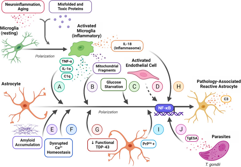
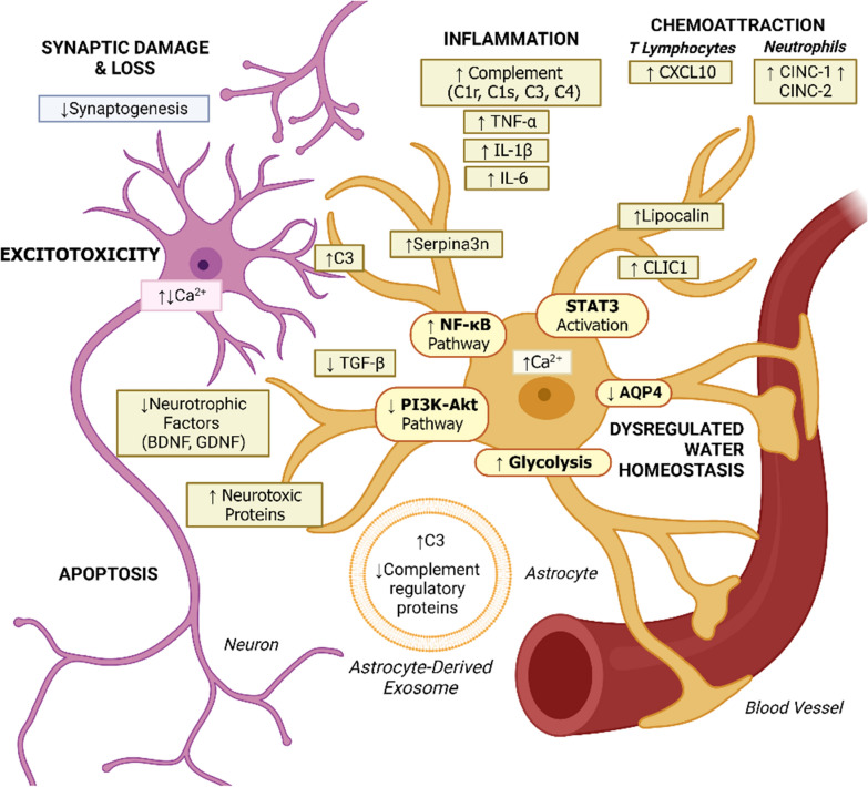
In the contexts of aging, injury, or neuroinflammation, activated microglia signaling with TNF-α, IL-1α, and C1q induces a neurotoxic astrocytic phenotype, classified as A1, A1-like, or neuroinflammatory reactive astrocytes. In contrast to typical astrocytes, which promote neuronal survival, support synapses, and maintain blood-brain barrier integrity, these reactive astrocytes downregulate supportive functions and begin to secrete neurotoxic factors, complement components like C3, and chemokines like CXCL10, which may facilitate recruitment of immune cells across the BBB into the CNS. The proportion of pro-inflammatory reactive astrocytes increases with age through associated microglia activation, and these pro-inflammatory reactive astrocytes are particularly abundant in neurodegenerative disorders. As the identification of astrocyte phenotypes progress, their molecular and cellular effects are characterized in a growing array of neuropathologies.

Keywords: A1 astrocytes; Activated astrocytes; Neurodegeneration; Neuroinflammation; Neurotoxic reactive astrocytes.


Conflict of interest statement

Not applicable.

Figures

Fig. 1
Fig. 1
Mechanisms of neurotoxic A1-like (C3+) reactive astrocyte polarization. a Neuroinflammation and the presence of misfolded, cytotoxic, or pathological proteins activates microglia, inducing an M1 phenotype that secretes TNF-α, IL-1α, and C1q [66, 76]. This combination of inflammatory signals is the primary mechanism of inducing neurotoxic astrocyte reactivity. b Microglial activation in response to pathogenic proteins is associated with excessive Drp1-Fis1-binding-induced mitochondrial fission, and subsequent release of mitochondrial fragments, a process that may work in conjunction with TNF-α, IL-1α, and C1q signaling to efficiently induce neuroinflammatory phenotype polarization [52]. c Prolonged glucose starvation induces astrocyte polarization to a pro-inflammatory phenotype that specifically displays significant upregulation of unfolded protein response genes [62]. d Activated endothelial cells induce astrocytic upregulation of C3, the extracellular matrix remodeling protein Decorin, and phagocytic functions [122]. e AD-associated amyloid accumulation is associated with activation of the NF-κB pathway in astrocytes, which adopt a C3 + neurotoxic phenotype [70, 74]. f In a TMT-intoxication model of AD, an influx of Ca2+ enters astrocytes via voltage-gated ion channels, resulting in mitochondrial membrane depolarization, upregulation of ROS and NOS, and pro-inflammatory astrocyte-associated C3 expression [28]. g Astrocytes with ALS-associated loss of functional TDP-43 adopt a neuroinflammatory phenotype, a response that is correlated with a loss of oligodendrocytes and indirect motor neuron damage [100]. h Exposure to IL-18 processed by the activated microglial NLRP3 inflammasome induces a C3 + synaptotoxic astrocyte response [44]. i Prion-propagating PrPSc + astrocytes upregulate C3 but also undergo distinct transcriptional changes that are considered pan-reactive [38]. j Astrocytes exposed to the T. gondii excreted-secreted antigens (TgESAs) underwent polarization to a C3 + reactive phenotype via NF-κB pathway activation [51]
Fig. 2
Fig. 2
Molecular and cellular effects of A1-like C3 + astrocytes. Neurotoxic reactive astrocytes upregulation of the pro-inflammatory NF-κB signaling pathway results in increased complement component expression (C1r, C1s, C3, C4) and synaptic damage [43, 74, 75, 117, 142]. Other pro-inflammatory mediators expressed by A1-like astrocytes include IL-1β, TNF-α, and IL-6 [63]. Hyperphosphorylation and activation of the STAT pathway has been reported in inflammatory reactive astrocytes associated with injury response [144]. Inhibition of the anti-inflammatory PI3K-Akt pathway results in reduced TGF-β expression, and downregulation of synaptogenic factors and neurotrophic signals, including BDNF and GDNF, contribute to synapse loss and apoptosis [129, 135]. Increased glycolytic functions and secretion of astrocyte-derived exosomes containing increased concentrations of C3 have been identified in neuropathology-associated reactive astrocytes, specifically in the context of AD [34, 145]. Changes in expression of AQP4, resulting in dysregulated water homeostasis at the BBB, are another hallmark of A1-like astrocytes [60]. Other factors secreted by A1-like neurotoxic reactive astrocytes with unidentified functions include Serpina3n, Lipocalin, and CLIC1, with several neurotoxic factors yet to be identified [83, 129]. Chemoattractive signals for T lymphocytes, such as CXCL10, and neutrophils, such as CINC-1 and CINC-2, are also produced by neurotoxic astrocytes, potentially contributing to immune cell infiltration and establishing a pro-inflammatory environment [21, 41, 57, 75, 121]. Secreted C3 binds to the C3aR receptor on local neurons, resulting in subsequent dysregulation of intraneuronal Ca2+ homeostasis and excitotoxicity [74]

References

    1. Absinta M, Maric D, Gharagozloo M, Garton T, Smith MD, Jin J, Fitzgerald KC, Song A, Liu P, Lin JP, et al. A lymphocyte-microglia-astrocyte axis in chronic active multiple sclerosis. Nature. 2021;597:709–714. doi: 10.1038/s41586-021-03892-7. - DOI - PMC - PubMed
    1. Allen NJ, Barres BA. Neuroscience: Glia—more than just brain glue. Nature. 2009;457:675–677. doi: 10.1038/457675a. - DOI - PubMed
    1. Almer G, Hainfellner JA, Brücke T, Jellinger K, Kleinert R, Bayer G, Windl O, Kretzschmar HA, Hill A, Sidle K, et al. Fatal familial insomnia: a new Austrian family. Brain. 1999;122(Pt 1):5–16. doi: 10.1093/brain/122.1.5. - DOI - PubMed
    1. Aloisi F, Ria F, Adorini L. Regulation of T-cell responses by CNS antigen-presenting cells: different roles for microglia and astrocytes. Immunol Today. 2000;21:141–147. doi: 10.1016/s0167-5699(99)01512-1. - DOI - PubMed
    1. Anderson MA, Burda JE, Ren Y, Ao Y, O’Shea TM, Kawaguchi R, Coppola G, Khakh BS, Deming TJ, Sofroniew MV. Astrocyte scar formation aids central nervous system axon regeneration. Nature. 2016;532:195–200. doi: 10.1038/nature17623. - DOI - PMC - PubMed

Publication types

MeSH terms