Detecting obstructive coronary artery disease with machine learning: rest-only gated single photon emission computed tomography myocardial perfusion imaging combined with coronary artery calcium score and cardiovascular risk factors
- PMID: 36915324
- PMCID: PMC10006131
- DOI: 10.21037/qims-22-758
Detecting obstructive coronary artery disease with machine learning: rest-only gated single photon emission computed tomography myocardial perfusion imaging combined with coronary artery calcium score and cardiovascular risk factors
Abstract
Background: The rest-only single photon emission computed tomography (SPECT) myocardial perfusion imaging (MPI) has low diagnostic performance for obstructive coronary artery disease (CAD). Coronary artery calcium score (CACS) is strongly associated with obstructive CAD. The aim of this study was to investigate the performance of rest-only gated SPECT MPI combined with CACS and cardiovascular risk factors in diagnosing obstructive CAD through machine learning (ML).
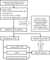
Methods: We enrolled 253 suspected CAD patients who underwent the 1-stop rest-only SPECT MPI and computed tomography (CT) scan due to stress test-related contraindications. Myocardial perfusion and wall motion were assessed using quantitative perfusion SPECT + quantitative gated SPECT (QPS + QGS) automated quantification software. The Agatston algorithm was used to calculate CACS. The clinical data of patients, including cardiovascular risk factors, were collected. Based on feature selection and clinical experience, 8 factors were identified as modeling variables. Subsequently, patients were divided randomly into 2 groups: the training (70%) and test (30%) groups. The performance of 8 supervised ML algorithms was evaluated in the training and test groups.
Results: Obstructive CAD was diagnosed by coronary angiography in 94 (37.2%, 94/253) patients. In the training group, the area under the receiver operator characteristic (ROC) curve (AUC) of the random forest was the highest, and the AUCs of Logistic, extreme gradient boosting (XGBoost), support vector machine (SVM), and adaptive boosting (AdaBoost) were all above 0.9. In the test group, the AUC of recursive partitioning and regression trees (Rpart) was the highest (0.911). Rpart and Naïve Bayes had the highest accuracy (0.840). Rpart had a sensitivity and specificity of 0.851 and 0.821, respectively; Naïve Bayes had a sensitivity and specificity of 0.809 and 0.893, respectively. Next was Logistic, with an accuracy of 0.827, a sensitivity of 0.872, and a specificity of 0.750. The random forest and XGBoost algorithms also had high accuracy, which was 0.813 for each algorithm.
Conclusions: Rest-only SPECT MPI combined with CACS and cardiovascular risk factors using an ML algorithm to detect obstructive CAD is feasible. Among the algorithms validated in the test group, Rpart, Naïve Bayes, XGBoost, Logistic, and random forest are all highly accurate for diagnosing obstructive CAD. The application of ML in resting MPI and CACS may be used for screening obstructive CAD.
Keywords: Machine learning (ML); coronary artery calcium score (CACS); coronary artery disease (CAD); myocardial perfusion imaging (MPI); single photon emission computed tomography (SPECT).
2023 Quantitative Imaging in Medicine and Surgery. All rights reserved.
Conflict of interest statement
Conflicts of Interest: All authors have completed the ICMJE uniform disclosure form (available at https://qims.amegroups.com/article/view/10.21037/qims-22-758/coif). The authors have no other conflicts of interest to declare.
Figures


References
-
- Klocke FJ, Baird MG, Lorell BH, Bateman TM, Messer JV, Berman DS, et al. ACC/AHA/ASNC guidelines for the clinical use of cardiac radionuclide imaging--executive summary: a report of the American College of Cardiology/American Heart Association Task Force on Practice Guidelines (ACC/AHA/ASNC Committee to Revise the 1995 Guidelines for the Clinical Use of Cardiac Radionuclide Imaging). Circulation 2003;108:1404-1418. 10.1161/01.CIR.0000080946.42225.4D - DOI - PubMed
-
- Germano G, Kiat H, Kavanagh PB, Moniel M, Mazzanti M, Su H-T, Van Train KF, Berman DS. Automatic quantification of ejection fraction from gated myocardial perfusion SPECT. J Nucl Med 1995;36:2138-2147. - PubMed
-
- Chua T, Kiat H, Germano G, Maurer G, Train KV, Friedman J, Berman D. Gated technetium-99m sestamibi for simultaneous assessment of stress myocardial perfusion, postexercise regional ventricular function and myocardial viability: Correlation with echocardiography and rest thallium-201 scintigraphy. J Am Coll Cardiol 1994;23:1107-1114. 10.1016/0735-1097(94)90598-3 - DOI - PubMed
LinkOut - more resources
Full Text Sources
Miscellaneous
