. 2023 Mar 1;9(3):e14179.
doi: 10.1016/j.heliyon.2023.e14179. eCollection 2023 Mar.

Antioxidants prevent particulate matter-induced senescence of lung fibroblasts

Affiliations

Antioxidants prevent particulate matter-induced senescence of lung fibroblasts

Sein Jin et al. Heliyon. .

Abstract

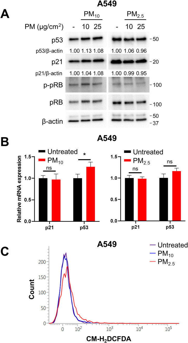
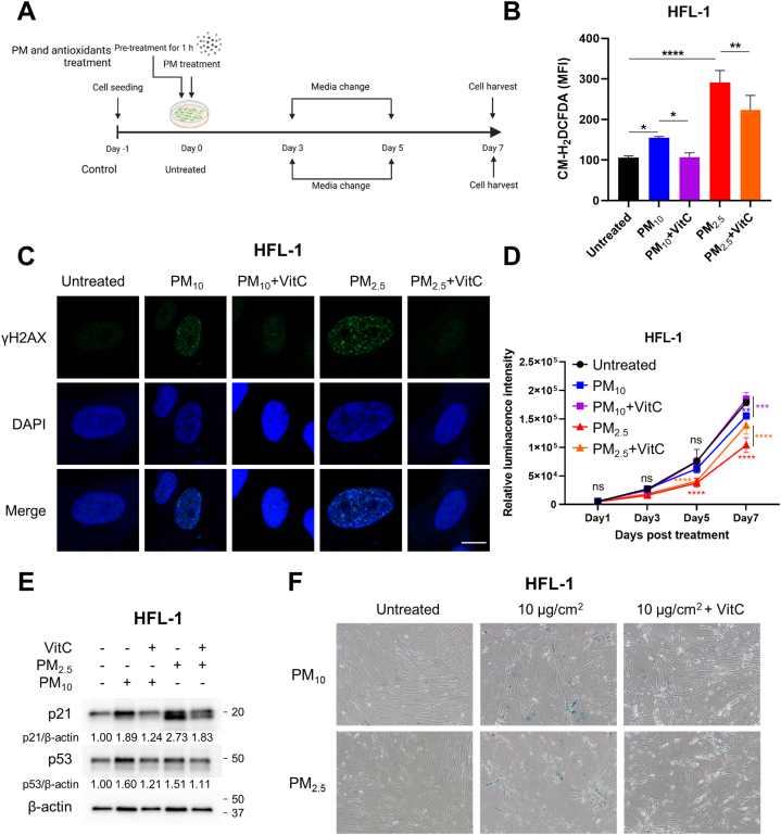
Particulate matter (PM) contributes to human diseases, particularly lung disease; however, the molecular mechanism of its action is yet to be determined. Herein, we found that prolonged PM exposure induced the cellular senescence of normal lung fibroblasts via a DNA damage-mediated response. This PM-induced senescence (PM-IS) was only observed in lung fibroblasts but not in A549 lung adenocarcinoma cells. Mechanistic analysis revealed that reactive oxygen species (ROS) activate the DNA damage response signaling axis, increasing p53 phosphorylation, ultimately leading to cellular senescence via an increase in p21 expression without affecting the p16-pRB pathway. A549 cells, instead, were resistant to PM-IS due to the PM-induced ROS production suppression. Water-soluble antioxidants, such as vitamin C and N-Acetyl Cysteine, were found to alleviate PM-IS by suppressing ROS production, implying that antioxidants are a promising therapeutic intervention for PM-mediated lung pathogenesis.

Keywords: Antioxidants; Cellular senescence; DNA damage Response; Particulate matter; Reactive oxygen species.


Conflict of interest statement

The authors declare no conflict of interest.

Figures

Fig. 1
Fig. 1
Sublethal and prolonged exposure to particulate matter (PM)10 and PM2.5 inhibits the proliferation of lung fibroblasts. IMR-90, HFL-1, and WI-38 cells were treated with 10 or 25 μg/cm2 of PM10 or PM2.5, while control groups were untreated. Cell proliferation was measured on days 1, 3, 5, and 7 after PM treatment. Cell proliferation was analyzed using CellTiter-Glo (A) and hemocytometer (B). Data are presented as the mean ± standard deviation (SD) of three independent experiments. Statistical analyses were calculated by two-way ANOVA. Significances were determined by Tukey's HSD (ns = not significant; *P < 0.05, **P < 0.01, ***P < 0.001, and ****P < 0.0001).
Fig. 2
Fig. 2
PM treatment promotes the cellular senescence of lung fibroblasts. (A–C) Cells were treated with the indicated concentration of PM10 or PM2.5, while control groups were untreated. After 7 days of PM treatment, SA-β-gal staining was performed, and the cells were observed under a microscope. (D–E) In RT-qPCR experiments for SASP gene expression analysis, 10 μg/cm2 of PM10 or PM2.5 was treated for 7 days, while control groups were untreated for the same days. Relative fold changes of mRNA expression are presented as the mean ± SD of four independent experiments (ns = not significant; *p < 0.05, **p < 0.01, and ***p < 0.001).
Fig. 3
Fig. 3
PM induces p21 mRNA expression by activating the p53–p21 signaling axis. (A) Cells were treated with 10 or 25 μg/cm2 of PM10 or PM2.5 for the indicated days, while control groups were untreated for 7 days. Harvested cells were analyzed using anti-p53, -p21, -p-pRB, and -pRB antibodies. The asterisks denote non-specific bands, which were suspected to be β-actin bands remaining after the extensive stripping and washing. (B) HFL-1 and IMR-90 cells were treated with 10 μg/cm2 of PM10 or PM2.5 for 7 days, while control groups were untreated for the same days. mRNA expression levels of senescence driver genes were determined via RT-qPCR analysis. Relative fold changes of mRNA expression are presented as the mean ± SD of three independent experiments (ns = not significant; *p < 0.05, **p < 0.01, and ***p < 0.001). Images of Western blot raw data are presented in Fig. S3.
Fig. 4
Fig. 4
Prolonged exposure to PM causes DNA damage in lung fibroblasts. Cells were treated with 10 μg/cm2 of PM10 or PM2.5 for 7 days, while control groups were untreated for the same days. (A) γH2AX and the nucleus were stained with the anti-γH2AX antibody and 4′,6-diamidino-2-phenylindole dihydrochloride (DAPI), respectively. Stained cells were observed under a confocal microscope. The scale bars are 10 μm. (B) DNA damage response (DDR) signaling pathway was analyzed using anti-p-ATM, -ATM, -p-p53, and -p53 antibodies. Images of Western blot raw data are presented in Fig. S4.
Fig. 5
Fig. 5
PM treatment produces excessive reactive oxygen species (ROS) in lung fibroblasts. Cells were treated with 10 μg/cm2 of PM10 or PM2.5 for 7 days, while control groups were untreated for the same days. (A) ROS were detected via chloromethyl derivative of H2DCFDA (CM-H2DCFDA) staining-based (10 μM) flow cytometry analysis. (B) Data are presented as the mean ± SD of RFI of three independent experiments. Statistical analyses were performed by one-way ANOVA. Significances were calculated by Tukey's HSD (**P < 0.01, ***P < 0.001, and ****P < 0.0001).
Fig. 6
Fig. 6
A549 cells are resistant to PM-induced cellular senescence (PM-IS) by suppressing excessive ROS production. A549 cells were treated with 10 or 25 μg/cm2 of PM10 or PM2.5 for 7 days, while control groups were untreated for the same days. (A) Expression levels of senescence driver proteins were determined via immunoblotting. (B) p21 and p53 mRNA expression levels were determined via RT-qPCR analysis after treatment with 10 μg/cm2 of PM. Relative fold changes are presented as the mean ± SD of three independent experiments. Statistical analyses were performed by Student-t-test (ns = not significant; *P < 0.05). (C) ROS production was analyzed via CM-H2DCFDA staining-based (10 μM) flow cytometry after treatment with 10 μg/cm2 of PM. Images of Western blot raw data are presented in Fig. S5.
Fig. 7
Fig. 7
Vitamin C (VitC) prevents PM-IS of lung fibroblasts by removing PM-induced ROS. (A) Schematic diagram of the antioxidant treatment experiment. Cells were pre-incubated with 100 μM VitC for 1 h, followed by treatment with 10 μg/cm2 of PM10 or PM2.5 for the indicated days, while control groups were untreated for 7 days. (B) ROS production was analyzed via CM-H2DCFDA staining-based (10 μM) flow cytometry. Data are presented as the mean ± SD of RFI from four independent experiments. Statistical analysis was performed by one-way ANOVA. Significances were calculated by Tukey's HSD (*P < 0.05, **P < 0.01, and ****P < 0.0001). (C) γH2AX and nucleus were stained with the anti-γH2AX antibody and DAPI, respectively, followed by confocal microscope analysis. The scale bar is 10 μm. (D) Cell proliferation was measured using CellTiter-Glo on days 1, 3, 5, and 7 after PM treatment. Data are presented as the mean ± SD of three independent experiments. Statistical analysis was performed by two-way ANOVA. Significances were calculated by Tukey's HSD (ns = not significant; **P < 0.01, ***P < 0.001, and ****P < 0.0001). (E) Senescence driver protein levels were determined via immunoblotting using anti-p53 and -p21 antibodies. (F) SA-β-gal staining was performed, and the cells were observed under a microscope. Images of Western blot raw data are presented in Fig. S6.
Fig. 8
Fig. 8
Schematic diagram of PM-IS mechanism. Exposure to PM10 and PM2.5 causes DNA double-strand break (DSB) by producing excessive ROS. DSB activates the DDR signaling pathway, ATM–p53–p21, which induces cellular senescence by inhibiting the cyclin-dependent kinases (CDKs). VitC prevents PM-IS by removing excess ROS.

References

    1. Collaborators G.B.D.R.F. Global burden of 87 risk factors in 204 countries and territories, 1990-2019: a systematic analysis for the Global Burden of Disease Study 2019. Lancet. 2020;396(10258):1223–1249. doi: 10.1016/S0140-6736(20)30752-2. - DOI - PMC - PubMed
    1. Zhu C., Maharajan K., Liu K., Zhang Y. Role of atmospheric particulate matter exposure in COVID-19 and other health risks in human: a review. Environ. Res. 2021;198 doi: 10.1016/j.envres.2021.111281. - DOI - PMC - PubMed
    1. Pfeffer P.E., Mudway I.S., Grigg J. Air pollution and asthma: mechanisms of harm and considerations for clinical interventions. Chest. 2021;159(4):1346–1355. doi: 10.1016/j.chest.2020.10.053. - DOI - PubMed
    1. Lee P.H., Park S., Lee Y.G., Choi S.M., An M.H., Jang A.S. The impact of environmental pollutants on barrier dysfunction in respiratory disease. Allergy Asthma Immunol. Res. 2021;13(6):850–862. doi: 10.4168/aair.2021.13.6.850. - DOI - PMC - PubMed
    1. Winterbottom C.J., Shah R.J., Patterson K.C., Kreider M.E., Panettieri R.A., Jr., Rivera-Lebron B., Miller W.T., Litzky L.A., Penning T.M., Heinlen K., Jackson T., Localio A.R., Christie J.D. Exposure to ambient particulate matter is associated with accelerated functional decline in idiopathic pulmonary fibrosis. Chest. 2018;153(5):1221–1228. doi: 10.1016/j.chest.2017.07.034. - DOI - PMC - PubMed