

Large intra-abdominal venous malformations in associated with inferior vena cava aneurysm
- PMID: 36915604
- PMCID: PMC10006310
- DOI: 10.1016/j.radcr.2023.01.085
Large intra-abdominal venous malformations in associated with inferior vena cava aneurysm
Abstract
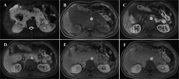
Intra-abdominal venous malformations and inferior vena cava aneurysms are rare and difficult to diagnose because of their nonspecific clinical symptoms. These vascular anomalies are important entities due to the risk of thrombosis or rupture. According to the classification of International Society for the Study of Vascular Anomalies, venous malformations are classified as low-flow vascular anomalies, showing absence of arterial and early venous enhancement and slow gradual filling with contrast on delayed venous imaging. Phleboliths related to thrombosis and calcifications, are the key finding of venous malformations. In this article, we report an exceptional case of large intra-abdominal venous malformations in associated with an inferior vena cava aneurysm.
Keywords: Inferior vena cava aneurysm; Venous malformations.
© 2023 The Authors.
Figures


References
-
- Loose D. Surgical management of venous malformations. Phlebology. 2007;22(6):276–282. - PubMed
-
- Hussein A., Malguria N. Imaging of vascular malformations. Radiol Clin. 2020;58(4):815–830. - PubMed
-
- Olivieri B., White C.L., Restrepo R., McKeon B., Karakas S.P., Lee E.Y., et al. Low-flow vascular malformation pitfalls: from clinical examination to practical imaging evaluation—part 2, venous malformation mimickers. Am J Roentgenol. 2016;206(5):952–962. - PubMed
-
- Montero-Baker M.F., Branco B.C., Leon L.L., Jr., Labropoulos N., Echeverria A., Mills J.L., Sr., et al. Management of inferior vena cava aneurysm. J Cardiovasc Surg (Torino) 2015;56(5):769–774. - PubMed
-
- Gradman W.S., Steinberg F. Aneurysm of the inferior vena cava: case report and review of the literature. Ann Vasc Surg. 1993;7(4):347–353. - PubMed
Publication types
LinkOut - more resources
Full Text Sources
