. 2023 Mar;95(3):e28673.
doi: 10.1002/jmv.28673.

Distinct in vitro and in vivo neutralization profiles of monoclonal antibodies elicited by the receptor binding domain of the ancestral SARS-CoV-2

Affiliations

Distinct in vitro and in vivo neutralization profiles of monoclonal antibodies elicited by the receptor binding domain of the ancestral SARS-CoV-2

Hyung J Kwon et al. J Med Virol. 2023 Mar.

Abstract

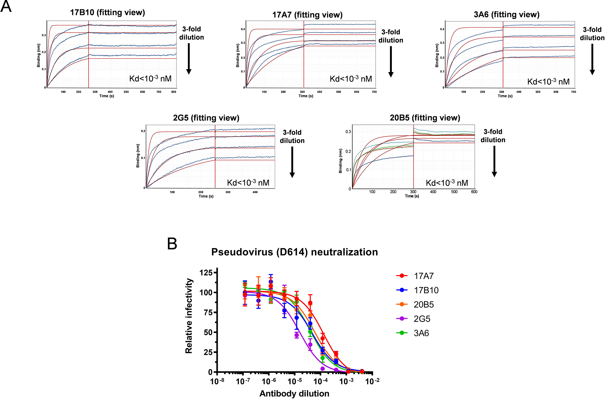
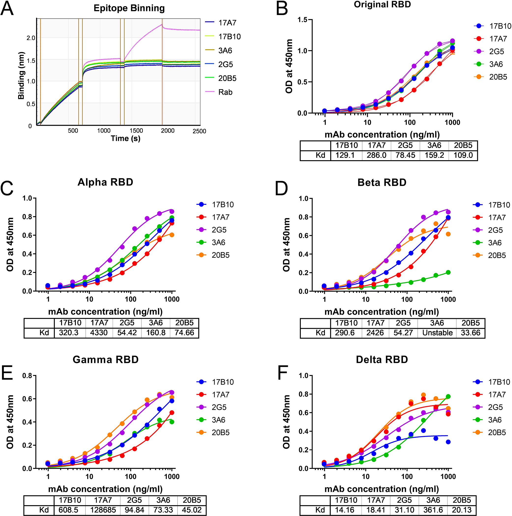
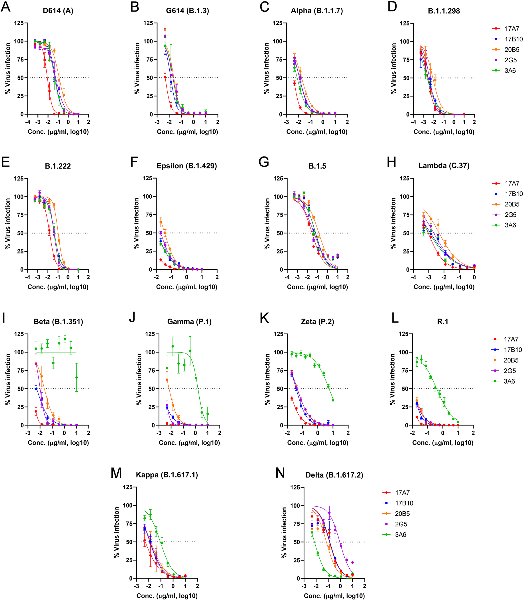
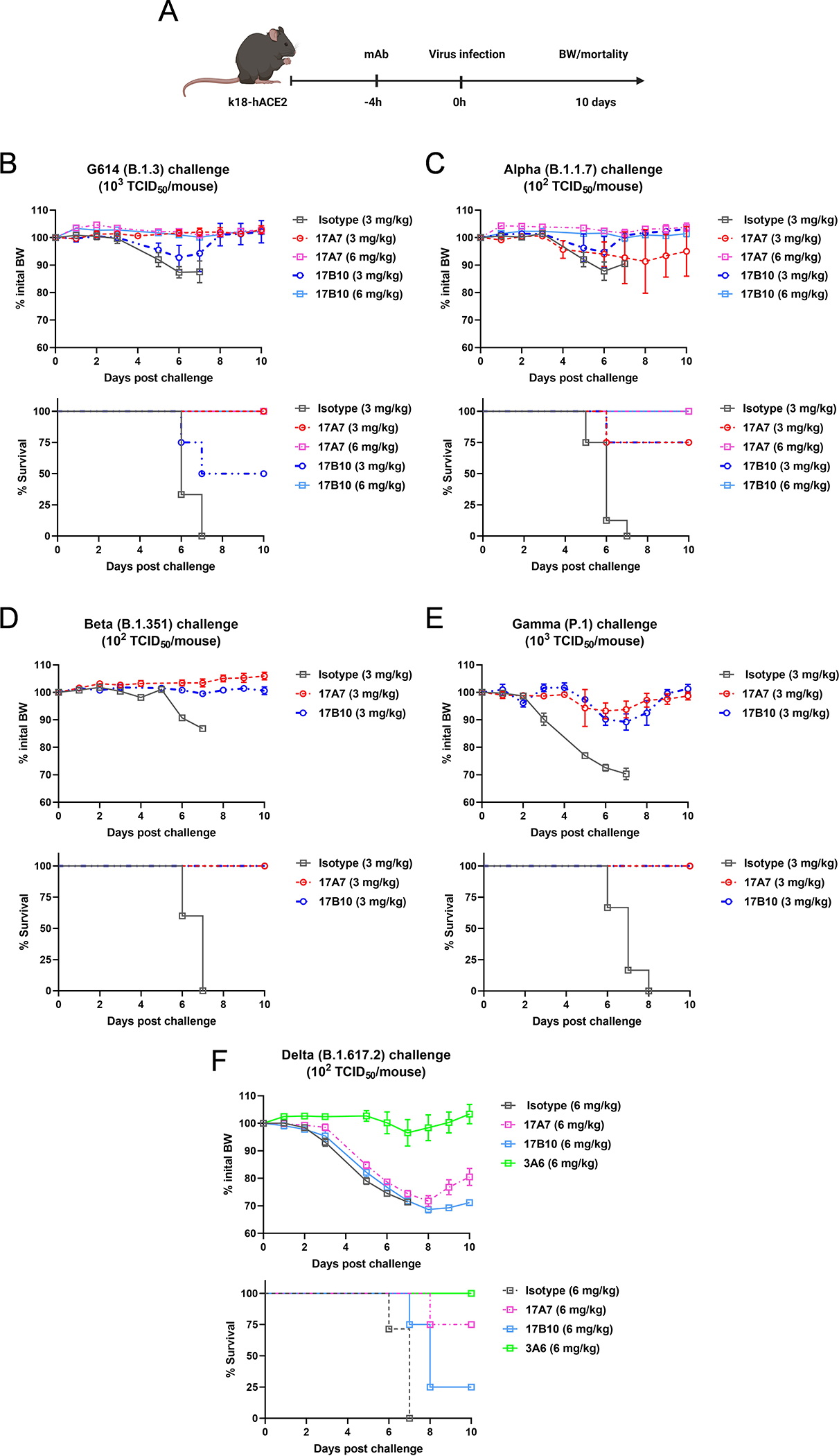
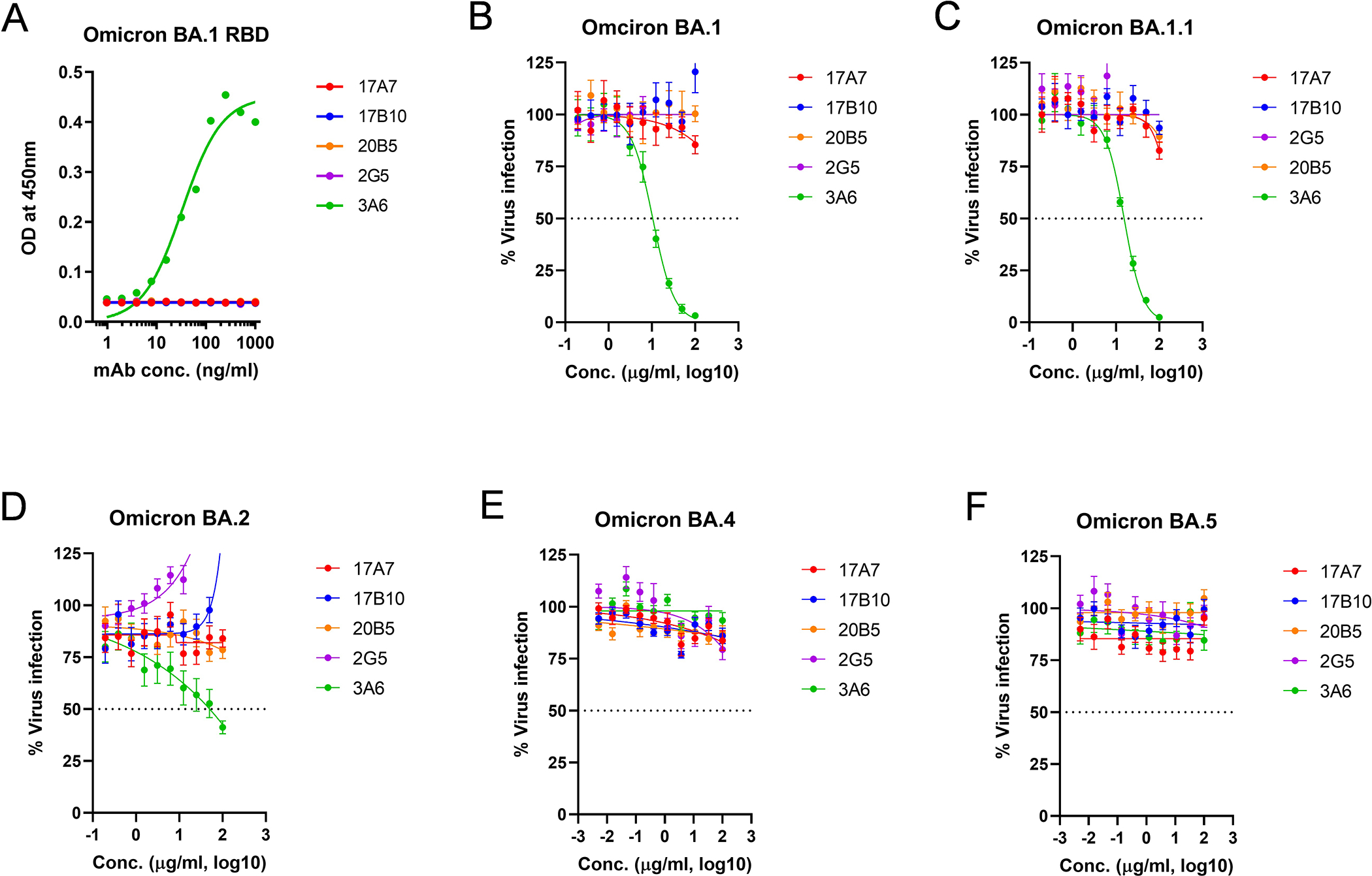
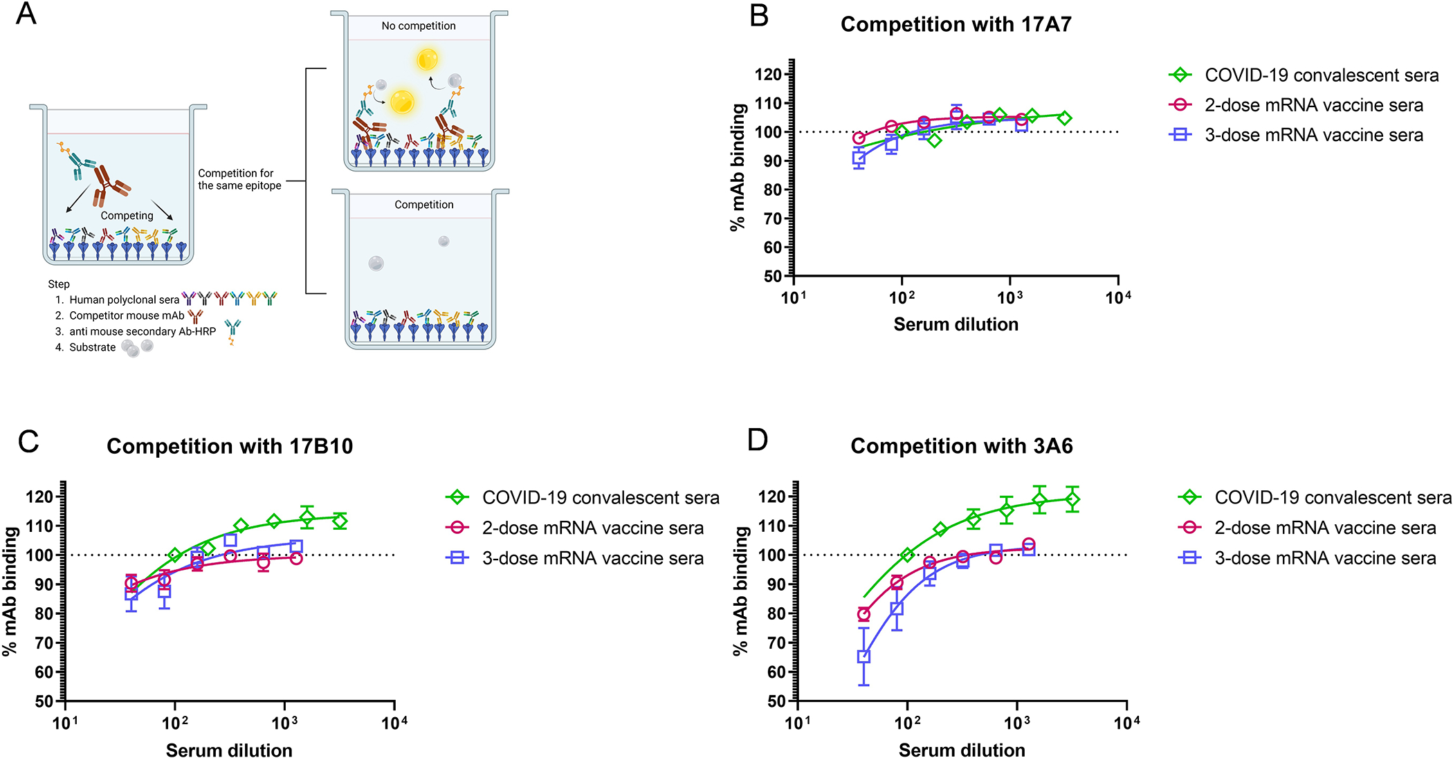
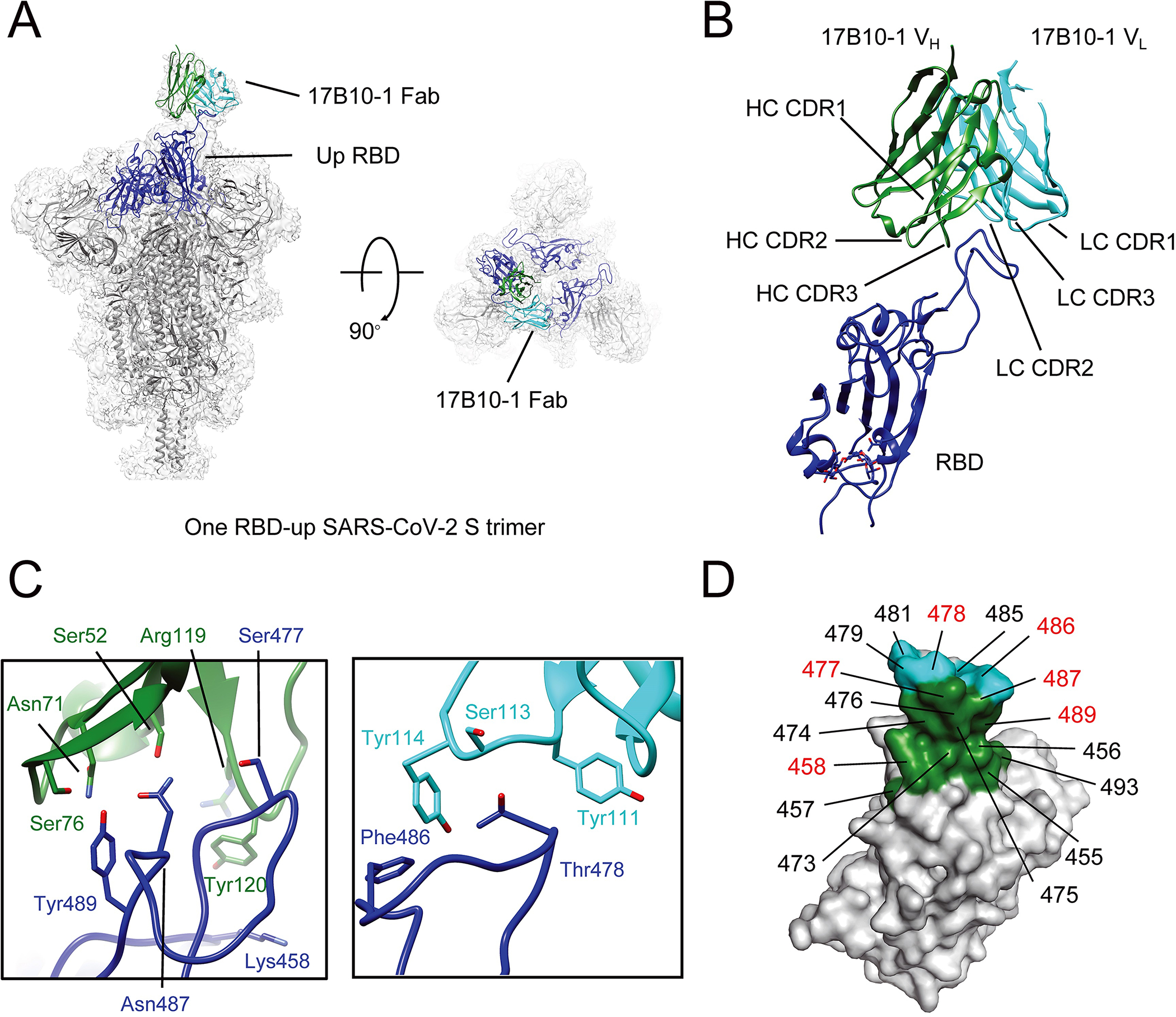
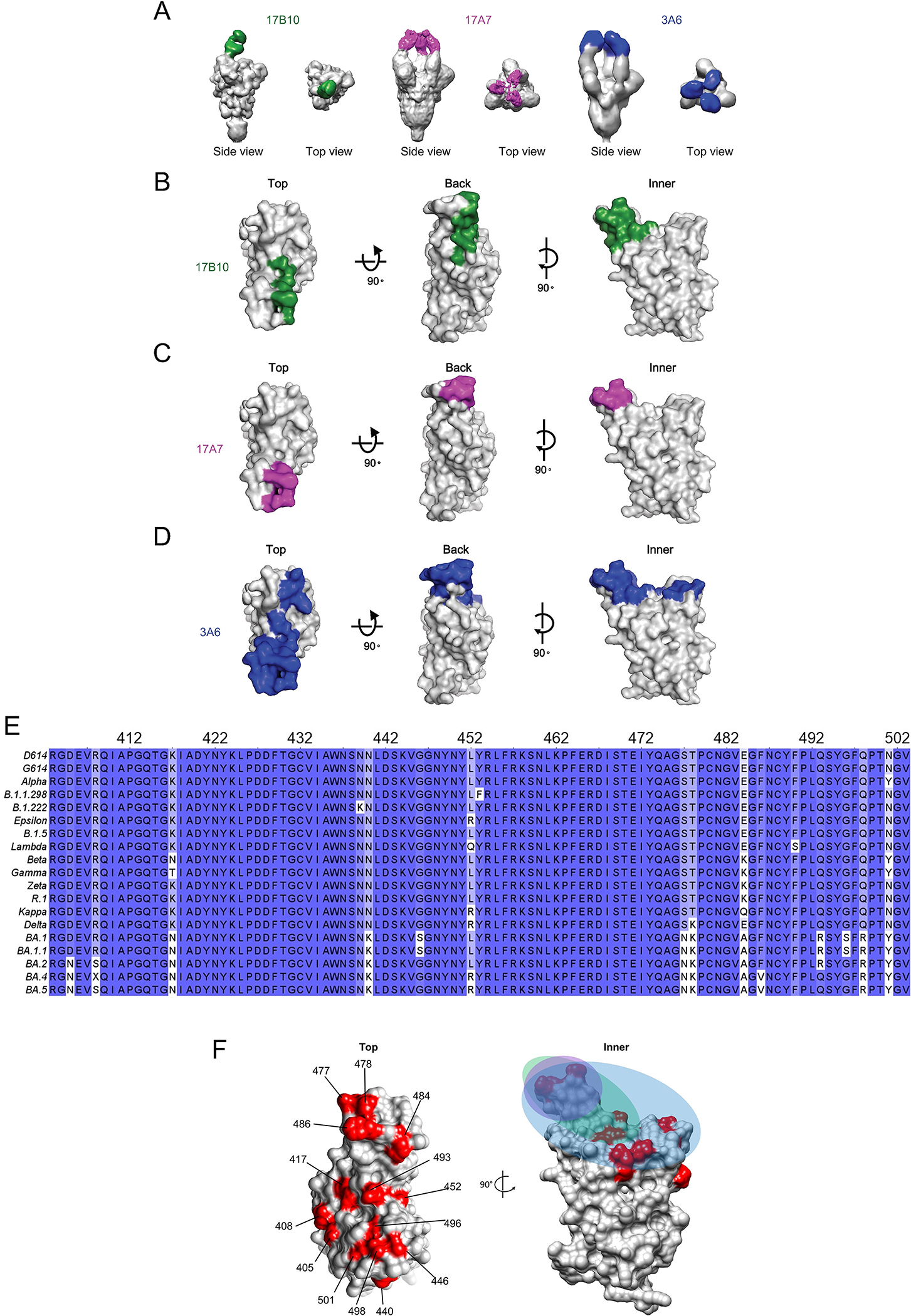
Broadly neutralizing antibodies against severe acute respiratory syndrome coronavirus 2 (SARS-CoV-2) variants are sought to curb coronavirus disease 2019 (COVID-19) infections. Here we produced and characterized a set of mouse monoclonal antibodies (mAbs) specific for the ancestral SARS-CoV-2 receptor binding domain (RBD). Two of them, 17A7 and 17B10, were highly potent in microneutralization assay with 50% inhibitory concentration (IC50 ) ≤135 ng/mL against infectious SARS-CoV-2 variants, including G614, Alpha, Beta, Gamma, Delta, Epsilon, Zeta, Kappa, Lambda, B.1.1.298, B.1.222, B.1.5, and R.1. Both mAbs (especially 17A7) also exhibited strong in vivo efficacy in protecting K18-hACE2 transgenic mice from the lethal infection with G614, Alpha, Beta, Gamma, and Delta viruses. Structural analysis indicated that 17A7 and 17B10 target the tip of the receptor binding motif in the RBD-up conformation. A third RBD-reactive mAb (3A6) although escaped by Beta and Gamma, was highly effective in cross-neutralizing Delta and Omicron BA.1 variants in vitro and in vivo. In competition experiments, antibodies targeting epitopes similar to these 3 mAbs were rarely enriched in human COVID-19 convalescent sera or postvaccination sera. These results are helpful to inform new antibody/vaccine design and these mAbs can be useful tools for characterizing SARS-CoV-2 variants and elicited antibody responses.

Keywords: RBD-reactive antibody; SARS-CoV-2; cross-neutralization; neutralizing antibody; receptor binding domain (RBD); structural analysis.


Conflict of interest statement

CONFLICT OF INTEREST DISCLOUSRE

The authors declare no competing interests.

Figures

Figure 1.
Figure 1.
Mouse mAb clones with high affinity to the RBD of the original Wuhan-Hu-1 strain. Mouse hybridomas were generated using the original RBD as the immunogen. Five mAb clones (17B10, 17A7, 2G5, 3A6 and 20B5) were selected and evaluated for RBD binding by bio-layer interferometry (BLI). (A) Representative BLI sensorgrams of individual mAbs binding to immobilized RBD. All five mAbs were 3-fold diluted from the initial concentration of 15 μg/mL and collected data were applied to a built-in 1:2 global curve-fitting model with Kd values shown. (B) Pseudovirus-based neutralization against SARS-CoV-2 spike (D614).
Figure 2.
Figure 2.
Distinct binding profiles to RBD variants. (A) Epitope binning of 17B10 mAb with competitive mAbs (17A7, 2G5, 3A6 or 20B5) for RBD binding. Spike-specific rabbit polyclonal Ab (Rab) was used as control. (B-F) ELISA binding to different RBD variants: (B) original RBD; (C) Alpha RBD; D) Beta RBD; (E) Gamma RBD; (F) Delta RBD. OD at 450 nm (mean± SD, n= 4 replicates per mAb dilution) were plotted by nonlinear regression curve fit (Specific binding with Hill slope) with individual Kd values shown.
Figure 3.
Figure 3.
RBD-reactive mAbs cross-neutralizing multiple SARS-CoV-2 variants. Five mAb clones (17B10, 17A7, 2G5, 3A6 and 20B5) were assessed for neutralizing potential against live infectious SARS-CoV-2 strains, including (A) the ancestral D614 (A), (B) G614(B.1.3), (C) Alpha (B.1.1.7), (D) B.1.1.298, (E) B.1.222, (F) Epsilon (B.1.429), (G) B.1.5, (H_ Lambda (C.37), (I) Beta (B.1.351), (J) Gamma (P.1), (K) Zeta (P.2), (L) R.1, (M) Kappa (B.1.617.1) and (N) Delta (B.1.617.2). The % virus infection verse the wells that contained virus only were plotted by nonlinear regression. Data are expressed as mean± SD, n= 8 replicates per mAb concentration. Estimated IC50 values of individual mAbs are summarized in Table 1.
Figure 4.
Figure 4.
Efficacy of selected RBD mAbs in protecting K18-hACE2 mice against lethal challenges of SARS-CoV-2 and variants. Naïve K18-hACE2 mice were injected intraperitoneally with RBD-reactive 17A7, 17B10 or 3A6mAbs or a nonspecific mouse isotype control. Recipient mice were then challenged intranasally with a pre-determined lethal dose of live infectious SARS-CoV-2. (A) Schematic timelines of the passive transfer and challenge in K18-hACE2 mice. Body weight (BW) drop and mortality of infected mice were monitored for up to 10 days after a lethal challenge of (B) G614 (B.1.3), (C) Alpha (B.1.1.7), (D) Beta (B.1.351), (E) Gamma (P.1) or (F) Delta (B.1.617.2). % BW drops (mean ± s.e.m. of 4–6 mice/group) and % cumulative survivals (n=4–6 mice/group) are shown.
Figure 5.
Figure 5.
ELISA binding and cross-neutralization of RBD-reactive mAbs against Omicron subvariants. (A) Omicron BA.1 RBD ELISA. The OD at 450 nm (mean± SD, n= 4 replicates per mAb dilution) was plotted by nonlinear regression curve fit (Specific binding with Hill slope). Microneutralization against live infectious Omicron subvariants including (B) Omicron BA.1, (C) Omicron BA.1.1, (D) Omicron BA.2, (E) Omicron BA.4 and (F) Omicron BA.5. The % virus infection verse the wells that contained virus only (mean± SD, n= 8 replicates per mAb concentration) were plotted by 4-parameter logistic curve fitting. Estimated IC50 values of individual mAbs are summarized in Table 1.
Figure 6.
Figure 6.
Competition of 17A7, 17B10 and 3A6 mAbs with human COVID-19 convalescent sera or mRNA post-vaccination sera for binding to immobilized original RBD. (A) Schematic illustration of competition between mouse mAbs and human COVID-19 specific polyclonal sera. The % of residual (B) 17A7, (C) 17B10 or (D) 3A6 after competing with human COVID-19 convalescent sera or post-vaccination sera as compared to the wells that contained only mAbs were plotted by nonlinear regression. Data are expressed as mean ± s.e.m (n = 6–8 sera/group).
Figure 7.
Figure 7.
Cryo-EM structures of 17B10 Fab in complex of a full-length SARS-CoV-2 Spiks (G614) trimer. (A) One 17B10–1 Fab bound to one spike trimer in the one-RBD-up (2.8Å) conformation resolved by Cryo-EM. The EM density is colored in gray, and the structures are represented in ribbon diagram with the RBD in blue and the rest in dark gray. The heavy chain of 17B10–1 is shown in green and the light chain in cyan. (B) Close-up view of the interactions between 17B10–1 Fab and the RBD of the S trimer in the one-RBD-up conformation. (C) Zoom-in views of the binding interface between the heavy (green) or light chain (cyan) of 17B10–1 and the tip of one RBD-up conformation. (D) The footprints on the RBD interacting with the heavy chain (green) or light chain (cyan) of 17B10–1 with the major contacting residues highlighted in red.
Figure 8.
Figure 8.
17B10, 17A7 and 3A6 Fab bound to different regions of RBD. The 3D map of 17B10-Spike complex was produced by low-pass filter 17B10-S complex cryo-EM map to 10Å. The 3D maps of 17A7-S and 3A6-S complexes were produced by negative stain EM with a resolution at 11Å and 13Å, respectively. (A) Top view and side view of 3D reconstruction of Fab-spike (gray) complexes. (B-D) Binding footprints of Fabs of three neutralizing antibodies, 17B10 (B), 17A7(C) and 3A6 (D) shading on the RBD in surface representation. The RBD surface (gray) was represented in three views (top, back and inner). The Fabs are colored as 17B10: green; 17A7: magenta; 3A6: blue. (E) Sequence alignment showing multiple mutations in the RBD region (aa 403–503) among SARS-CoV-2 variants. (F) The positions on the RBD being mutated in Omicron subvariants highlighted in red with the colored footprints of 17B10 (green), 17A7 (magenta) and 3A6 (blue).

References

    1. Walls AC et al. Structure, Function, and Antigenicity of the SARS-CoV-2 Spike Glycoprotein. Cell 181, 281–292 e286 (2020). 10.1016/j.cell.2020.02.058 - DOI - PMC - PubMed
    1. Wang Q et al. Structural and Functional Basis of SARS-CoV-2 Entry by Using Human ACE2. Cell 181, 894–904 e899 (2020). 10.1016/j.cell.2020.03.045 - DOI - PMC - PubMed
    1. Shang J et al. Structural basis of receptor recognition by SARS-CoV-2. Nature 581, 221–224 (2020). 10.1038/s41586-020-2179-y - DOI - PMC - PubMed
    1. Wrapp D et al. Cryo-EM structure of the 2019-nCoV spike in the prefusion conformation. Science 367, 1260–1263 (2020). 10.1126/science.abb2507 - DOI - PMC - PubMed
    1. Yuan Y et al. Cryo-EM structures of MERS-CoV and SARS-CoV spike glycoproteins reveal the dynamic receptor binding domains. Nature communications 8, 15092 (2017). 10.1038/ncomms15092 - DOI - PMC - PubMed

Publication types

Supplementary concepts