. 2023 Mar 14;80(4):86.
doi: 10.1007/s00018-023-04743-6.

AAV-Net1 facilitates the trans-differentiation of supporting cells into hair cells in the murine cochlea

Affiliations

AAV-Net1 facilitates the trans-differentiation of supporting cells into hair cells in the murine cochlea

Liyan Zhang et al. Cell Mol Life Sci. .

Abstract

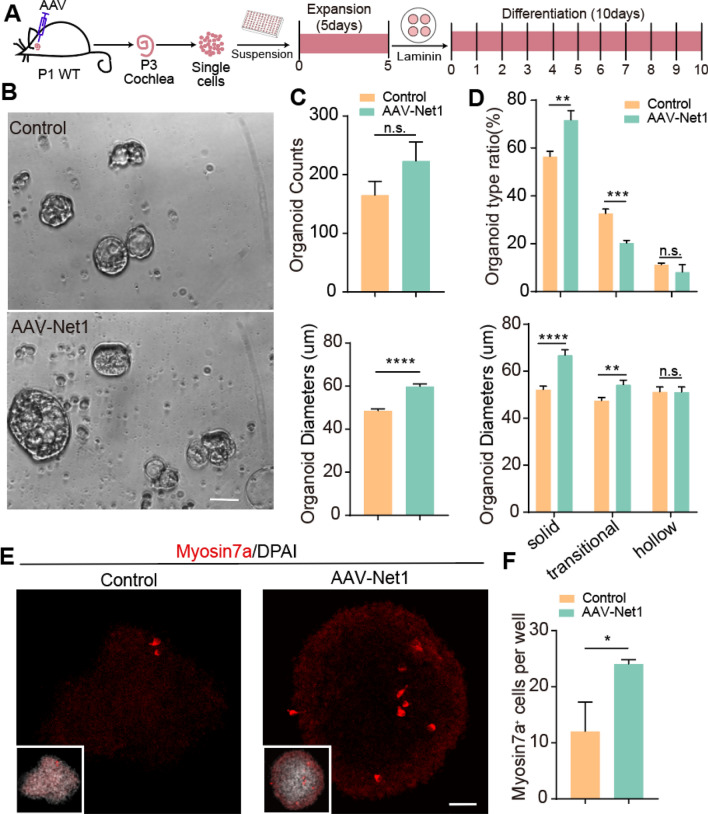
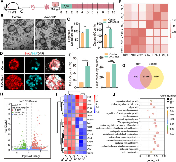
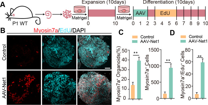
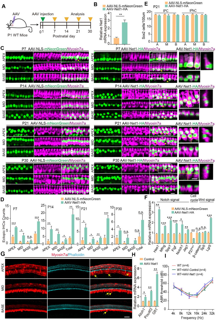
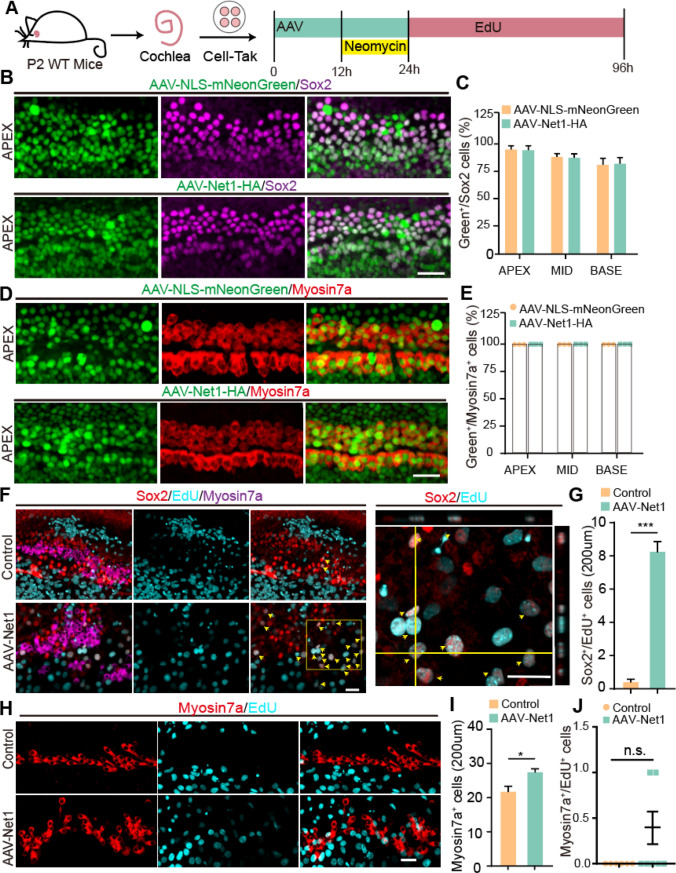
Mechanosensitive hair cells (HCs) in the cochlear sensory epithelium are critical for sound detection and transduction. Mammalian HCs in the cochlea undergo cytogenesis during embryonic development, and irreversible damage to hair cells postnatally is a major cause of deafness. During the development of the organ of Corti, HCs and supporting cells (SCs) originate from the same precursors. In the neonatal cochlea, damage to HCs activates adjacent SCs to act as HC precursors and to differentiate into new HCs. However, the plasticity of SCs to produce new HCs is gradually lost with cochlear development. Here, we delineate an essential role for the guanine nucleotide exchange factor Net1 in SC trans-differentiation into HCs. Net1 overexpression mediated by AAV-ie in SCs promoted cochlear organoid formation and HC differentiation under two and three-dimensional culture conditions. Also, AAV-Net1 enhanced SC proliferation in Lgr5-EGFPCreERT2 mice and HC generation as indicated by lineage tracing of HCs in the cochleae of Lgr5-EGFPCreERT2/Rosa26-tdTomatoloxp/loxp mice. We further found that the up-regulation of Wnt/β-catenin and Notch signaling in AAV-Net1-transduced cochleae might be responsible for the SC proliferation and HC differentiation. Also, Net1 overexpression in SCs enhanced SC proliferation and HC regeneration and survival after HC damage by neomycin. Taken together, our study suggests that Net1 might serve as a potential target for HC regeneration and that AAV-mediated gene regulation may be a promising approach in stem cell-based therapy in hearing restoration.

Keywords: AAV; Cochlea; Hair cell regeneration; Net1; Organoid.


Conflict of interest statement

Jieyu Qi had filed a patent on the use of AAV-ie for gene therapy in the inner ear. The authors declare no other competing interests.

Figures

Fig. 1
Fig. 1
AAV-Net1 promotes cochlear organoid formation and HC regeneration in two-dimensional culture assay. A Illustration of the experimental design. All mice were injected with the indicated AAVs at a dose of 7.5E10 GCs in the left ears. B Representative bright-field images of cochlear organoids overexpressing AAV-control and AAV-Net1 after expansion. Scale bar, 50 μm. C Cochlear organoid counts per cell and average organoid diameters. D The proportions and average diameters of three organoid morphologies after 5 days of expansion. E Representative confocal images of cochlear organoids overexpressing AAV-control or AAV-Net1 showing newly formed HCs in cochlear organoids after 10 days of differentiation. Scale bar, 40 μm. F The proportion of Myosin7a+ organoids after 10 days of differentiation. The controls here refer to AAV-NLS-mNeonGreen. NLS, nuclear localization sequence. Data are presented as the mean ± SEM. The p-value was calculated by two-tailed unpair Student’s t-test. *p < 0.05, **p < 0.01, ***p < 0.001, ****p < 0.0001, n.s., no significance
Fig. 2
Fig. 2
AAV-Net1 administration promotes cochlear SC expansion in optimized three-dimensional culture assay. A Experimental design for cochlear organoid expansion. Corresponding AAVs were introduced at a dose of 2E10 GCs into every well. B Representative bright-field images of cochlear organoids overexpressing AAV-control or AAV-Net1 after expansion. Scale bar, 100 μm. C Average organoid diameters and cochlear organoid counts per cell. D Representative confocal images of cochlear organoids immunostained with Sox2 after 10 days of expansion. Sox2 (red) marked SCs. EdU incorporation (cyan) was analyzed after 1 h EdU treatment on day 10 of expansion. Scale bar, 10 μm. E The proportion of EdU+ organoids and the proportion of EdU+ cells in the organoids at day 10 of expansion. F The cluster analysis of all replicated samples of AAV-control and AAV-Net1. G The amount of gene expression between AAV-control and AAV-Net1 in the venn diagram. H The number of differential genes between AAV-control and AAV-Net1 in the volcano Plot. I The changes of AAV-control and AAV-Net1 differential genes in heatmap. J The GO analysis of differential genes in AAV-control and AAV-Net1. The controls here refer to AAV-NLS-mNeonGreen. Data are presented as the mean ± SEM. The p-value was calculated by two-tailed unpaired Student’s t-test. *p < 0.05, **p < 0.01, ****p < 0.0001
Fig. 3
Fig. 3
AAV-Net1 administration promotes HC production in optimized three-dimensional culture assay. A Experimental design for cochlear organoid differentiation after expansion. Corresponding AAVs were introduced at a dose of 2E10 GCs into every well. B Representative confocal images of cochlear organoids immunostained with Myosin7a after 10 days of differentiation. Myo7a (red) marked HCs. EdU incorporation (cyan) was also analyzed. Scale bar, 50 μm. C The proportions of Myosin7a+ organoids and Myosin7a+ cells after 10 days of differentiation. D Myosin7a+/ EdU+ cell counts. The controls here refer to AAV-NLS-mNeonGreen. Data are presented as the mean ± SEM. The p-value was calculated by two-tailed unpaired Student’s t-test. **p < 0.01
Fig. 4
Fig. 4
AAV-Net1 promotes the proliferation of Sox2+ SCs and Lgr5+ progenitors via Wnt/β-catenin signaling. A Illustration of the experimental design. All mice were injected with corresponding AAVs at a dose of 7.5E10 GCs in the left ears. B EdU was stained (cyan) in cochleae transduced by AAV-control and AAV-Net1, respectively. Sox2 (red) marked SCs. Magnified images and orthogonal views are shown to the right. Arrows indicate the Sox2+/EdU+ SCs. Scale bars, 40 µm and 20 μm, respectively. C Sox2+/EdU+ SC counts. D Representative images of Lgr5-EGFP (green) signals in AAV-injected cochleae and the contralateral cochleae. Myosin7a (magenta) marked HCs. Scale bar, 50 μm. E Immunosignal intensity of Lgr5-EGFP in cochleae transduced with AAV-control and AAV-Net1 in the DC region and IPC region. F Relative Lgr5 mRNA expression in cochleae transduced with AAV-control and AAV-Net1. A, M, and B refer to the apical, middle, and basal turns of the cochlea. G Illustration of the experimental design. Corresponding AAVs were introduced at a dose of 2E10 GCs into every well. H Representative bright-field images of cochlear organoids overexpressing AAV-control and AAV-Net1 after expansion. Scale bar, 40 μm. I Cochlear organoid counts per cell. J The proportions of the three organoid morphologies after 5 days of expansion. The controls in D refer to Lgr5-EGFPCreERT2 mice, and the controls in the other panels refer to AAV-NLS-mNeonGreen. Data are presented as the mean ± SEM. The p-value was calculated by two-tailed unpair Student’s t-test. *p < 0.05, **p < 0.01, ***p < 0.001, ****p < 0.0001, n.s., no significance
Fig. 5
Fig. 5
AAV-Net1 transduction in SCs results in increased HC numbers in postnatal cochleae. A Illustration of the experimental design. All mice were injected with corresponding AAVs at a dose of 7.5E10 GCs in the left ears. B Relative Net1 mRNA expression in cochleae transduced with AAV-NLS-mNeonGreen and AAV-Net1-HA. C Extra IHCs (arrows) were seen in the apical (APEX), middle (MID), and basal (BASE) turns of cochleae transduced with AAV-NLS-mNeonGreen and AAV-Net1-HA. Myosin7a (magenta) marked the HCs. Magnified images and orthogonal views are shown to the right. Scale bar, 10 μm. D Ectopic IHC counts in P7, P14, P21, and P30 cochleae transduced with AAV-NLS-mNeonGreen (control) and AAV-Net1-HA. E Sox2 counts in P21 cochleae transduced with AAV-NLS-mNeonGreen and AAV-Net1-HA. A, M, B refer to the apical, middle, and basal turns of the cochlea. F Relative mRNA expression in cochleae transduced with AAV-NLS-mNeonGreen and AAV-Net1-HA, respectively. G Representative confocal images of Mosin7a staining (red) and phalloidin (cyan) in cochleae transduced at P14. Myosin7a (red) marked HCs. phalloidin (cyan) marked HC stereocilia. The yellow arrow indicated the ectopic IHCs in the cochleae. All mice were injected with corresponding AAVs at a dose of 7.5E10 GCs in the left ears. Scale bar, 40 μm. H Relative Atoh1, Pou4f3 and Gfi1 mRNA expression in the AAV-Net1 transduced cochleae at P14. All P1 mice were injected with corresponding AAVs at a dose of 7.5E10 GCs in the left ears. I The P1 wildtype mice were injected with AAV-control and AAV-Net1 and the ABR measurement was performed at P30. Corresponding AAVs were introduced at a dose of 2E10 GCs into every left ear. AAV-NLS-mNeonGreen refers to the control. Data are presented as the mean ± SEM. The p-value was calculated by two-tailed unpaired Student’s t-test. *p < 0.05, **p < 0.01, ***p < 0.001, n.s., no significance
Fig. 6
Fig. 6
AAV-Net1 facilities SC-to-HC conversion in postnatal cochleae. A Illustration of the experimental design. All mice were injected with the indicated AAVs at a dose of 7.5E10 GCs in the left ears. B Lineage tracing images of cochlear Lgr5+ SCs in Lgr5CreER/+-Rosa26-tdTomatoloxp/loxp mice injected with AAV-control or AAV-Net1. Arrows and stars indicated tdTomato + OHCs and IHCs, respectively. Scale bar, 10 μm. C The magnified images and orthogonal views from the (B). Scale bar, 10 μm. D Myosin7a+/tdTomato+ hair cells count in Lgr5CreER/+-Rosa26-tdTomatoloxp/loxp mice cochleae transduced with AAV-NLS-mNeonGreen (control) and AAV-Net1-HA. The proportions of Myosin7a+ organoids and Myosin7a+ cells after 10 days of differentiation. AAV-NLS-mNeonGreen refers to control AAV. Data are presented as the mean ± SEM. The p-value was calculated by one-way ANOVA. *p < 0.05, **p < 0.01, ***p < 0.001
Fig. 7
Fig. 7
AAV-Net1 facilities SC reprogramming after neomycin exposure in postnatal cochleae. A Illustration of the experimental design. Corresponding AAVs were introduced at a dose of 3E10 GCs into every well. B Representative confocal images of NLS-mNeonGreen/Net1-HA fluorescence (green) and Sox2 staining (magenta) in the apical turns (APEX) of cochlea transduced at P2 with the indicated AAV at a dose of 3E10 GCs per well. Scale bar, 30 μm. C The ratio of AAV-positive SCs (green+/Sox2+) in the apical (APEX), middle (MID), and basal (BASE) turns of the cochlea. D Representative confocal images of NLS-mNeonGreen/Net1-HA fluorescence (green) and Myosin7a (red) staining in the apical turns (APEX) of cochleae transduced at P2 with the corresponding AAV at a dose of 3E10 GCs per well. Scale bar, 30 μm. E The ratio of AAV-positive HCs (green+/Myosin7a+) in the apical (APEX), middle (MID), and basal (BASE) turns of the cochlea. F EdU was stained (cyan) in cochleae transduced by AAV-NLS-mNeonGreen and AAV-Net1. Sox2 (red) marked SCs, and Myosin7a (magenta) marked HCs. Magnified images and their orthogonal views are shown to the right. Arrows indicate the Sox2+/EdU+ SCs. Scale bars, 30 μm. G The number of Sox2+/EdU+ SCs per 200 µm corresponding to (F). H Representative confocal images of NLS-mNeonGreen/Net1-HA fluorescence (green) and Sox2 Myosin7a (red) staining in cochleae transduced at P2 with the indicated AAVs at a dose of 3E10 GCs per well. Myosin7a (red) marked HCs. Scale bar, 30 μm. I The number of Myosin7a+ cells per 200 µm corresponding to (H). J The Myosin7a+/EdU+ cell counts in cochleae transduced with AAV-NLS-mNeonGreen and AAV-Net1-HA, respectively. AAV-NLS-mNeonGreen refers to control AAVs. Data are presented as the mean ± SEM. The p-value was calculated by two-tailed unpaired Student’s t-test. *p < 0.05, ***p < 0.001, n.s., no significance

References

    1. Kelley MW. Regulation of cell fate in the sensory epithelia of the inner ear. Nat Rev Neurosci. 2006;7(11):837–849. doi: 10.1038/nrn1987. - DOI - PubMed
    1. Fekete DM, Muthukumar S, Karagogeos D. Hair cells and supporting cells share a common progenitor in the avian inner ear. J Neurosci. 1998;18(19):7811–7821. doi: 10.1523/JNEUROSCI.18-19-07811.1998. - DOI - PMC - PubMed
    1. Driver EC, Sillers L, Coate TM, et al. The Atoh1-lineage gives rise to hair cells and supporting cells within the mammalian cochlea. Dev Biol. 2013;376(1):86–98. doi: 10.1016/j.ydbio.2013.01.005. - DOI - PMC - PubMed
    1. White PM, Doetzlhofer A, Lee YS, et al. Mammalian cochlear supporting cells can divide and trans-differentiate into hair cells. Nature. 2006;441(7096):984–987. doi: 10.1038/nature04849. - DOI - PubMed
    1. Chai R, Kuo B, Wang T, et al. Wnt signaling induces proliferation of sensory precursors in the postnatal mouse cochlea. Proc Natl Acad Sci USA. 2012;109(21):8167–8172. doi: 10.1073/pnas.1202774109. - DOI - PMC - PubMed

LinkOut - more resources