Review
. 2023 Jun 8:14:217-241.
doi: 10.1146/annurev-chembioeng-101121-084508. Epub 2023 Mar 14.

Outsmarting Pathogens with Antibody Engineering

Affiliations
Review

Outsmarting Pathogens with Antibody Engineering

Ahlam N Qerqez et al. Annu Rev Chem Biomol Eng. .

Abstract

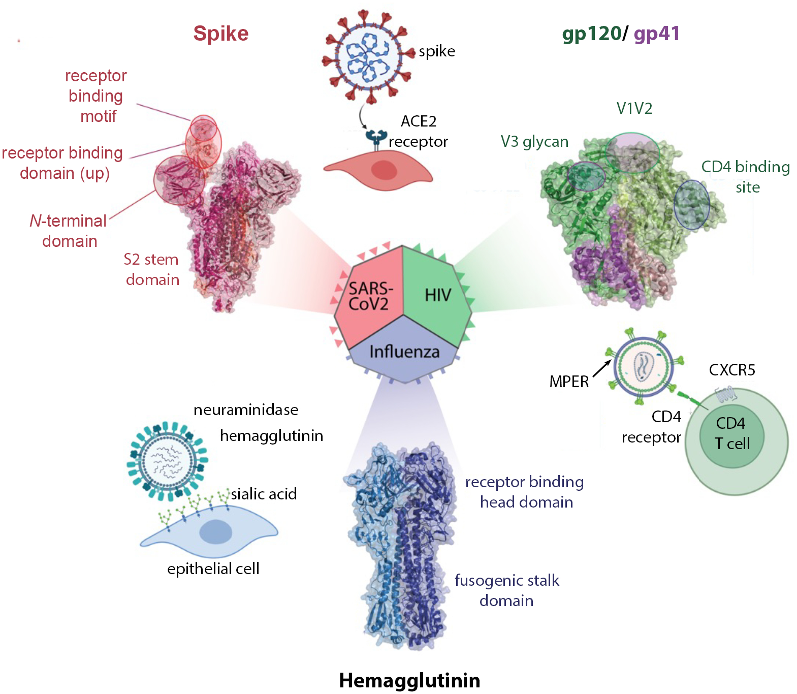
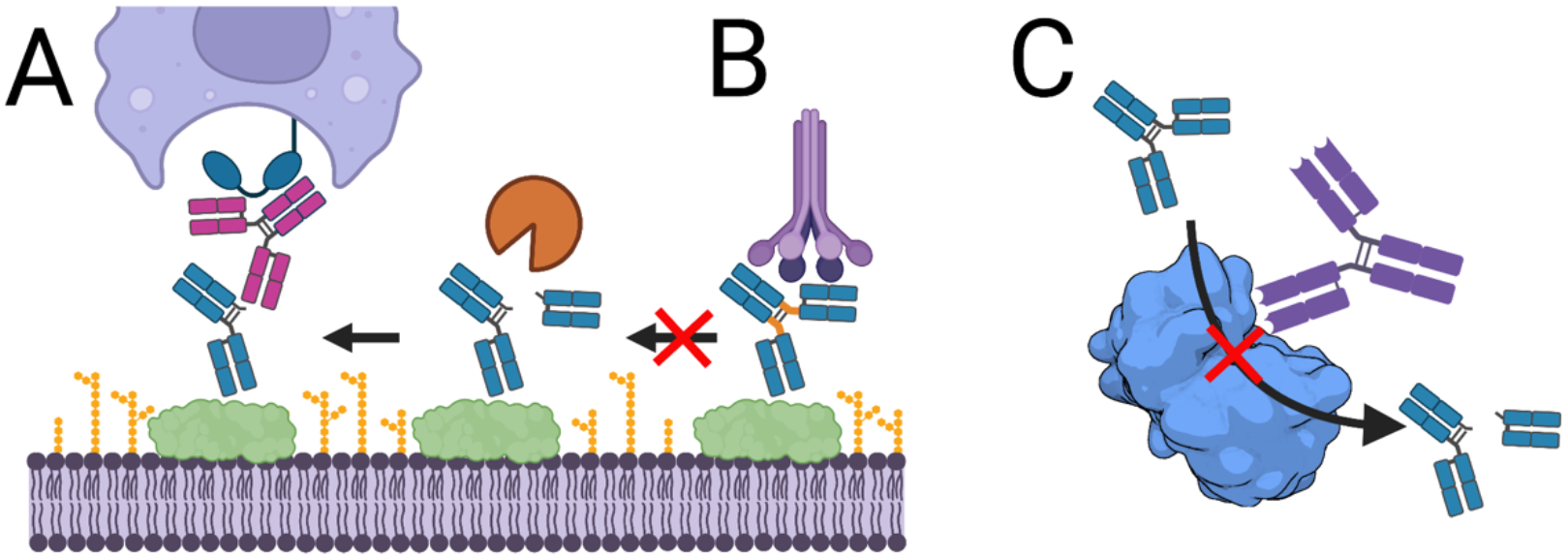
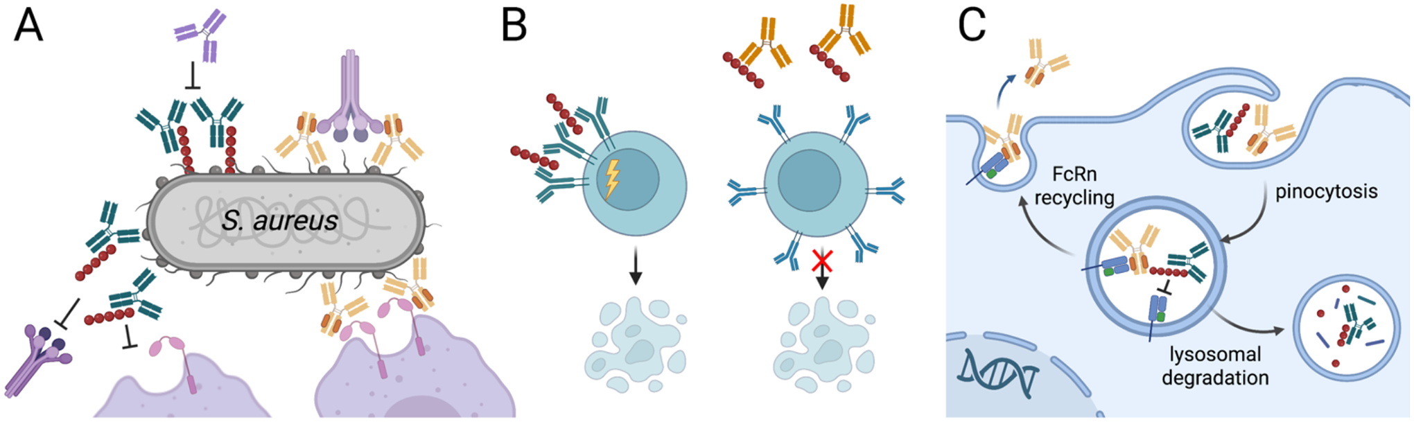
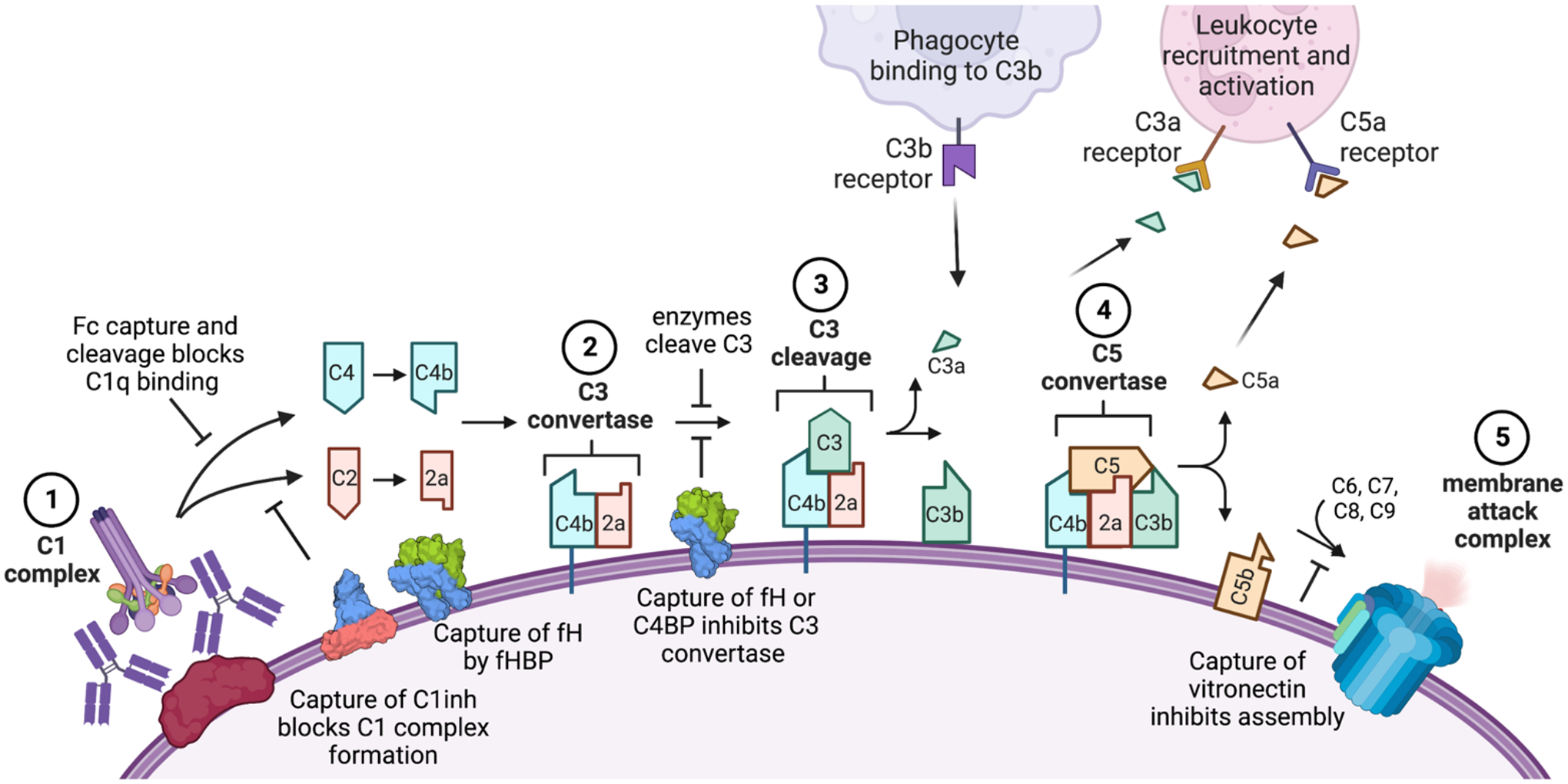
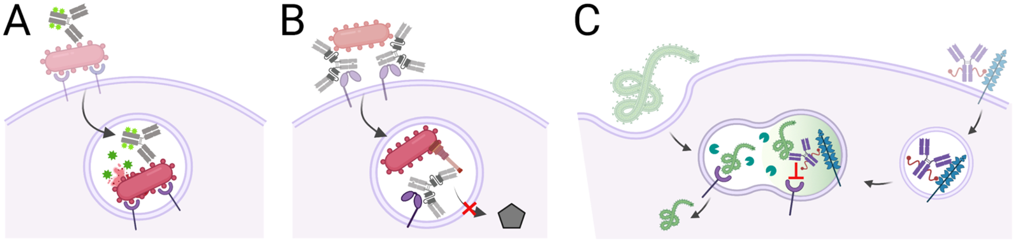
There is growing interest in identifying antibodies that protect against infectious diseases, especially for high-risk individuals and pathogens for which no vaccine is yet available. However, pathogens that manifest as opportunistic or latent infections express complex arrays of virulence-associated proteins and are adept at avoiding immune responses. Some pathogens have developed strategies to selectively destroy antibodies, whereas others create decoy epitopes that trick the host immune system into generating antibodies that are at best nonprotective and at worst enhance pathogenesis. Antibody engineering strategies can thwart these efforts by accessing conserved neutralizing epitopes, generating Fc domains that resist capture or degradation and even accessing pathogens hidden inside cells. Design of pathogen-resistant antibodies can enhance protection and guide development of vaccine immunogens against these complex pathogens. Here, we discuss general strategies for design of antibodies resistant to specific pathogen defense mechanisms.

Keywords: Fc engineering; antibody–drug conjugate; bispecific antibody; immune evasion; passive immunization; vaccines.


Figures

Figure 1.
Figure 1.. Key domains and antibody-targeted vulnerable sites in viral fusogen proteins from SARS-CoV-2, HIV-1, and influenza.
For each virus, the cellular receptors are shown as well as structure of the fusogen with key domains and epitopes identified. Structures for influenza hemagglutinin H3 from PDB 4FNK, for HIV-1 envelope PDB 5CJX, for SARS-CoV-2 spike from PDB 7SXT.
Figure 2.
Figure 2.. Antibodies that resist pathogenic protease activities to sustain recognition by host immune proteins, such as the C1q component of complement and CD32a Fc receptor found on phagocytes.
A, Antibodies recognizing hinge epitopes exposed after cleavage restore Fc functions to cleaved antibodies. B, Antibodies with engineered hinge regions no longer serve as suitable substrates for pathogenic proteases. C, Antibodies that block the activity of pathogenic proteases by directly blocking access to the active site (shown) or non-competitive (allosteric) mechanisms that bind an alternate enzyme epitope protect antibody functions. Shown is the structure of LasB (PDB 3DBK).
Figure 3.
Figure 3.. Antibodies that resist capture by Fc binding protein
A. S. aureus protein A disrupts antibody responses in multiple ways, which can be restored by Fc domains with reduced protein A affinity (most IgG3 allotypes or engineered IgG1 domains) or antibodies that bind protein A to block Fc capture. A, Membrane-bound protein A can block antibody Fc binding to the low affinity Fc receptors CD16b and CD32a, block the Fc hexamerization required for efficient recruitment of C1q and activation of complement and shield the bacterial surface from recognition by antibody Fab domains. Secreted protein A can B, crosslink- VH3 domains to trigger B cell receptor activation and apoptotic collapse and C, block antibody recycling by Fc/FcRn binding to reduce antibody half-life. Unmodified antibodies shown in green, Fc engineered antibodies shown in yellow, anti-protein A antibodies shown in orange.
Figure 4.
Figure 4.. Microbial disruption of the classical complement cascade by recruiting inhibitors or degrading complement proteins.
Key steps of the classical pathway of antibody activation shown: (1) the complement proteins C1q and then C1r and C1s bind the microbial surface or IgG/ IgM to form the C1 complex; (2) this cleaves C2 and C4 to produce C4b and C2a which form the C3 convertase; (3) this cleaves C3 to release C3a and deposit C3b covalently on the cell surface; (4) when C3b levels are high, it joins the C3 convertase to form the C5 convertase and deposit C5b on the surface; (5) components C6, C7, C8 and C9 join C5b to form the membrane attack complex and lyse the target cell. The lectin pathway follows a similar cascade but is initiated by the mannose binding lectin complex which recruits C1q, while the alternate pathway results from spontaneous C3 cleavage and C3b deposition to join the cascade at the C3 convertase step using an alternate C3b/Bb complex. Many of these steps can be inhibited by pathogen components, including proteins that bind or cleave the antibody Fc to inhibit C1q recruitment (e.g., protein A, staphylokinase); proteins that recruit host complement regulators (e.g., the Neisseria factor H binding protein [fHBP] which recruits factor H [fH] and B. pertussis Vag8 which recruits C1 inhibitor [C1inh]) and enzymes that degrade complement components (e.g., staphylokinase depletion of C3 away from the microbial surface). Engineering efforts to overcome these strategies include use of Hexabodies whose altered Fc domains favor hexamerization and C1q binding, antibodies altered to resist capture by Fc binding proteins or cleavage by bacterial proteases and antibodies that target microbial evasion proteins to block their functions.
Figure 5.
Figure 5.. Antibodies that target intracellular pathogens.
A, Antibody-antibiotic conjugates bind bacterial surface antigens and are internalized with the bacteria by bacterial or immune-mediated mechanisms into endosomal compartments. In this environment, the antibiotic is released to kill co-localized bacteria. B, Bi-specific antibody uses one binding site to bind the Psl antigen on the P. aeruginosa surface and mediate phagocytosis. Once in the endosome, that other antibody binding site blocks Type III secretion to support endosome acidification and bacterial killing. C, Antibodies block Ebola-receptor interactions in the endo-lysosome by hijacking the mannose-6-phosphate receptor to mediate antibody transfer to an endo-lysosome which may already contain Ebola virions. Once co-localized, the antibody blocks glycoprotein-receptor interactions and viral escape into the cytosol.

References

    1. Setliff I, Shiakolas AR, Pilewski KA, Murji AA, Mapengo RE, et al. 2019. High-Throughput Mapping of B Cell Receptor Sequences to Antigen Specificity. Cell 179: 1636–46 e15 - PMC - PubMed
    1. Pedrioli A, Oxenius A. 2021. Single B cell technologies for monoclonal antibody discovery. Trends Immunol 42: 1143–58 - PubMed
    1. Ngwuta JO, Chen M, Modjarrad K, Joyce MG, Kanekiyo M, et al. 2015. Prefusion F-specific antibodies determine the magnitude of RSV neutralizing activity in human sera. Sci Transl Med 7: 309ra162 - PMC - PubMed
    1. Zhu Q, McLellan JS, Kallewaard NL, Ulbrandt ND, Palaszynski S, et al. 2017. A highly potent extended half-life antibody as a potential RSV vaccine surrogate for all infants. Sci Transl Med 9: eaaj1928 - PubMed
    1. Bergeron HC, Tripp RA. 2022. Breakthrough therapy designation of nirsevimab for the prevention of lower respiratory tract illness caused by respiratory syncytial virus infections (RSV). Expert Opin Investig Drugs 31: 23–29 - PubMed

Publication types