. 2023 Mar;11(3):e006092.
doi: 10.1136/jitc-2022-006092.

Profiling specific cell populations within the inflammatory tumor microenvironment by oscillating-gradient diffusion-weighted MRI

Affiliations

Profiling specific cell populations within the inflammatory tumor microenvironment by oscillating-gradient diffusion-weighted MRI

Emily Hoffmann et al. J Immunother Cancer. 2023 Mar.

Abstract

Background: The inflammatory tumor microenvironment (TME) is formed by various immune cells, being closely associated with tumorigenesis. Especially, the interaction between tumor-infiltrating T-cells and macrophages has a crucial impact on tumor progression and metastatic spread. The purpose of this study was to investigate whether oscillating-gradient diffusion-weighted MRI (OGSE-DWI) enables a cell size-based discrimination between different cell populations of the TME.

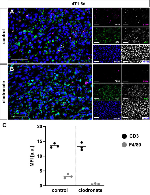
Methods: Sine-shaped OGSE-DWI was combined with the Imaging Microstructural Parameters Using Limited Spectrally Edited Diffusion (IMPULSED) approach to measure microscale diffusion distances, here relating to cell sizes. The accuracy of IMPULSED-derived cell radii was evaluated using in vitro spheroid models, consisting of either pure cancer cells, macrophages, or T-cells. Subsequently, in vivo experiments aimed to assess changes within the TME and its specific immune cell composition in syngeneic murine breast cancer models with divergent degrees of malignancy (4T1, 67NR) during tumor progression, clodronate liposome-mediated depletion of macrophages, and immune checkpoint inhibitor (ICI) treatment. Ex vivo analysis of IMPULSED-derived cell radii was conducted by immunohistochemical wheat germ agglutinin staining of cell membranes, while intratumoral immune cell composition was analyzed by CD3 and F4/80 co-staining.

Results: OGSE-DWI detected mean cell radii of 8.8±1.3 µm for 4T1, 8.2±1.4 µm for 67NR, 13.0±1.7 for macrophage, and 3.8±1.8 µm for T-cell spheroids. While T-cell infiltration during progression of 4T1 tumors was observed by decreasing mean cell radii from 9.7±1.0 to 5.0±1.5 µm, increasing amount of intratumoral macrophages during progression of 67NR tumors resulted in increasing mean cell radii from 8.9±1.2 to 12.5±1.1 µm. After macrophage depletion, mean cell radii decreased from 6.3±1.7 to 4.4±0.5 µm. T-cell infiltration after ICI treatment was captured by decreasing mean cell radii in both tumor models, with more pronounced effects in the 67NR tumor model.

Conclusions: OGSE-DWI provides a versatile tool for non-invasive profiling of the inflammatory TME by assessing the dominating cell type T-cells or macrophages.

Keywords: T-lymphocytes; immunotherapy; macrophages; translational medical research; tumor microenvironment.


Conflict of interest statement

Competing interests: None declared.

Figures

Figure 1
Figure 1
OGSE-DWI is able to differentiate between cancer cells, macrophages, and T-cells using in vitro spheroids. (A) 67NR tumor spheroid after WGA staining of cell membranes (red) and nuclei (blue); scale bar indicates 200 µm. (B) For MR imaging, the spheroid was embedded in agarose gel-solidified cell culture medium. (C) Localization of the spheroid was performed using T2-weighted imaging. Scale bar indicates 1.5 mm. (D) Corresponding diffusion-weighted image of the spheroid (b=0.25 ms/µm2, fOGSE=50 Hz). Scale bar indicates 1.5 mm. (E) IMPULSED-derived cell radii (black) differed significantly between cancer cells (4T1, 67NR) and immune cells, with larger cell radii for macrophages and smaller radii for T-cells, validated by immunohistochemical WGA staining (gray). Statistical significance was determined by two-sided t-tests (**p<0.01, ****p<0.0001). Each dot indicates mean cell radius of one spheroid. Horizontal lines represent group means. Figure partially created using Biorender.com. IMPULSED, Imaging Microstructural Parameters Using Limited Spectrally Edited Diffusion; OGSE-DWI, oscillating-gradient diffusion-weighted MRI; WGA, wheat germ agglutinin.
Figure 2
Figure 2
Capability of OGSE-DWI to monitor immune cell infiltrate during tumor progression. (A) T2-weighted and diffusion-weighted MR images (b=0.125 ms/µm2, fOGSE=80 Hz) of 4T1 tumors on day three, six, and nine. During progression, tumor volume increased and tumors exhibited central necrotic and hemorrhagic areas, identified as hypointense areas in the tumor center. ROIs for IMPULSED-based cell size analysis are indicated with dashed white circles. (B) T2-weighted and diffusion-weighted MR images (b=0.125 ms/µm2, fOGSE=80 Hz) of 67NR tumors on day three, six, and nine. During progression, tumor volume increased with only minor signs of necrosis, but generally homogeneous appearance. ROIs for IMPULSED-based cell size analysis are indicated with dashed white circles. (C) Mean IMPULSED-derived cell radii (black) decreased during progression of 4T1 tumors. WGA staining of cell membranes (gray) displayed similar trends. (D) Mean IMPULSED-derived cell radii (black) increased during progression of 67NR tumors. WGA staining of cell membranes (gray) displayed similar trends. Statistical significance was determined by one-way ANOVAs with Tukey’s post hoc analysis or Kruskal-Wallis test, depending on normal distribution of data (**p<0.01, ***p<0.001, ****p<0.0001). Each dot indicates mean cell radius of one tumor. Horizontal lines represent group means. ANOVAs, one-way analyses of variance; IMPULSED, Imaging Microstructural Parameters Using Limited Spectrally Edited Diffusion; OGSE-DWI, oscillating-gradient diffusion-weighted MRI; ROI, region of interest; WGA, wheat germ agglutinin.
Figure 3
Figure 3
Immunohistochemical analysis of the immune cell infiltrate during tumor progression. (A–C) Representative 4T1 tumor sections after 3-channel stainings of nuclei (blue), F4/80 (violet, macrophages), and CD3 (green, T-cells) with correlating merged images. (D) Mean fluorescence intensity (MFI) of immunohistochemical CD3 and F4/80 expression in 4T1 tumors showed a balanced ratio of macrophages and T-cells on day three, with increasing share of T-cells on day six and day nine. (E–G) Representative 67NR tumor sections after 3-channel stainings of nuclei (blue), F4/80 (violet, macrophages), and CD3 (green, T-cells) with correlating merged images. (H) MFI of immunohistochemical CD3 and F4/80 expression in 67NR tumors revealed overall lower immune cell content compared with 4T1 tumors. During progression, the content of macrophages increased, while the amount of T-cells remained consistently low. Scale bars represent 50 µm. Each dot indicates mean MFI of one tumor. Horizontal lines represent group means.
Figure 4
Figure 4
OGSE-DWI is capable of detecting clodronate liposome-mediated depletion of macrophages. (A) T2-weighted and diffusion-weighted MR images (b=0.125 ms/µm2, fOGSE=80 Hz) of 4T1 tumors after 6 days of clodronate liposome treatment compared with untreated controls. ROIs for IMPULSED-based cell size analysis are indicated with dashed white circles. (B) IMPULSED-derived cell radii (black) were significantly lower after treatment. Immunohistochemical WGA staining of cell membranes (gray) displayed a similar trend. Statistical significance was determined by Mann-Whitney U test (*p<0.05). Each dot indicates mean cell radius of one tumor. Horizontal lines represent group means. IMPULSED, Imaging Microstructural Parameters Using Limited Spectrally Edited Diffusion; OGSE-DWI, oscillating-gradient diffusion-weighted MRI; ROI, region of interest; WGA, wheat germ agglutinin.
Figure 5
Figure 5
Immunohistochemical analysis of clodronate liposome-mediated depletion of macrophages. (A, B) Representative clodronate liposome-treated 4T1 tumor sections compared with untreated controls after 3-channel stainings of nuclei (blue), F4/80 (violet, macrophages), and CD3 (green, T-cells) with correlating merged images. (C) Mean fluorescence intensity (MFI) of immunohistochemical CD3 and F4/80 expression revealed successful depletion of macrophages, along with an approximately constant content of T-cells after clodronate liposome treatment. Scale bars represent 50 µm. Each dot indicates mean MFI of one tumor. Horizontal lines represent group means.
Figure 6
Figure 6
OGSE-DWI enables early response assessment to ICI treatment. (A–D) T2-weighted and diffusion-weighted MR images (b=0.125 ms/µm2, fOGSE=80 Hz) of 4T1 and 67NR tumors after ICI treatment compared with untreated controls. ICI-treated tumors exhibited increasing necrosis, especially in the 4T1 model. ROIs for IMPULSED-based cell size analysis are indicated with dashed white circles. (E–H) IMPULSED-derived cell radii (black) were significantly lower after ICI treatment in both tumor models and at all time points. Stronger reductions in mean cell size after treatment were found in 67NR tumors. Ex vivo immunohistochemical WGA staining of cell membranes (gray) validated decreasing radii after treatment, showing more pronounced effects in 67NR tumors. Statistical significance was determined by two-sided t-tests or Mann-Whitney U tests, depending on normal distribution of data (*p<0.05, **p<0.01). Each dot indicates mean cell radius of one tumor. Horizontal lines represent group means. ICI, immune checkpoint inhibitor; IMPULSED, Imaging Microstructural Parameters Using Limited Spectrally Edited Diffusion; OGSE-DWI, oscillating-gradient diffusion-weighted MRI; WGA, wheat germ agglutinin.
Figure 7
Figure 7
Immunohistochemical analysis of the intratumoral immune cell infiltrate after ICI treatment. (A–D) Representative ICI-treated 4T1 tumor sections compared with untreated controls after 3-channel stainings of nuclei (blue), F4/80 (violet, macrophages), and CD3 (green, T-cells) with correlating merged images. (E) Mean fluorescence intensity (MFI) of immunohistochemical CD3 and F4/80 expression in ICI-treated 4T1 tumors showed increasing intratumoral content of T-cells with slightly increasing content of macrophages on day six and day nine. (F–I) Representative ICI-treated 67NR tumor sections compared with untreated controls after 3-channel stainings of nuclei (blue), F4/80 (violet, macrophages), and CD3 (green, T-cells) with correlating merged images. (J) Mean fluorescence intensity (MFI) of immunohistochemical CD3 and F4/80 expression in ICI-treated 67NR tumors showed excessive increase of intratumoral content of T-cells with slightly increasing content of macrophages on day six and day nine. Scale bars represent 50 µm. Each dot indicates mean MFI of one tumor. Horizontal lines represent group means. ICI, immune checkpoint inhibitor.

References

    1. Giraldo NA, Sanchez-Salas R, Peske JD, et al. . The clinical role of the Tme in solid cancer. Br J Cancer 2019;120:45–53. 10.1038/s41416-018-0327-z - DOI - PMC - PubMed
    1. Salemme V, Centonze G, Cavallo F, et al. . The crosstalk between tumor cells and the immune microenvironment in breast cancer: implications for immunotherapy. Front Oncol 2021;11:610303. 10.3389/fonc.2021.610303 - DOI - PMC - PubMed
    1. Heinrich EL, Walser TC, Krysan K, et al. . The inflammatory tumor microenvironment, epithelial mesenchymal transition and lung carcinogenesis. Cancer Microenviron 2012;5:5–18. 10.1007/s12307-011-0089-0 - DOI - PMC - PubMed
    1. García-Martínez E, Gil GL, Benito AC, et al. . Tumor-infiltrating immune cell profiles and their change after neoadjuvant chemotherapy predict response and prognosis of breast cancer. Breast Cancer Res 2014;16:488. 10.1186/s13058-014-0488-5 - DOI - PMC - PubMed
    1. Shen L, Zhou Y, He H, et al. . Crosstalk between macrophages, T cells, and iron metabolism in tumor microenvironment. Oxid Med Cell Longev 2021;2021:8865791. 10.1155/2021/8865791 - DOI - PMC - PubMed

Publication types