Review
. 2023 Mar 14;11(1):15.
doi: 10.1038/s41413-023-00249-w.

The roles of bone remodeling in normal hematopoiesis and age-related hematological malignancies

Affiliations
Review

The roles of bone remodeling in normal hematopoiesis and age-related hematological malignancies

Hengwei Zhang et al. Bone Res. .

Abstract

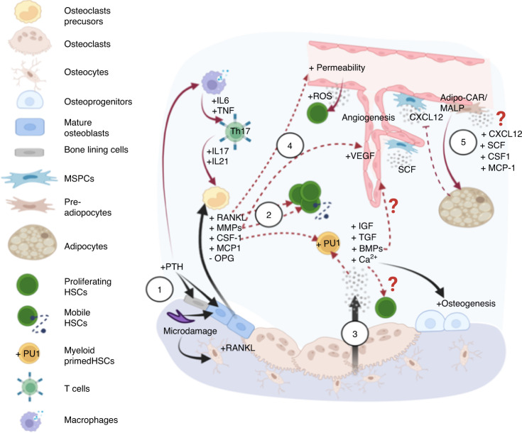
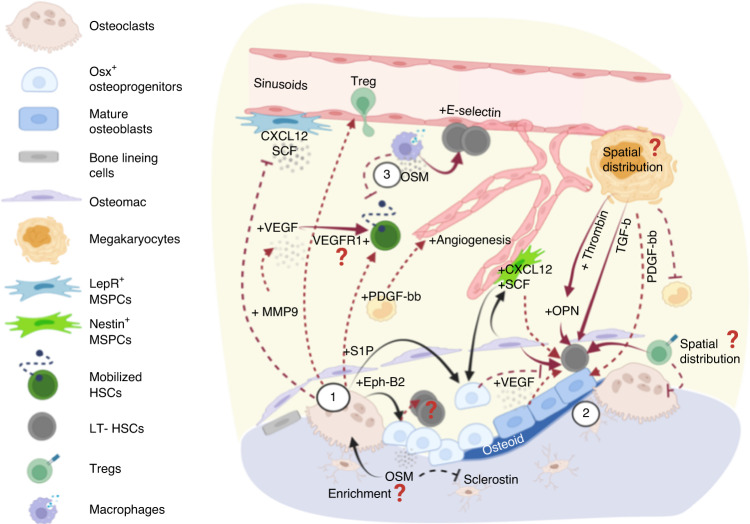
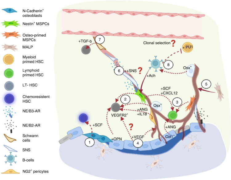
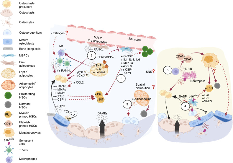
Prior research establishing that bone interacts in coordination with the bone marrow microenvironment (BMME) to regulate hematopoietic homeostasis was largely based on analyses of individual bone-associated cell populations. Recent advances in intravital imaging has suggested that the expansion of hematopoietic stem cells (HSCs) and acute myeloid leukemia cells is restricted to bone marrow microdomains during a distinct stage of bone remodeling. These findings indicate that dynamic bone remodeling likely imposes additional heterogeneity within the BMME to yield differential clonal responses. A holistic understanding of the role of bone remodeling in regulating the stem cell niche and how these interactions are altered in age-related hematological malignancies will be critical to the development of novel interventions. To advance this understanding, herein, we provide a synopsis of the cellular and molecular constituents that participate in bone turnover and their known connections to the hematopoietic compartment. Specifically, we elaborate on the coupling between bone remodeling and the BMME in homeostasis and age-related hematological malignancies and after treatment with bone-targeting approaches. We then discuss unresolved questions and ambiguities that remain in the field.


Conflict of interest statement

The authors declare no competing interests.

Figures

Fig. 1
Fig. 1
Factors involved in the initiation of bone remodeling and their crosstalk with the hematopoietic compartment. (1) Homeostatic bone remodeling occurs in response to microdamage and systemic Ca2+ needs. The nucleating event involves RANKL expression triggered in traumatized osteocytes or by PTH stimulation of bone-lining cells/osteoblasts and immune cells expressing PTH receptors. These lead to production of RANKL and other factors to promote osteoclastogenesis and osteoclast adhesion, respectively. (2) These bone-remodeling cascades exert functional effects on HSCs in the microenvironment via direct stimulation and alterations to the stem cell niche. Examples include myeloid differentiation bias, HSC expansion and mobilization from MCP-1, CSF-1, RANKL and MMPs released from activated osteoblasts. (3) During bone resorption, growth factors and calcium stored in the bone are released to promote osteodifferentiation, which can promote angiogenesis and impact hematopoiesis directly. (4) Global changes to the bone marrow microenvironment can also occur due to RANKL-increased vascular permeability and subsequently elevated ROS levels in HSPCs. (5) Additional changes are mediated by adipocytes and adipocyte-primed progenitors, including a recently identified MALP population that largely overlaps with LepR+ perivascular MSPCs, that release factors important to stem cell maintenance and regeneration (CXCL12 and SCF), myeloid differentiation (CSF-1), and osteoclastogenesis (MCP-1). The overlap in the regulation of homeostatic bone remodeling and hematopoiesis within the same microenvironment leads to several questions to be addressed. The first question pertains to the regulatory mechanisms by which MALP affects HSCs and the bone compartment. The second relates to the need to understand how extracellular calcium modulates HSC dormancy and proliferation. It is also unclear whether the angiogenic effects of growth factors are specific to sinusoidal or arteriolar vessels that are known to exert distinct hematopoietic supports. (In all figures, black solid arrows indicate interactions within the bone compartment; red solid arrows indicate interactions within the hematopoietic compartment; red dashed arrows indicate interactions between the bone and hematopoietic compartments)
Fig. 2
Fig. 2
Factors involved in the reversal stage of bone remodeling and their crosstalk with the hematopoietic compartment. (1) Osteoclasts secrete several coupling factors to promote osteodifferentiation during the reversal stage of bone remodeling and that also target hematopoietic cells. S1P acts through S1P1 receptors that are highly expressed on hematopoietic cells to regulate cell trafficking, which is crucial for cell egress after treatment with mobilizing agents (e.g., G-CSF or GM-CSF). Osteoclast-derived Ephrin-B2 acts on EphB4-expressing osteolineage cells, leading to the subsequent expansion of long-term HSCs mediated via mechanisms that remain to be elucidated. Osteoclasts also recruit regulatory T cells, which may constitute immune-privileged sites that promote HSC survival. In addition, MMP9 secreted from osteoclasts modulates the bone marrow microenvironment in several ways, including the release of VEGF from extracellular matrix to promote angiogenesis and the degradation/shedding of CXCL12 and SCF to promote HSC mobilization. (2) In a bone remodeling unit, osteolineage cells, osteomacs, and megakaryocytes (MKs) work collaboratively to promote osteogenesis. Each unit produces abundant factors such as thrombin-cleaved, activated OPN, and MK-derived TGF-β, which regulate HSC dormancy. (3) Moreover, oncostatin M (OSM) secreted by osteolineage cells (e.g., MSPCs and osteoblasts) or immune cells (e.g., macrophages) plays pleiotropic roles to promote both remodeling (inducing RANKL expression) and osteodifferentiation (suppressing sclerostin expression). Importantly, OSM induces CXCL12 to inhibit cell mobilization and boost E-selectin-mediated HSC self-renewal/expansion. Overall, the reversal stage involves functionally diverse signaling pathways that promote dormancy, mobilization, and expansion. It will be insightful to understand whether HSC proliferation in such microenvironment is mediated by self-renewal-based expansion or loss of stemness, and whether active bone remodeling leads to enrichment in MK distribution, OPN/OSM concentration, and accumulation of regulatory T cells that are associated with the self-renewal potency and survival of HSCs
Fig. 3
Fig. 3
Factors involved in the bone formation stage and their crosstalk with the hematopoietic compartment. (1) N-cadherin+ osteoblasts have been shown to be spatially associated with chemoresistant HSCs via SCF regulation. (2) Osteolineage cells maintain phenotypic LT-HSCs via IL-18 and Nestin+/Osterix+/NG2+ cell-derived angiogenin (ANG). (3) In contrast, angiogenin from mature osteoblasts and LepR+/osteolectin+ osteo-committed MSPCs maintain lymphoid-primed HSCs and common lymphoid progenitors. Periarteriolar Nestin+ stromal cells secrete SCF and CXCL12 to maintain lymphoid primed or unbiased HSCs. (4) Osteogenesis impacts the bone marrow microenvironment in several ways. Osteoblast-derived VEGF boosts angiogenesis via VEGFR2, which also promotes the repopulating capacity of HSCs. (5) In addition, osteogenesis is associated with the enrichment of the arteriole-connecting capillaries (Type H vessels), which has been shown to be associated with MALP and Osterix+ populations. (6) Moreover, the SNS has been found to be spatially associated with arterioles, which can enhance Nestin+ MSPC-derived CXCL12 secretion through the action of β3-adrenergic receptors and modulate cell motility following circadian cycles. (7) Schwann cells on nerve axons also contribute to HSC quiescence by activating latent TGF-β. (8) During bone formation, Osterix+ osteoprogenitors and osteoblasts are critical for B lymphopoiesis, and pro-B cells secrete acetylcholine to retain HSCs/HSPCs in the bone marrow and suppress the expansion of myeloid cell progenitors. Several questions remain to be answered. For example, do different stages of bone remodeling affect perivascular stromal composition, cytokine gradient, and SNS regulation? Does bone remodeling also shape the spatial landscape of VEGF/VEGFR1+ (Fig. 2) and VEGF/VEGFR2+ signaling to regulate HSCs differently? Importantly, whether distinct hematopoietic clones exhibit specific tropism toward a given microenvironment during bone remodeling/modeling cycles remains to be addressed
Fig. 4
Fig. 4
Skeletal aging and its impact on the hematopoietic compartment. Aging of osteolineage cells leads to an overall inflammatory microenvironment (left panel) and acquisition of the senescence-associated secretory phenotype (SASP, right panel). (Left 1) In general, aged osteoprogenitors employ a myeloid-promoting program, attributed to telomere dysfunction that manifests as upregulation of G-CSF, IL-3, MIP-3a levels in bone-associated stromal cells, and aging of bone cells that overexpress CSF-1, IL-1, CCL5, etc. (2) Aged MSPCs are also primed for adipocyte differentiation, synergistically promoting myelopoiesis. Specifically, the preadipocyte (Pref-1+ or MALP) population is expanded with upregulated RANKL expression. Adipocytes further secrete leptins, DPP4, IL-6, TNF, and CXCL1/CXCL2 to promote osteoclastogenesis while inhibiting osteodifferentiation, which promotes the proinflammatory immune phenotype. (3) In contrast, adiponectins promote osteoprogenitors and have been shown to protect HSCs from inflammatory insult and enhance self-renewal. Hypothetically, leptin- and adiponectin-secreting adipocytes exhibit spatial associations with bone resorption and formation sites, respectively, and may contribute to differential HSC responses. (Right 4) Multiple cell types, including osteoblasts, osteoprogenitors, osteocytes, lymphocytes, and myeloid cells, undergo senescence and are associated with the overproduction of IL-6, IL-1, MMPs and other proinflammatory cytokines that promote bone resorption and myeloid bias. (5) Reduced phagocytic capability of macrophages leads to the accumulation of senescent neutrophils, an increase in IL-1 level and induced platelet bias. Whether increased megakaryopoiesis causes myeloid bias and HSC accumulation remains unclear. Overall, aging is associated with contraction of Type H vessels, and arterioles, depleting niche factors that support lymphopoiesis and possibly negatively impacting the SNS. Although important, it remains unclear whether effects of estrogen deficiency on bone remodeling kinetics cause substantial variability in aged HSC niches between sexes
Fig. 5
Fig. 5
Crosstalk between skeletal aging and age-associated myeloid diseases. (1) MSPC senescence augments the proinflammatory microenvironment (Fig. 4) in MDS/leukemia. (2) An adipogenic propensity promotes the survival of leukemia blasts, although it has been found that (3) in AML, Runx2+ and Osterix+ osteoprogenitors are expanded via BMP signaling to enhance leukemia engraftment. (4) In addition, growth factors released during bone resorption have been shown to drive SASP acquisition (via IGF) and activate inflammasome NLRP3 (via DAMPs and calcium), which is overexpressed in MDS patients. (5) Coupling factors in the reversal stage may play roles in disease progression. S1P/S1PR3 participates in leukemogenesis, myeloid bias and promotes LSC differentiation. In contrast, (6) OPN has been found to drive cell dormancy in B-cell acute lymphoblastic lymphoma; however, its role in MDS and AML still needs to be elucidated. (7) The bone-forming zone may promote leukemia cell expansion and maintenance of leukemia-initiating cells. For example, hyperactive Wnt pathways in osteoblasts have been associated with a differentiation blockade and excessive blast numbers mediated through Jagged1/Notch signaling. Higher ATP levels in the osteoblastic zone sustain leukemia-initiating cells via ATP-P2X7 signaling. Notably, this process involves the influx of calcium ions; however, whether different levels of extracellular calcium at distinct stages of bone remodeling exert differential impacts on leukemic cells remains to be investigated. (8) Distinct subsets in the sinusoidal niche can support the proliferation (VLA-4/CD98) or dormancy (CD44/E-selectin) of leukemia stem cells. (9) CXCL12 from Tie2+ periarteriolar stroma has been shown to promote cell proliferation, while CXCL12 from Prx1+ MSPCs drives chemoresistance. While both endosteal and vascular niches can support tumor proliferation and dormancy, sex effects (Fig. 4) and whether distinct tumor subclones exhibit tropism toward a given niche remain to be elucidated
Fig. 6
Fig. 6
Crosstalk between the components of aged bone and age-associated multiple myeloma. Multiple myeloma is an age-associated disease that is closely related with bone cells. Myeloma cells establish a tumor microenvironment with osteoblastic and osteoclastic cells and generation of bone matrix, which creates a vicious cycle that promotes myeloma cell expansion on bone lesions. The contribution of bone cells and senescent cells under aging conditions to the tumor microenvironment and maintenance of myeloma cells are described as follows: (1) Most cell compartments in the tumor microenvironment, except the osteoblast compartment, support tumors. Mesenchymal progenitors directly promote the proliferation, migration, and adhesion of myeloma cells through the action IL-6, survivin, CD44, VLAs, syndecan 1 and MCP-1 or indirectly affect myeloma cells through osteoclastogenesis and angiogenesis. (2) An increase in the number of adipocytes support MM through their release of adipokines, such as leptin, adipsin, visfatin and resistin. (3) Osteoblasts have been identified as the only cells that inhibit myeloma cells through Decorin. (4) However, osteoblasts indirectly promote myeloma cells through the activation of osteoclasts. Osteoclasts resorb bone matrix, release numerous tumorigenic factors, such as TGF-β, IGF-1, FGF, PDGFs and BMPs, and increase the extracellular calcium level. (5) The direct mechanism by which osteoclasts affect myeloma cells involves the secretion of soluble factors, such as osteopontin, MIP-1a, IL-6, Annexin II, BAFF and APRIL. Although senescent mesenchymal progenitors and adipocytes have been reported in MM, the mechanisms underlying their senescence are still not clear, particularly in the early stages of MM. (6) In MM, an increased number of senescent cells in the tumor environment has been observed, but the contribution of increased senescent cells remains unclear. Senescent cells inhibit osteoblast differentiation but promote osteoclast differentiation through SASP factors
Fig. 7
Fig. 7
Cell senescence in SMM. The expression of the cell senescence marker gene CDKN2A in CD138+ myeloma cells and CD138-expressing nonmyeloma cells was measured by RNA sequencing. Black bars represent clinically diagnosed SMM patients, and white bars represent age-paired healthy donors

References

    1. Morrison SJ, Scadden DT. The bone marrow niche for hematopoietic stem cells. Nature. 2014;505:327–334. doi: 10.1038/nature12984. - DOI - PMC - PubMed
    1. Spradling A, Drummond-Barbosa D, Kai T. Stem cells find their niche. Nature. 2001;414:98–104. doi: 10.1038/35102160. - DOI - PubMed
    1. Levesque JP, Helwani FM, Winkler IG. The endosteal ‘osteoblastic’ niche and its role in hematopoietic stem cell homing and mobilization. Leukemia. 2010;24:1979–1992. doi: 10.1038/leu.2010.214. - DOI - PubMed
    1. Kunisaki Y, Frenette PS. Influences of vascular niches on hematopoietic stem cell fate. Int. J. Hematol. 2014;99:699–705. doi: 10.1007/s12185-014-1580-4. - DOI - PubMed
    1. Schepers K, Campbell TB, Passegue E. Normal and leukemic stem cell niches: insights and therapeutic opportunities. Cell Stem Cell. 2015;16:254–267. doi: 10.1016/j.stem.2015.02.014. - DOI - PMC - PubMed