. 2023 Mar 14;14(1):1411.
doi: 10.1038/s41467-023-36997-w.

Genetic architecture of spatial electrical biomarkers for cardiac arrhythmia and relationship with cardiovascular disease

William J Young  1   2 Jeffrey Haessler  3 Jan-Walter Benjamins  4 Linda Repetto  5 Jie Yao  6 Aaron Isaacs  7   8 Andrew R Harper  9   10 Julia Ramirez  1   11   12 Sophie Garnier  13   14 Stefan van Duijvenboden  1   11 Antoine R Baldassari  15 Maria Pina Concas  16 ThuyVy Duong  17 Luisa Foco  18 Jonas L Isaksen  19 Hao Mei  20 Raymond Noordam  21 Casia Nursyifa  22 Anne Richmond  23 Meddly L Santolalla  24   25 Colleen M Sitlani  26 Negin Soroush  27 Sébastien Thériault  28   29 Stella Trompet  21   30 Stefanie Aeschbacher  31 Fariba Ahmadizar  27   32 Alvaro Alonso  33 Jennifer A Brody  26 Archie Campbell  34   35   36 Adolfo Correa  37 Dawood Darbar  38 Antonio De Luca  39 Jean-François Deleuze  40   41   42 Christina Ellervik  43   44   45 Christian Fuchsberger  18   46   47 Anuj Goel  9   10 Christopher Grace  9   10 Xiuqing Guo  6   48   49 Torben Hansen  22 Susan R Heckbert  26   50 Rebecca D Jackson  51 Jan A Kors  52 Maria Fernanda Lima-Costa  53 Allan Linneberg  54   55 Peter W Macfarlane  56 Alanna C Morrison  57 Pau Navarro  23 David J Porteous  36   58 Peter P Pramstaller  18   59 Alexander P Reiner  50   60 Lorenz Risch  61   62   63 Ulrich Schotten  7 Xia Shen  5   64   65 Gianfranco Sinagra  39 Elsayed Z Soliman  66 Monika Stoll  8   67   68 Eduardo Tarazona-Santos  24 Andrew Tinker  1   69 Katerina Trajanoska  70 Eric Villard  13   14 Helen R Warren  1   69 Eric A Whitsel  15   71 Kerri L Wiggins  26 Dan E Arking  17 Christy L Avery  15 David Conen  28 Giorgia Girotto  16   72 Niels Grarup  22 Caroline Hayward  73 J Wouter Jukema  30   74   75 Dennis O Mook-Kanamori  76   77 Morten Salling Olesen  78 Sandosh Padmanabhan  79 Bruce M Psaty  26   50   80 Cristian Pattaro  18 Antonio Luiz P Ribeiro  81   82 Jerome I Rotter  6   48   83 Bruno H Stricker  27 Pim van der Harst  4   84 Cornelia M van Duijn  85   86 Niek Verweij  4 James G Wilson  87   88 Michele Orini  2   11 Philippe Charron  13   14   89   90 Hugh Watkins  9   10 Charles Kooperberg  3 Henry J Lin  6   48   49 James F Wilson  5   23 Jørgen K Kanters  19 Nona Sotoodehnia  91 Borbala Mifsud  1   92 Pier D Lambiase  2   11 Larisa G Tereshchenko  93   94 Patricia B Munroe  95   96
Affiliations

Genetic architecture of spatial electrical biomarkers for cardiac arrhythmia and relationship with cardiovascular disease

William J Young et al. Nat Commun. .

Abstract

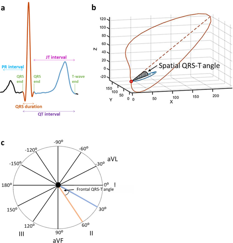
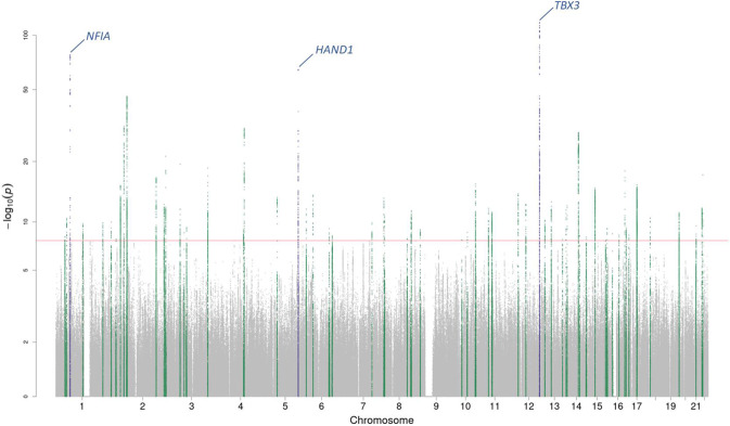
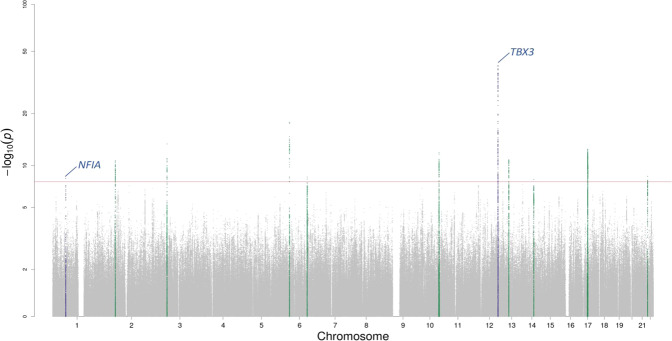
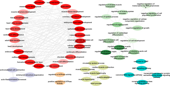
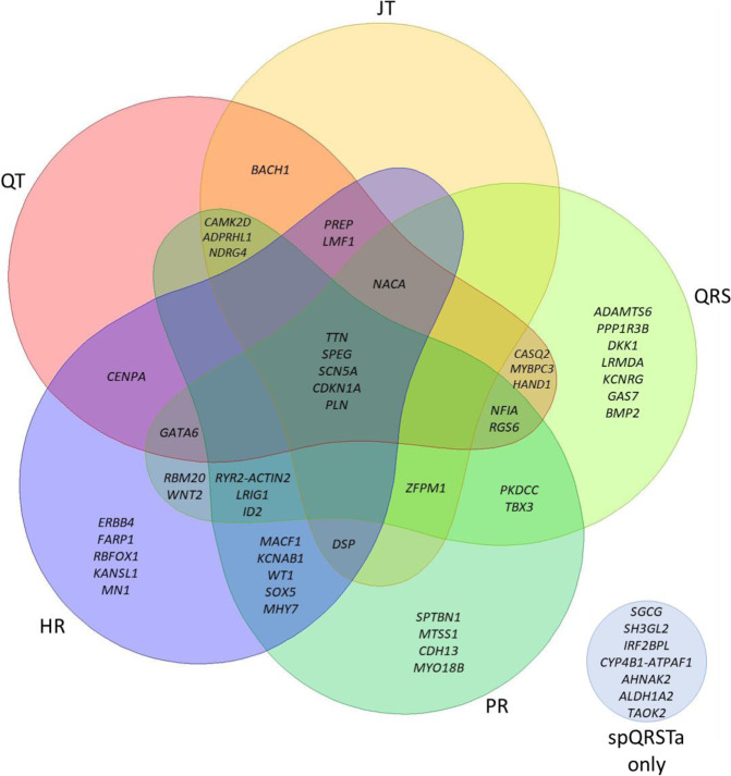
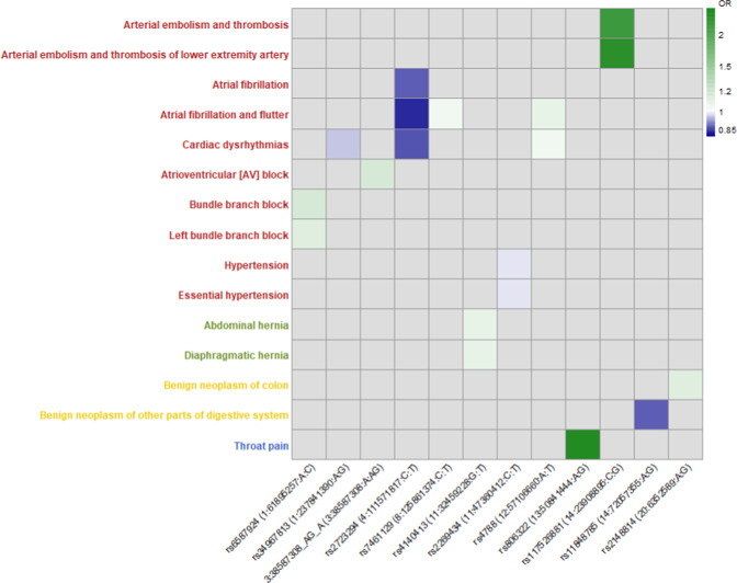
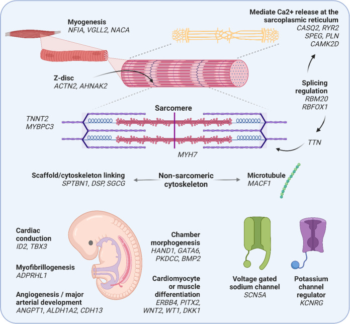
The 3-dimensional spatial and 2-dimensional frontal QRS-T angles are measures derived from the vectorcardiogram. They are independent risk predictors for arrhythmia, but the underlying biology is unknown. Using multi-ancestry genome-wide association studies we identify 61 (58 previously unreported) loci for the spatial QRS-T angle (N = 118,780) and 11 for the frontal QRS-T angle (N = 159,715). Seven out of the 61 spatial QRS-T angle loci have not been reported for other electrocardiographic measures. Enrichments are observed in pathways related to cardiac and vascular development, muscle contraction, and hypertrophy. Pairwise genome-wide association studies with classical ECG traits identify shared genetic influences with PR interval and QRS duration. Phenome-wide scanning indicate associations with atrial fibrillation, atrioventricular block and arterial embolism and genetically determined QRS-T angle measures are associated with fascicular and bundle branch block (and also atrioventricular block for the frontal QRS-T angle). We identify potential biology involved in the QRS-T angle and their genetic relationships with cardiovascular traits and diseases, may inform future research and risk prediction.


Conflict of interest statement

B.M.P serves on the Steering Committee of the Yale Open Data Access Project funded by Johnson & Johnson. D.C has received speaker fees from BMS/Pfizer and Servier, and consultation fees from Roche Diagnostics and Trimedics. U.S received consultancy fees or honoraria from Università della Svizzera Italiana (USI, Switzerland), Roche Diagnostics (Switzerland), EP Solutions Inc. (Switzerland), Johnson & Johnson Medical Limited, (United Kingdom), Bayer Healthcare (Germany). D.O.M.-K is a part time research consultant at Metabolon, Inc. U.S is co-founder and shareholder of YourRhythmics BV, a spin-off company of the University Maastricht. The remaining authors declare no competing interests.

Figures

Fig. 1
Fig. 1. Graphical representation of the spQRSTa and fQRSTa alongside a single electrocardiogram lead signal.
a Single lead electrocardiogram (ECG) signal with classical measures QRS duration and the QT interval labelled. The dark orange (estimates ventricular depolarization time) and blue (ventricular repolarization time) shaded sections of the signal represent the regions used to calculate the QRS and T-wave axes respectively with multiple ECG leads. b The spatial QRS-T angle (spQRSTa) mean is the angle between the mean amplitude of QRS and T-wave spatial loops. These spatial loops can be constructed from the resting 12-lead ECG using a standardised transformation, to produce representative X, Y and Z vectors that can be plotted over time. c The frontal QRS-T angle (fQRSTa) is the absolute difference between QRS and T-wave axes in the frontal plane only.
Fig. 2
Fig. 2. Manhattan plot for the spQRSTa multi-ancestry meta-analysis.
Manahattan plot for the spatial QRS-T angle (spQRSTa) meta-analysis. Two-sided P-values are plotted on the -log10 scale (Y-axis). The red horizontal line indicates genome-wide significance (P < 5 × 10−8). Variants within the boundaries of loci previously reported for the spatial QRS-T angle are labelled with the candidate gene and colored blue. Variants at previously unreported loci are green.
Fig. 3
Fig. 3. Manhattan plot for the fQRSTa multi-ancestry meta-analysis.
Manahattan plot for the frontal QRS-T angle (fQRSTA) meta-analysis. Two-sided P-values are plotted on the -log10 scale (Y-axis). The red horizontal line indicates genome-wide significance (P < 5 × 10−8). Variants within the boundaries of loci previously reported for the spatial QRS-T angle are labelled with the candidate gene and colored blue. Variants at previously unreported loci are green.
Fig. 4
Fig. 4. Significant GO biological processes from spQRSTa DEPICT multi-ancestry findings.
All significant (false discovery rate <0.01) multi-ancestry spatial QRS-T angle (spQRSTa) gene-ontology (GO) biological processes from Data-driven Expression-Prioritization Integration for Complex Traits (DEPICT) software were analyzed using the Reduce and Visualize Gene Ontology (REVIGO) web application to remove redundant terms and cluster related nodes. Highly similar GO terms are linked by edges where the line width indicates the degree of similarity. Within each cluster, the colour gradient represents differences in the DEPICT gene-set enrichment two-sided P-values, with lighter gradients reflecting smaller enrichment P-values (therefore more significant) compared with other nodes in the same cluster.
Fig. 5
Fig. 5. Overlap of multi-ancestry spQRSTa loci with ECG measures.
Venn diagram showing spatial QRS-T angle (spQRSTa) multi-ancestry loci where a lead variant reported for another electrocardiographic ECG measure maps within the locus boundaries. For this figure, ECG measures shown are PR interval (cardiac conduction), QRS duration (ventricular depolarization), QT and JT intervals (ventricular repolarization) and heart rate (HR). Overlap was declared if a lead variant for these ECG measures mapped to within ±500 kb or r2 > 0.1 of a lead variant at a spQRSTa locus. Some loci overlap with other ECG traits (not visualised here but presented in Supplementary Data 15). At seven spQRSTa loci, no overlap was observed with any ECG trait (blue circle bottom right).
Fig. 6
Fig. 6. Significant associations observed in phenome-wide association study of lead and conditionally independent spQRSTa variants.
X-axis: Lead variant (RsID [Chromosome: Position (hg19): Allele1: Allele2]) or conditionally independent variant from the spatial QRS-T angle (spQRSTa) European ancestry meta-analysis that had a significant association with a clinical phenotype in UK Biobank. Y-axis: Phenotype derived from hospital episode statistics, with colour coding for each major group (circulatory system; red, digestive system; green, neoplasms; yellow, respiratory; blue). Odds ratios (OR) are color coded according to decreasing (blue) or increasing (green) odds. 3:38587306:A:G was a conditionally independent variant at the SCN5A locus.
Fig. 7
Fig. 7. Illustration of candidate genes at spQRSTa multi-ancestry loci and their potential function.
Candidate genes at spatial QRS-T angle (spQRSTa) loci are grouped according to potential roles in embryonic development, cardiac structure and function. RYR2 and ACTN2 are candidate genes from the same locus. A summary of the bioinformatic evidence for each gene is presented in Supplementary Data 14. Created using BioRender.com.

Similar articles

  • Sex hormones and reproductive factors with cardiac arrhythmia and ECG indices: a mendelian randomization study.
    Wang X, Wei Z, Zuo Z, Sun Y, Guo X, Tong Y, Liu G, Xu D, Zhang Z. Wang X, et al. BMC Cardiovasc Disord. 2024 Nov 20;24(1):659. doi: 10.1186/s12872-024-04335-7. BMC Cardiovasc Disord. 2024. PMID: 39567890 Free PMC article.
  • Spatial QRS-T Angle and Cognitive Decline in Older Subjects.
    Mahinrad S, Ferguson I, Macfarlane PW, Clark EN, Stott DJ, Ford I, Mooijaart SP, Trompet S, van Heemst D, Jukema JW, Sabayan B. Mahinrad S, et al. J Alzheimers Dis. 2019;67(1):279-289. doi: 10.3233/JAD-180633. J Alzheimers Dis. 2019. PMID: 30584139
  • Genetic analyses of the electrocardiographic QT interval and its components identify additional loci and pathways.
    Young WJ, Lahrouchi N, Isaacs A, Duong T, Foco L, Ahmed F, Brody JA, Salman R, Noordam R, Benjamins JW, Haessler J, Lyytikäinen LP, Repetto L, Concas MP, van den Berg ME, Weiss S, Baldassari AR, Bartz TM, Cook JP, Evans DS, Freudling R, Hines O, Isaksen JL, Lin H, Mei H, Moscati A, Müller-Nurasyid M, Nursyifa C, Qian Y, Richmond A, Roselli C, Ryan KA, Tarazona-Santos E, Thériault S, van Duijvenboden S, Warren HR, Yao J, Raza D, Aeschbacher S, Ahlberg G, Alonso A, Andreasen L, Bis JC, Boerwinkle E, Campbell A, Catamo E, Cocca M, Cutler MJ, Darbar D, De Grandi A, De Luca A, Ding J, Ellervik C, Ellinor PT, Felix SB, Froguel P, Fuchsberger C, Gögele M, Graff C, Graff M, Guo X, Hansen T, Heckbert SR, Huang PL, Huikuri HV, Hutri-Kähönen N, Ikram MA, Jackson RD, Junttila J, Kavousi M, Kors JA, Leal TP, Lemaitre RN, Lin HJ, Lind L, Linneberg A, Liu S, MacFarlane PW, Mangino M, Meitinger T, Mezzavilla M, Mishra PP, Mitchell RN, Mononen N, Montasser ME, Morrison AC, Nauck M, Nauffal V, Navarro P, Nikus K, Pare G, Patton KK, Pelliccione G, Pittman A, Porteous DJ, Pramstaller PP, Preuss MH, Raitakari OT, Reiner AP, Ribeiro ALP, Rice KM, Risch L, Schlessinger D, Schotten U, Schurmann C, Shen X… See abstract for full author list ➔ Young WJ, et al. Nat Commun. 2022 Sep 1;13(1):5144. doi: 10.1038/s41467-022-32821-z. Nat Commun. 2022. PMID: 36050321 Free PMC article.
  • Multi-ancestry GWAS of the electrocardiographic PR interval identifies 202 loci underlying cardiac conduction.
    Ntalla I, Weng LC, Cartwright JH, Hall AW, Sveinbjornsson G, Tucker NR, Choi SH, Chaffin MD, Roselli C, Barnes MR, Mifsud B, Warren HR, Hayward C, Marten J, Cranley JJ, Concas MP, Gasparini P, Boutin T, Kolcic I, Polasek O, Rudan I, Araujo NM, Lima-Costa MF, Ribeiro ALP, Souza RP, Tarazona-Santos E, Giedraitis V, Ingelsson E, Mahajan A, Morris AP, Del Greco M F, Foco L, Gögele M, Hicks AA, Cook JP, Lind L, Lindgren CM, Sundström J, Nelson CP, Riaz MB, Samani NJ, Sinagra G, Ulivi S, Kähönen M, Mishra PP, Mononen N, Nikus K, Caulfield MJ, Dominiczak A, Padmanabhan S, Montasser ME, O'Connell JR, Ryan K, Shuldiner AR, Aeschbacher S, Conen D, Risch L, Thériault S, Hutri-Kähönen N, Lehtimäki T, Lyytikäinen LP, Raitakari OT, Barnes CLK, Campbell H, Joshi PK, Wilson JF, Isaacs A, Kors JA, van Duijn CM, Huang PL, Gudnason V, Harris TB, Launer LJ, Smith AV, Bottinger EP, Loos RJF, Nadkarni GN, Preuss MH, Correa A, Mei H, Wilson J, Meitinger T, Müller-Nurasyid M, Peters A, Waldenberger M, Mangino M, Spector TD, Rienstra M, van de Vegte YJ, van der Harst P, Verweij N, Kääb S, Schramm K, Sinner MF, Strauch K, Cutler MJ, Fatkin D, London B, Olesen M, Roden DM, Benjamin Shoemaker M, Gustav Smith… See abstract for full author list ➔ Ntalla I, et al. Nat Commun. 2020 May 21;11(1):2542. doi: 10.1038/s41467-020-15706-x. Nat Commun. 2020. PMID: 32439900 Free PMC article.
  • The spatial QRS-T angle: implications in clinical practice.
    Voulgari C, Pagoni S, Tesfaye S, Tentolouris N. Voulgari C, et al. Curr Cardiol Rev. 2013 Aug;9(3):197-210. doi: 10.2174/1573403x113099990031. Curr Cardiol Rev. 2013. PMID: 23909632 Free PMC article. Review.

Cited by

  • The impact of common and rare genetic variants on bradyarrhythmia development.
    Weng LC, Rämö JT, Jurgens SJ, Khurshid S, Chaffin M, Hall AW, Morrill VN, Wang X, Nauffal V, Sun YV, Beer D, Lee S, Nadkarni GN, Duong T, Wang B, Czuba T, Austin TR, Yoneda ZT, Friedman DJ, Clayton A, Hyman MC, Judy RL, Skanes AC, Orland KM, Treu TM, Oetjens MT, Alonso A, Soliman EZ, Lin H, Lunetta KL, van der Pals J, Issa TZ, Nafissi NA, May HT, Leong-Sit P, Roselli C, Choi SH; FinnGen; Million Veteran Program; Regeneron Genetics Center; Khan HR, Knight S, Karlsson Linnér R, Bezzina CR, Ripatti S, Heckbert SR, Gaziano JM, Loos RJF, Psaty BM, Smith JG, Benjamin EJ, Arking DE, Rader DJ, Shah SH, Roden DM, Damrauer SM, Eckhardt LL, Roberts JD, Cutler MJ, Shoemaker MB, Haggerty CM, Cho K, Palotie A, Wilson PWF, Ellinor PT, Lubitz SA. Weng LC, et al. Nat Genet. 2025 Jan;57(1):53-64. doi: 10.1038/s41588-024-01978-2. Epub 2025 Jan 2. Nat Genet. 2025. PMID: 39747593 Free PMC article.
  • Genomic and molecular evidence that the lncRNA DSP-AS1 modulates Desmoplakin expression.
    Foco L, De Bortoli M, Fabiola Del Greco M, Frommelt LS, Volani C, Riekschnitz DA, Motta BM, Fuchsberger C, Delerue T, Völker U, Huan T, Gögele M, Winkelmann J, Dörr M, Levy D, Waldenberger M, Teumer A, Pramstaller PP, Rossini A, Pattaro C. Foco L, et al. medRxiv [Preprint]. 2025 Mar 31:2025.03.29.25324867. doi: 10.1101/2025.03.29.25324867. medRxiv. 2025. Update in: Hum Genet. 2025 Jul 30. doi: 10.1007/s00439-025-02761-x. PMID: 40236443 Free PMC article. Updated. Preprint.
  • Latent profiles of global electrical heterogeneity: the Hispanic Community Health Study/Study of Latinos.
    Tereshchenko LG, Haq KT, Howell SJ, Mitchell EC, Martínez J, Hyde J, Briceno G, Pena J, Pocius E, Khan A, Soliman EZ, Lima JAC, Kapadia SR, Misra-Hebert AD, Kattan MW, Kansal MM, Daviglus ML, Kaplan R. Tereshchenko LG, et al. Eur Heart J Digit Health. 2024 Jul 8;5(5):611-621. doi: 10.1093/ehjdh/ztae048. eCollection 2024 Sep. Eur Heart J Digit Health. 2024. PMID: 39318685 Free PMC article.
  • Sex hormones and reproductive factors with cardiac arrhythmia and ECG indices: a mendelian randomization study.
    Wang X, Wei Z, Zuo Z, Sun Y, Guo X, Tong Y, Liu G, Xu D, Zhang Z. Wang X, et al. BMC Cardiovasc Disord. 2024 Nov 20;24(1):659. doi: 10.1186/s12872-024-04335-7. BMC Cardiovasc Disord. 2024. PMID: 39567890 Free PMC article.
  • Applying multimodal AI to physiological waveforms improves genetic prediction of cardiovascular traits.
    Zhou Y, Khasentino J, Yun T, Biradar MI, Shreibati J, Lai D, Schwantes-An TH, Luben R, McCaw ZR, Engmann J, Providencia R, Schmidt AF, Munroe PB, Yang H, Carroll A, Khawaja AP, McLean CY, Behsaz B, Hormozdiari F. Zhou Y, et al. Am J Hum Genet. 2025 Jul 3;112(7):1562-1579. doi: 10.1016/j.ajhg.2025.05.015. Epub 2025 Jun 20. Am J Hum Genet. 2025. PMID: 40543505 Free PMC article.

References

    1. Fishman GI, et al. Sudden cardiac death prediction and prevention: report from a national heart, lung, and blood institute and heart rhythm society workshop. Circulation. 2010;122:2335–2348. doi: 10.1161/CIRCULATIONAHA.110.976092. - DOI - PMC - PubMed
    1. Joukar S. A comparative review on heart ion channels, action potentials and electrocardiogram in rodents and human: extrapolation of experimental insights to clinic. Lab. Anim. Res. 2021;37:25. doi: 10.1186/s42826-021-00102-3. - DOI - PMC - PubMed
    1. Oehler A, Feldman T, Henrikson CA, Tereshchenko LG. QRS-T angle: a review. Ann. Noninvasive Electrocardiol. 2014;19:534–542. doi: 10.1111/anec.12206. - DOI - PMC - PubMed
    1. Plonsey R. A contemporary view of the ventricular gradient of Wilson. J. Electrocardiol. 1979;12:337–341. doi: 10.1016/S0022-0736(79)80001-1. - DOI - PubMed
    1. Young, W. et al. Comparisons of the spatial QRS-T angle with intra-cardiac markers of depolarization and repolarization. Comput. Cardiol. 2020 Computing in Cardiology, Rimini, Italy, pp. 1–4 (2020).

Publication types