Review
. 2023 Mar 14;8(1):114.
doi: 10.1038/s41392-023-01378-8.

Metabolic landscape in cardiac aging: insights into molecular biology and therapeutic implications

Affiliations
Review

Metabolic landscape in cardiac aging: insights into molecular biology and therapeutic implications

Saiyang Xie et al. Signal Transduct Target Ther. .

Abstract

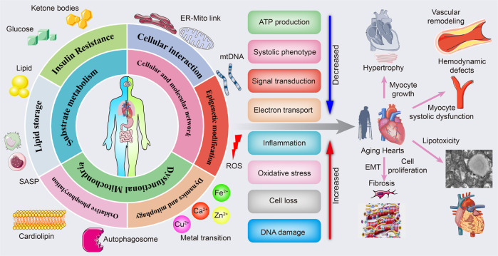
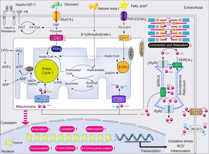
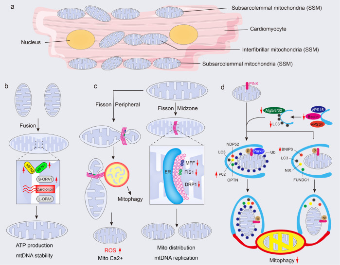
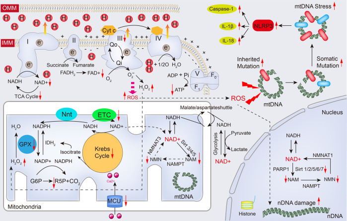
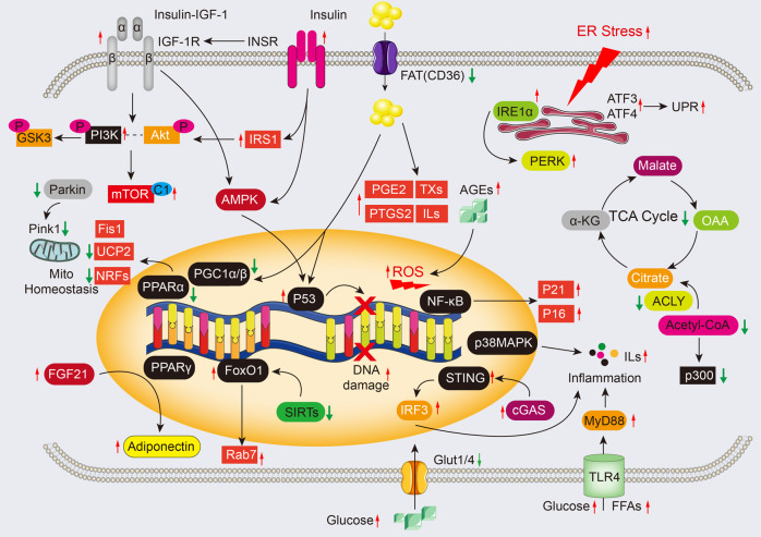
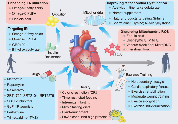
Cardiac aging is evident by a reduction in function which subsequently contributes to heart failure. The metabolic microenvironment has been identified as a hallmark of malignancy, but recent studies have shed light on its role in cardiovascular diseases (CVDs). Various metabolic pathways in cardiomyocytes and noncardiomyocytes determine cellular senescence in the aging heart. Metabolic alteration is a common process throughout cardiac degeneration. Importantly, the involvement of cellular senescence in cardiac injuries, including heart failure and myocardial ischemia and infarction, has been reported. However, metabolic complexity among human aging hearts hinders the development of strategies that targets metabolic susceptibility. Advances over the past decade have linked cellular senescence and function with their metabolic reprogramming pathway in cardiac aging, including autophagy, oxidative stress, epigenetic modifications, chronic inflammation, and myocyte systolic phenotype regulation. In addition, metabolic status is involved in crucial aspects of myocardial biology, from fibrosis to hypertrophy and chronic inflammation. However, further elucidation of the metabolism involvement in cardiac degeneration is still needed. Thus, deciphering the mechanisms underlying how metabolic reprogramming impacts cardiac aging is thought to contribute to the novel interventions to protect or even restore cardiac function in aging hearts. Here, we summarize emerging concepts about metabolic landscapes of cardiac aging, with specific focuses on why metabolic profile alters during cardiac degeneration and how we could utilize the current knowledge to improve the management of cardiac aging.


Conflict of interest statement

The authors declare no competing interests.

Figures

Fig. 1
Fig. 1
The link between cardiac aging and metabolic pathology. Metabolic drivers of cardiac aging. Mitochondrial substrates metabolic disturbance including lipid storage and insulin resistance; dysfunctional mitochondria with impaired oxidative phosphorylation, mitochondria dynamics, and mitophagy; cellular and molecular network. All of which drive reduced ATP production, systolic phenotype, signal transduction, and electron transport, along with increased inflammation, oxidative stress, cell death, and DNA damage. Finally, various pathological alterations, including myocyte growth-induced cardiac hypertrophy, endothelium–mesenchymal transition and cell proliferation-mediated cardiac fibrosis, lipid deposition-induced comparative cardiac lipotoxicity, and insufficient energy-mediated myocyte systolic dysfunction and hemodynamic disorder, contribute to cardiac aging, thus the failing heart. The online resource inside this figure was quoted or modified from Servier Medical Art
Fig. 2
Fig. 2
Metabolic substates utilization and excitation-contraction coupling in the aged heart. In the aging heart, myocardial lipids catabolism and glucose oxidation are reduced due to insulin resistance, but glycolysis dominates the energy source. Also, the relative contribution of ketone body utilization to ATP production is enhanced. The mismatch between FA oxidation and uptake results in the accumulation of toxic lipid intermediates, leading to impairment of ATP production and formation of high-energy phosphates. Furthermore, several metabolic intermediates interfere with metabolism and course oxidative stress, RCD, and inflammation by post-translational modifications and serving as chromatin-modifying enzymes in cardiac aging. Krebs cycle and excitation-contraction coupling are stimulated by Ca2+ that mainly sources from sarcoplasmic reticulum (SR), while the transport of Ca2+ is disturbed in the aging heart. Red arrows indicate alterations that occur in the aging heart (see text for details). α-KG α-ketoglutarate, ADP adenosine diphosphate, ATP adenosine triphosphate, β-OX fatty acid β-oxidation, CPT1 carnitine O-palmitoyltransferase 1, ETC electron transport chain, FADH2 reduced flavin adenine dinucleotide, FAT fatty acid translocase (also known as CD36), FA-CoA fatty acyl-CoA ester, Glut4 glucose transporter 4 (also known as SLC2A4), G6P glucose-6-phosphate, IGF-1 Insulin-like growth factor 1, LTCC L-type Ca2+ channel, MFN1/2 Mitofusin1/2, MPC1/2 mitochondrial pyruvate carrier1/2, MCU mitochondrial Ca2+ uniporter protein, NADH nicotinamide adenine dinucleotide, NCLX mitochondrial Na+/Ca2+ exchanger protein, NCX1 Na+/Ca2+ exchanger 1, OPA1 dynamin-like guanosine triphosphatase, PCr phosphocreatine, PDH Pyruvate dehydrogenase, RCD regulated cell death, RYR2 ryanodine receptor 2, SERCA sarcoplasmic/endoplasmic reticulum Ca2+ ATPase, TAG triacylglycerol. The online resource inside this figure was quoted or modified from Servier Medical Art
Fig. 3
Fig. 3
Mitochondrial dynamics and mitophagy in the aged heart. a A schematic of a cardiomyocyte to highlight the location of subsarcolemmal (SSM) and interfibrillar mitochondria (IFM). b Mitofusins (MFN1 and MFN2) in the OMM, belongs to proteins of the dynamin-related family of large GTPases, synergizing with OPA1 in the IMM to regulate mitochondria fusion. In the aging heart, long OPA1 (L-OPA1) is cleaved to generate the short form of OPA1 (S-OPA1), and the latter cooperates with cardiolipin to promote the fusion of mitochondria, by which compensates the ATP production and maintains mtDNA stability. c Fission is predominantly orchestrated by the DRP1. DRP1 binds to OMM receptors MFF and mitochondrial FIS1, promoting the midzone fission to course mitochondria distribution with mtDNA replication. Also, DRP1 induces peripheral fission and enables damaged material to be destined for mitophagy. The latter results in higher ROS generation and enhanced mitochondria Ca2+. d As a serine/threonine-protein kinase, PINK1 serves as the sensor that detects impaired mitochondria and leads to the proteolytic cleavage of PINK1 by mitochondrial proteases. Uncleaved PINK1 plays a role in activating parkin through direct phosphorylation of the parkin Ub-like (UBL) domain, as well as phosphorylation of ubiquitin. This, in turn, recruits autophagy receptors such as p62, OPTN, and NDP52, promoting the recruitment of LC3 and subsequent engulfment of damaged mitochondria by autophagosomes. Also, autophagy receptors such as BNIP3, NIX, and FUNDC1 regulate ubiquitin-independent mitophagy by recruiting LC3 and facilitating the engulfment of damaged mitochondria by autophagosomes. Red arrows indicate alterations that occur in the aging heart (see text for details). Atg autophagy-related protein, ATP adenosine triphosphate, BNIP3 bcl-2 19-kDa interacting protein 3, DRP1 dynamin-related protein 1, ER endoplasmic reticulum, FIS1 fission 1 protein, FUNDC1 FUN14 domain containing 1, IMM inner mitochondrial membrane, MFF mitochondrial fission factor, MFN mitofusin, mtDNA mitochondria DNA, NDP52 Nuclear dot protein 52 kDa, OMM outer mitochondrial membrane, OPA1 optic atrophy 1, OPTN optineurin; ROS reactive oxygen species, VPS (also known as PI3KR4) phosphoinositide 3-kinase regulatory subunit 4. The online resource inside this figure was quoted or modified from Servier Medical Art
Fig. 4
Fig. 4
Altered NAD(H), ROS and mitochondrial DNA during cardiac aging. The electron transport chain (ETC) is a collection of four enzyme complexes (complexes I-IV) and a large protein complex (complex V) responsible for synthesizing ATP in the mitochondrial inner membrane. It generates ATP from ADP, Pi, and Mg2+ using an electrochemical gradient of protons created by the electron transport chain. As electrons pass down the redox potential gradient from NADH or FADH2 to oxygen, hydrogen ions are actively transported from the matrix to the cytosolic side of the inner membrane by complexes I, III, and IV. Complex I oxidizes NADH, which leads to sequential electron flow to coenzyme Q, complex III, cytochrome c, and ultimately to cytochrome oxidase (complex IV), where oxygen is reduced to water. However, during cardiac aging, the decline in respiration favors the relative reduction of complexes I and III, leading to increased ROS production. This increased ROS contributes to the impairment of the Krebs cycle and reduced ATP production. Furthermore, the cytoplasmic and nuclear NAD+ pools probably equilibrate by diffusion through the nuclear pore. However, the mitochondrial membrane is impermeable to both NAD+ and NADH. Reducing equivalents generated by glycolysis are transferred to the mitochondrial matrix via the malate/aspartate shuttle. In addition, different NAD+-consuming enzymes lead to the generation of nicotinamide, which is recycled via the NAD+ salvage pathway. Different forms of the NMNAT enzyme and sirtuins are localized in different compartments. Of note, a proportion of ROS is involved in reduction reaction by mitochondrial antioxidant system GPX device. While overburden of ROS leads to mtDNA mutation and damage to promote activation of NLRP3 inflammasome. Red arrows indicate alterations that occur in the aging heart (see text for details). ADP adenosine diphosphate, ATP adenosine triphosphate, Cyt c cytochrome c, FAD flavin adenine dinucleotide, FADH2 reduced flavin adenine dinucleotide, F1/Fo F1/Fo ATP synthase, G6P glucose-6-phosphate, GPX glutathione peroxidase, IL-1β interleukin-1β, IL-18 interleukin-18, IDH2 isocitrate dehydrogenase 2, IMM inner mitochondrial membrane, MCU mitochondrial Ca2+ uniporter, mtDNA mitochondria DNA, NADH nicotinamide adenine dinucleotide, NADPH reduced nicotinamide adenine dinucleotide phosphate, NAM nicotinamide, nDNA nuclear DNA, NLRP3 NOD-like receptor family pyrin domain containing 3, NMN nicotinamide mononucleotide, NMNAT1/3 nicotinamide nucleotide adenylyltransferase 1/3, Nnt mitochondrial NAD(P) transhydrogenase, OMM outer mitochondrial membrane, PARP1 poly(ADP-ribose) polymerase, Pi phosphate ion, ROS reactive oxygen species, R5P ribulose 5-phosphate, TCA cycle tricarboxylic acid cycle. The online resource inside this figure was quoted or modified from Servier Medical Art
Fig. 5
Fig. 5
Molecular mechanisms and potential signaling for cardiac aging. The insulin/IGF-1 signaling pathway can activate signal transduction through the PI3K/Akt pathway, which in turn phosphorylates multiple targets, regulating the activity of the mTOR complex (mTORC1). IGF-1 also contributes to protein synthesis by activating the PI3K/Akt/mTOR and PI3K/Akt/GSK3β pathways, which can lead to endoplasmic reticulum (ER) stress or protein degradation via the ubiquitin-proteasome system (UPS). In the presence of ER stress, activated IRE1α and PERK can initiate proinflammatory and proapoptotic signaling pathways. Increased NAD+ might contribute to SASP and aging by disturbing AMPK and p53 activation, as well as enhancing p38MAPK and NF-kB activity in senescent cells. During cardiac aging, the maintenance of mitochondrial homeostasis is impaired, which can result in the release of mtDNA and the activation of the cGAS/STING/IRF3 pathway, leading to the production of inflammatory cytokines. The PINK1-Parkin pathway plays a crucial role in maintaining mitochondrial function and dynamics through mitophagy. In addition, ROS can activate the MAPK and PI3K/Akt signaling pathways, as well as increase levels of p53 and p21, which can promote apoptosis and inflammation in chondrocytes. The TLR4-MyD88 pathway can also be activated by circulating FFAs and glucose, leading to the production of proinflammatory factors. The online resource inside this figure was quoted or modified from Servier Medical Art
Fig. 6
Fig. 6
Potential therapeutic approach for cardiac aging. Metabolic therapies for the treatment of cardiac aging are aimed to improve insulin resistance, FA oxidation, mitochondria dysfunction, and ROS. Synthetic small molecular drugs targeting insulin resistance, sirtuin activation, and ROS clearance are verified to delay cardiac aging in animal model. Moreover, both dietary advice and exercise training are beneficial for cardiac aging in the elderly. The online resource inside this figure was quoted or modified from Servier Medical Art

References

    1. Zeng Y, Hesketh T. The effects of China’s universal two-child policy. Lancet. 2016;388:1930–1938. doi: 10.1016/S0140-6736(16)31405-2. - DOI - PMC - PubMed
    1. Mattson MP. Lifelong brain health is a lifelong challenge: from evolutionary principles to empirical evidence. Ageing Res. Rev. 2015;20:37–45. doi: 10.1016/j.arr.2014.12.011. - DOI - PMC - PubMed
    1. Mattson MP, Arumugam TV. Hallmarks of brain aging: adaptive and pathological modification by metabolic states. Cell Metab. 2018;27:1176–1199. doi: 10.1016/j.cmet.2018.05.011. - DOI - PMC - PubMed
    1. Evans MA, Sano S, Walsh K. Cardiovascular disease, aging, and clonal hematopoiesis. Annu. Rev. Pathol. 2020;15:419–438. doi: 10.1146/annurev-pathmechdis-012419-032544. - DOI - PMC - PubMed
    1. Lutz W, Sanderson W, Scherbov S. The coming acceleration of global population ageing. Nature. 2008;451:716–719. doi: 10.1038/nature06516. - DOI - PubMed

Publication types