Review
. 2023 Feb 15;15(1):127-143.
doi: 10.1007/s12551-023-01044-x. eCollection 2023 Feb.

Structure-function relationship and physiological role of apelin and its G protein coupled receptor

Affiliations
Review

Structure-function relationship and physiological role of apelin and its G protein coupled receptor

Subhashree Murali et al. Biophys Rev. .

Erratum in

Abstract

Apelin receptor (APJR) is a class A peptide (apelin) binding G protein-coupled receptor (GPCR) that plays a significant role in regulating blood pressure, cardiac output, and maintenance of fluid homeostasis. It is activated by a wide range of endogenous peptide isoforms of apelin and elabela. The apelin peptide isoforms contain distinct structural features that aid in ligand recognition and activation of the receptor. Site-directed mutagenesis and structure-based studies have revealed the involvement of extracellular and transmembrane regions of the receptor in binding to the peptide isoforms. The structural features of APJR activation of the receptor as well as mediating G-protein and β-arrestin-mediated signaling are delineated by multiple mutagenesis studies. There is increasing evidence that the structural requirements of APJR to activate G-proteins and β-arrestins are different, leading to biased signaling. APJR also responds to mechanical stimuli in a ligand-independent manner. A multitude of studies has focused on developing both peptide and non-peptide agonists and antagonists specific to APJR. Apelin/elabela-activated APJR orchestrates major signaling pathways such as extracellular signal-regulated kinase (ERKs), protein kinase B (PKB/Akt), and p70S. This review focuses on the structural and functional characteristics of apelin, elabela, APJR, and their interactions involved in the binding and activation of the downstream signaling cascade. We also focus on the diverse signaling profile of APJR and its ligands and their involvement in various physiological systems.

Keywords: Apelin receptor; Apelin; Elabela; GPCR; Signaling.


Conflict of interest statement

Conflict of interestThe authors declare no competing interests.

Figures

Fig. 1
Fig. 1
a Representation of crystal structure of APJR-AMG3054 complex (PDB: 5VBL). The modified peptide ligand is depicted in green (sticks) and the receptor is shown in orange (cartoon). b Amino acid sequence of apelin peptide isoforms. Residues of Apelin-36, Apelin-17, Apelin-13, and Pyro-glutamate (Pyr) Apelin-13 are indicated. Grey arrows show the site of cleavage by PCSK3/furin enzyme in the larger isoforms, Apelin-36 and Apelin-17 c Comparison between Apelin-17 and AMG3054. The latter is a modified cyclic peptide (cyclised between Glu10 and Lys13, indicated by dotted lines). The modified residues of AMG3054 are coloured in pink. hARG, homoarginine; CHA, cyclohexylalanine; OIC, Octahydroindole-2-carboxylic Acid; NLE, Norleucine; 4-Cl-PHE, 4-choloro-Phenylalanine
Fig. 2
Fig. 2
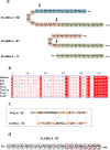
Conserved sequence of elabela isoforms. a Amino acid sequence of elabela-32, elabela-21 and elabela-11. Grey arrows depict the conserved cleavage site by PCSK3/Furin enzyme on larger isoforms elabela-32 and elabela-21. b Sequence alignment of elabela-54 pre-peptide in various organisms. The eight amino acids of the C-terminal region and the two cysteine residues show complete conservation, as highlighted in red. The alignment was performed using Clustal Omega (Sievers et al. 2011). c Comparison of Apelin-36 and Elabela-32 shows conserved residues (highlighted in orange) at similar positions in both the N- and C-terminal of the peptides. This further emphasizes their shared structure–function relationship inactivation of the receptor. d Structure–function relationship of elabela. The highlighted residues that predominantly fall in the C-terminal region are considered vital in the peptide’s function
Fig. 3
Fig. 3
Snake plot of APJR depicting the structure–function relationship of critical residues. The conserved amino acids of APJR are presented here as their single letter code with the amino acid position in superscript. They are highlighted based on their roles in ligand binding, G-protein activation, and structural stability. The disulfide bond formed between conserved cysteines is represented as dotted lines. ECL, extracellular loop; TM, transmembrane; ICL, intracellular loop
Fig. 4
Fig. 4
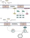
Overall signaling pathways mediated by APJR and its various ligands. a Classic G-protein-mediated activation pathway upon ligand binding. Gαq coupling to the activated receptor leads to the activation of PLCβ, which increases intracellular calcium that drives the phosphorylation of ERK, AKT, and production of NO. Gαi coupling to the receptor also mediates the ERK, AKT, and NOS pathways and inhibits cAMP production. b Desensitization pathway of APJR. Subsequent to activation, phosphorylation of the C-terminal chain of APJR recruits β-arrestins that drive the receptor’s endocytosis-mediated internalization drive the receptor’s endocytosis-mediated internalization. The internalized receptor is either recycled back to the membrane or directed to proteolysis. ERK, extracellular-signal-regulated kinase; NOS, nitric oxide synthase; NO, nitric oxide; MEK, mitogen-activated protein kinase; PLCβ. phospholipase C; PI3K, phosphoinositide 3-kinases; cAMP, cyclic AMP

References

    1. Abrisqueta M, Olivares C, Herraiz C, et al. Human melanocortin 1 receptor-mediated ubiquitination of nonvisual arrestins. Role of Mahogunin Ring Finger 1 E3 ligase. Biochim Biophys Acta Mol Cell Res. 2018;1865:76–94. doi: 10.1016/j.bbamcr.2017.09.013. - DOI - PubMed
    1. Adam F, Khatib A-M, Lopez JJ, et al. Apelin: an antithrombotic factor that inhibits platelet function. Blood. 2016;127:908–920. doi: 10.1182/blood-2014-05-578781. - DOI - PubMed
    1. Ahn KH, Nishiyama A, Mierke DF, Kendall DA. Hydrophobic residues in helix 8 of cannabinoid receptor 1 are critical for structural and functional properties. Biochemistry. 2010;49:502. doi: 10.1021/bi901619r. - DOI - PMC - PubMed
    1. Ashokan A, Aradhyam GK. Measurement of intracellular Ca2+ mobilization to study GPCR signal transduction. Methods Cell Biol. 2017;142:59–66. doi: 10.1016/bs.mcb.2017.07.002. - DOI - PubMed
    1. Ashokan A, Aradhyam GK. Effective purification of recombinant peptide ligands for GPCR research. Methods Cell Biol. 2017;142:111–118. doi: 10.1016/bs.mcb.2017.07.001. - DOI - PubMed

LinkOut - more resources