. 2023 Jan-Dec:32:9636897231160216.
doi: 10.1177/09636897231160216.

Comparison of the Cost and Effect of Combined Conditioned Medium and Conventional Medium for Fallopian Tube Organoid Cultures

Affiliations

Comparison of the Cost and Effect of Combined Conditioned Medium and Conventional Medium for Fallopian Tube Organoid Cultures

Yu-Hsun Chang et al. Cell Transplant. 2023 Jan-Dec.

Abstract

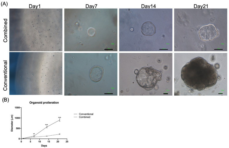
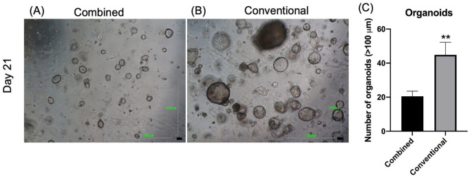
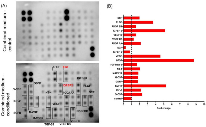
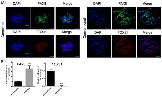
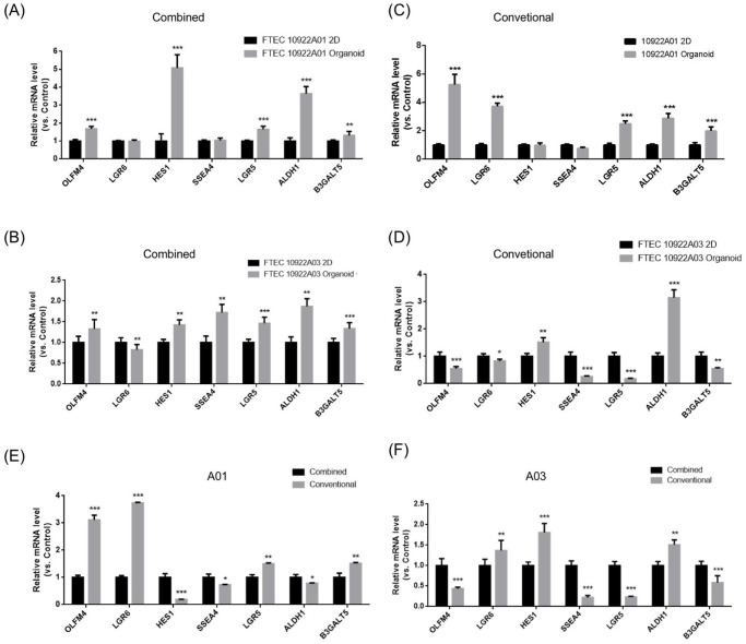
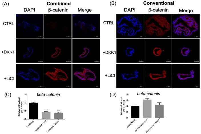
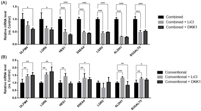
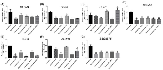
Fallopian tube epithelial cells (FTEC) are thought to be the cell of origin of high-grade serous ovarian carcinoma. FTEC organoids can be used as research models for the disease. Nevertheless, culturing organoids requires a medium supplemented with several expensive growth factors. We proposed that a combined conditioned medium based on the composition of the fallopian tubes, including epithelial, stromal, and endothelial cells could enhance FTEC organoid formation. We derived two primary culture cell lines from the fimbria portion of the fallopian tubes. The organoids were split into conventional or combined medium groups based on what medium they were grown in and compared. The number and size of the organoids were evaluated. Quantitative polymerase chain reaction (qPCR) and immunohistochemistry (IHC) were used to evaluate gene and protein expression (PAX8, FOXJ1, beta-catenin, and stemness genes). Enzyme-linked immunosorbent assay was used to measure Wnt3a and RSPO1 in both mediums. DKK1 and LiCl were added to the mediums to evaluate their influence on beta-catenin signaling. The growth factor in the combined medium was evaluated by the growth factor array. We found that the conventional medium was better for organoids regarding proliferation (number and size). In addition, WNT3A and RSPO1 concentrations were too low in the combined medium and needed to be added making the cost equivalent to the conventional medium. However, the organoid formation rate was 100% in both groups. Furthermore, the combined medium group had higher PAX8 and stemness gene expression (OLFM4, SSEA4, LGR5, B3GALT5) when compared with the conventional medium group. Wnt signaling was evident in the organoids grown in the conventional medium but not in the combined medium. PLGF, IGFBP6, VEGF, bFGF, and SCFR were found to be enriched in the combined medium. In conclusion, the combined medium could successfully culture organoids and enhance PAX8 and stemness gene expression. However, the conventional medium was a better medium for organoid proliferation. The expense of both mediums was comparable. The benefit of using a combined medium requires further exploration.

Keywords: Wnt/beta-catenin; endothelial cells; fallopian tube epithelial cells; medium; organoid; stromal cells.


Conflict of interest statement

The author(s) declared no potential conflicts of interest with respect to the research, authorship, and/or publication of this article.

Figures

Figure 1.
Figure 1.
Gross pictures and the proliferation rates of organoids cultured with different mediums from day 1 to 21. (A) Upper panel: combined medium, lower panel: conventional medium. Scale bar = 100 μm. (B) The proliferation rate of the organoids. The mean diameter (μm) of the largest organoids (n = 6 in each group) was calculated at each time point. **P < 0.01; ***P < 0.001 when compared with the combined medium group.
Figure 2.
Figure 2.
The number of large organoids (>100 μm) cultured in the different mediums after 21 days. Organoids were cultured in the combined medium (A) and conventional medium (B) after 21 days. Scale bar = 1000 μm. (C) The number of large organoids (>100 μm) in the combined and conventional medium groups (average count for six 40× fields). **P < 0.01.
Figure 3.
Figure 3.
Identification of growth factors from the combined medium. (A) The proteins in the combined medium were analyzed using a growth factor array. The boxes indicated higher protein expression including PLGF, IGFBP6, VEGF, bFGF, and SCFR, and lower protein expression including EGF and IGFBP2. (B) The quantification results of fold change in each protein were expressed as a histogram. PLGF: placental growth factor; IGFBP6: insulin growth factor binding protein-6; VEGF: vascular endothelial growth factor; bFGF: beta-fibroblast growth factor; SCFR: stem cell factor receptor; EGF: epidermal growth factor; IGFBP2: insulin growth factor binding protein-2; G-CSF: granulocyte colony-stimulating factor; IGF-2: insulin growth factor-2; GDNF: glial-derived neurotrophic factor; M-CSF: macrophage colony-stimulating factor; M-CSFR: macrophage colony-stimulating factor receptor; NT-4: Neurotrophin-4; TGF-β3: transforming growth factor-β3; EGF: epidermal growth factor; PDGF-AA: platelet-derived growth factor-AA; VEGFD: vascular endothelial growth factor D; VEGFR: vascular endothelial growth factor receptor; SCF: stem cell factor; PDGF-BB: platelet-derived growth factor-BB.
Figure 4.
Figure 4.
Immunohistochemistry of organoids cultured in different mediums. PAX8 and FOXJ1 staining in the organoids cultured in the combined (A) and conventional medium (B). Scale bar = 50 μm. (C) qRT-PCR showing PAX8 and FOXJ1 expression in the organoids cultured in both mediums. qRT-PCR: Quantitative reverse transcription-polymerase chain reaction. ***P < 0.001.
Figure 5.
Figure 5.
Quantitative mRNA expression of stemness genes in the 2D cultured cells and the organoids in two FTEC lines. Stemness markers (OFLM4, AXIN2, and LGR6), a Notch signaling gene (HES1), and stem cell markers (SSEA3, SSEA4, ALDH1, and LGR5) were quantified. (A) FTECA01 (combined medium, 2D vs organoids). (B) FTECA03 (combined medium, 2D vs organoids). (C) FTECA01 (conventional medium, 2D vs organoids). (D) FTECA03 (conventional medium, 2D vs organoids). (E) Combined vs conventional medium cultured FTECA01 organoids. (F) Combined vs conventional medium cultured FTECA03 organoids. FTEC: fallopian tube epithelium cell. *P < 0.05; **P < 0.01; ***P < 0.001.
Figure 6.
Figure 6.
The IHC and quantitative mRNA results for beta-catenin expression in organoids cultured in the different mediums and with or without DKK1 or LiCl. Beta-catenin IHC of organoids cultured in combined (A) or conventional medium (B) with or without DKK1 or LiCl. Scale bar = 50 μm. β-catenin entered the nucleus after adding DKK1 or LiCl for organoids grown in the conventional medium, but not in the combined medium. (C-D) qPCR revealed beta-catenin gene expression with or without DKK1 or LiCl in the organoids cultured in the combined (C) or conventional medium (D) for 7 days. IHC: immunohistochemistry; qPCR: Quantitative polymerase chain reaction. ***P < 0.01 when compared with the combined medium cultured organoids.
Figure 7.
Figure 7.
Quantitative mRNA expression of stemness genes in organoids cultured in different culture mediums with or without LiCl or DKK1. Stemness markers (OLFM4, LGR5, LGR6), a Notch signaling gene (HES1), and stem cell markers (B3GALT5, SSEA4, ALDH1) were evaluated by qPCR in the organoids cultured in the combined (A) and conventional mediums (B). qPCR: Quantitative polymerase chain reaction. *P < 0.05; **P < 0.01; ***P < 0.001 when compared with the control.
Figure 8.
Figure 8.
Quantitative mRNA expression of stemness genes in organoids cultured in combined and conventional culture medium with or without LiCl or DKK1 after 7 days of treatment. (A) OLFM4, (B) LGR6, (C) HES1, (D) SSEA4, (E) LGR5, (F) ALDH1, and (G) B3GALT5. *P < 0.05; **P < 0.01; ***P < 0.001 when compared with the combined medium cultured organoids.

Similar articles

Cited by

References

    1. Arora N, Talhouk A, McAlpine JN, Law MR, Hanley GE. Long-term mortality among women with epithelial ovarian cancer: a population-based study in British Columbia, Canada. BMC Cancer. 2018;18(1):1039. - PMC - PubMed
    1. Perrone MG, Luisi O, De Grassi A, Ferorelli S, Cormio G, Scilimati A. Translational theragnosis of ovarian cancer: where do we stand? Curr Med Chem. 2020;27(34):5675–715. - PubMed
    1. Shih IM, Wang Y, Wang TL. The origin of ovarian cancer species and precancerous landscape. Am J Pathol. 2021;191(1):26–39. - PMC - PubMed
    1. Wu NY, Fang C, Huang HS, Wang J, Chu TY. Natural history of ovarian high-grade serous carcinoma from time effects of ovulation inhibition and progesterone clearance of p53-defective lesions. Mod Pathol. 2020;33(1):29–37. - PubMed
    1. Chang Y-H, Chu T-Y, Ding D-C. Spontaneous transformation of a p53 and Rb-defective human fallopian tube epithelial cell line after long passage with features of high-grade serous carcinoma. Int J Mol Sci. 2022;23(22):13843. - PMC - PubMed

Publication types

LinkOut - more resources