Review
. 2023 Jun 1;12(2):CNS95.
doi: 10.2217/cns-2022-0020. Epub 2023 Mar 15.

H3G34-mutant diffuse hemispheric glioma with osseous metastases: a case report and literature review

Affiliations
Review

H3G34-mutant diffuse hemispheric glioma with osseous metastases: a case report and literature review

Nina Yu et al. CNS Oncol. .

Abstract

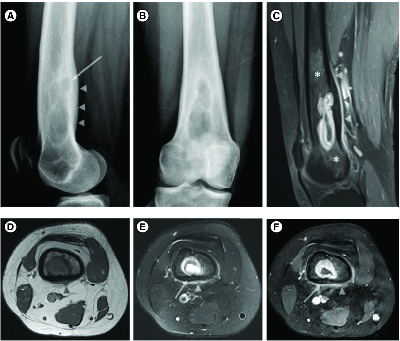
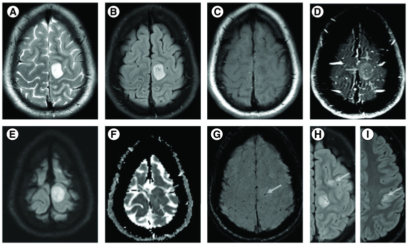
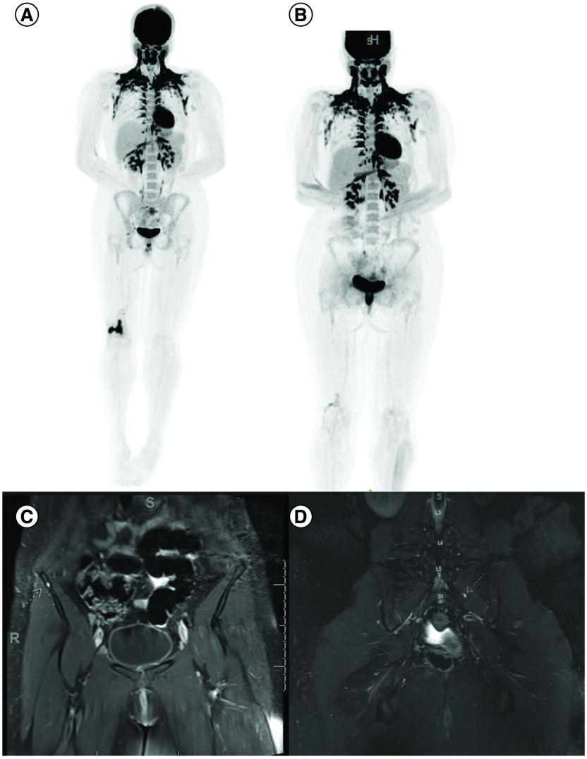
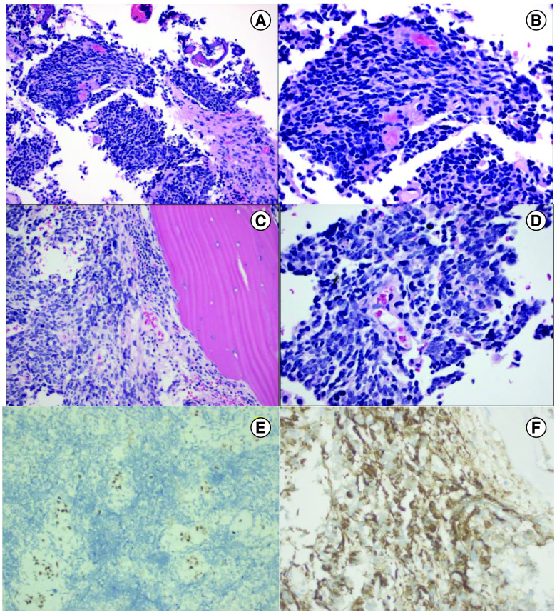
Aim: H3G34 diffuse hemispheric glioma is a CNS tumor that is difficult to diagnose and treat and accompanied with poor prognosis. It is becoming clear that extra CNS metastasis may present in a subset of patients with H3G34 gliomas, further complicating diagnosis and treatment. Materials & methods: We present a case of a 19-year-old female with a H3G34 mutant diffuse hemispheric glioma with osseous metastases. We then provide a literature review of the most recent understanding of H3G34 mutant malignancies. Conclusion: Given the stress that patients with H3G34 can experience and the poor prognosis, it is imperative to expand our knowledge and ascertain accurate diagnostic methodologies and targeted therapeutic approaches.

Keywords: H3G34-mutant; diffuse hemispheric glioma; osseous metastases.


Conflict of interest statement

The authors have no other relevant affiliations or financial involvement with any organization or entity with a financial interest in or financial conflict with the subject matter or materials discussed in the manuscript apart from those disclosed.

Figures

Figure 1.
Figure 1.. X-ray of distal right femur and coronal postcontrast fat suppressed images.
X-ray of distal right femur in lateral (A) and AP (B) views showing an ill-defined expansile lytic lesion in the right distal femoral shaft with endosteal scalloping and posterior cortical sclerotic reaction and thickening (arrow and arrowhead in (A), respectively). Coronal postcontrast fat suppressed sagittal (C) and axial (F) T1 WI and axial T1 (D) and fat suppressed T2 images of the right distal femur showing a rim enhancing lesion with central fluid signal. There is cortical thickening and invasion evidenced by loss of the normal low signal in the cortical bone (arrow in (E & F)), associated with endosteal scalloping and smooth periosteal/soft tissue extension (arrow heads in (C–F)). Note the surrounding enhancing marrow signal abnormality worrisome for lesion extension more than red marrow islands (asterisks in (C)).
Figure 2.
Figure 2.. MRI images of brain lesion.
MRI shows a well circumscribed cortical based 2.1 × 1.6 cm mass in the left peri-Rolandic region demonstrating hyperintense T2/FLAIR SI (A & B), hypointense T1-WI (C) and thick subtle incomplete ring enhancement on the postcontrast fat suppressed T1 WI (arrows) (D). DWI and ADC maps show internal foci of restricted diffusion (arrows) (E & F) and T2*-SWI images shows a punctate focus of intratumoral hemorrhage (arrow) (G). Axial FLAIR images and different axial levels showing similar but nonenhancing cortical/subcortical lesions in the left frontal and parietal lobes (arrows) (H & I). DWI: Diffusion weighted imaging.
Figure 3.
Figure 3.. Images showing multiple extra CNS lesions.
Intensely FDG-avid lytic lesion of the right distal femur (A) as well as lytic metastases involving the right popliteal region, right groin, right external and common iliac chains. Improvement in sacrum and right femoral FDG uptake after treatment (B). Note, physiological brown fat activation in the neck. Coronal contrast enhanced fat suppressed T1 WI showing corresponding punctate enhancing metastatic nodules in the right iliac bone (C) and left S1 level (D).
Figure 4.
Figure 4.. H&E histology images, and ATRX and GFAP images.
H&E 400X histology image from brain tissue showing small blue cell neoplasms composed of sheets of ribbons of cells with scant cytoplasm, hyperchromatic nuclei, and high nucleus/cytoplasm ratio (200× magnification in (A), 400× magnification in (B)). H&E image from bone biopsy showing small blue cell neoplasm involving bone, mitosis present (200× magnification in (C), 400× magnification in (D)). ATRX 200× image from brain tissue showing ATRX loss (200× magnification, (E)). GFAP 400X image from brain tissue showing GFAP positivity (400× magnification, (F)).

References

    1. Leske H, Dalgleish R, Lazar AJ, Reifenberger G, Cree IA. A common classification framework for histone sequence alterations in tumours: an expert consensus proposal. J. Pathol. 254(2), 109–120 (2021). - PubMed
    1. Crowell C, Mata-Mbemba D, Bennett J et al. Systematic review of diffuse hemispheric glioma, H3 G34-mutant: outcomes and associated clinical factors. Neurooncol. Adv. 4(1), vdac133 (2022). - PMC - PubMed
    1. Wang L, Shao L, Li H et al. Histone H3.3 G34-mutant diffuse gliomas in adults. Am. J. Surg. Pathol. 46(2), 249–257 (2022). - PubMed
    1. Lim KY, Won JK, Park CK et al. H3 G34-mutant high-grade glioma. Brain Tumor Pathol. 38(1), 4–13 (2021). - PubMed
    1. Schwartzentruber J, Korshunov A, Liu XY et al. Driver mutations in histone H3.3 and chromatin remodelling genes in paediatric glioblastoma. Nature 482(7384), 226–231 (2012). - PubMed