. 2023 Mar 23;66(6):3798-3817.
doi: 10.1021/acs.jmedchem.2c01227. Epub 2023 Mar 15.

Development of Fluorescent 4-[4-(3 H-Spiro[isobenzofuran-1,4'-piperidin]-1'-yl)butyl]indolyl Derivatives as High-Affinity Probes to Enable the Study of σ Receptors via Fluorescence-Based Techniques

Affiliations

Development of Fluorescent 4-[4-(3 H-Spiro[isobenzofuran-1,4'-piperidin]-1'-yl)butyl]indolyl Derivatives as High-Affinity Probes to Enable the Study of σ Receptors via Fluorescence-Based Techniques

Francesca Serena Abatematteo et al. J Med Chem. .

Abstract

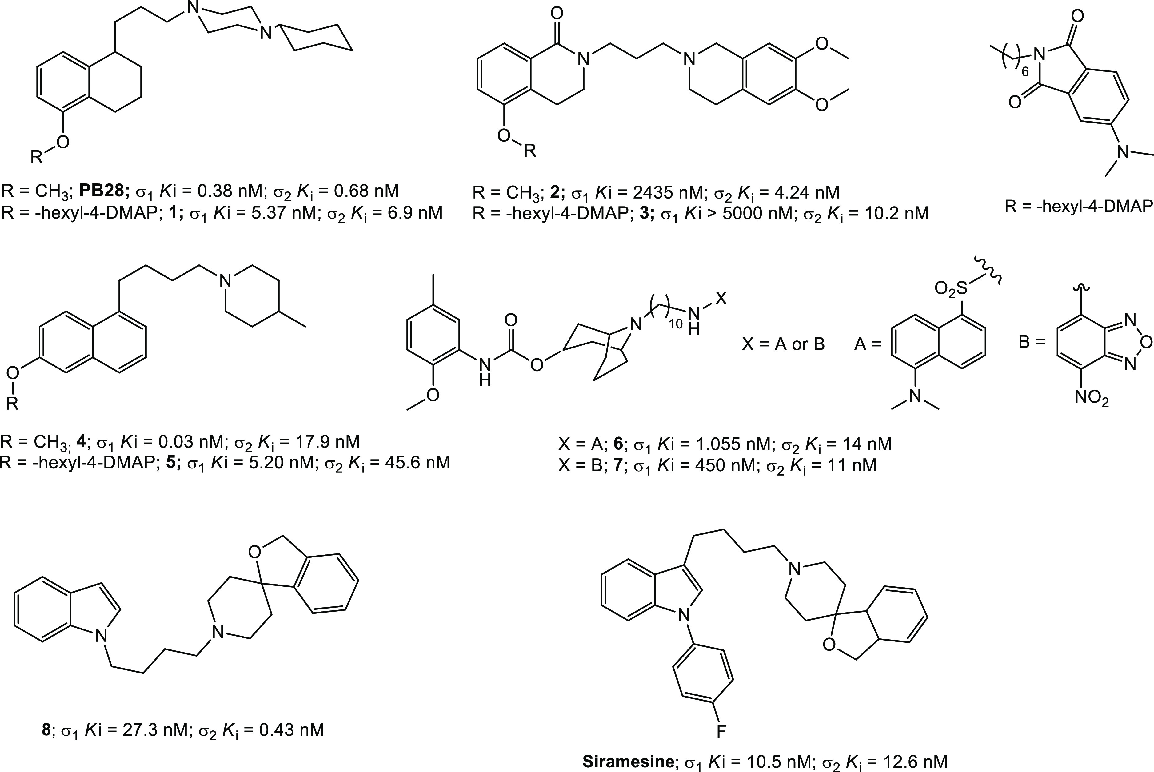
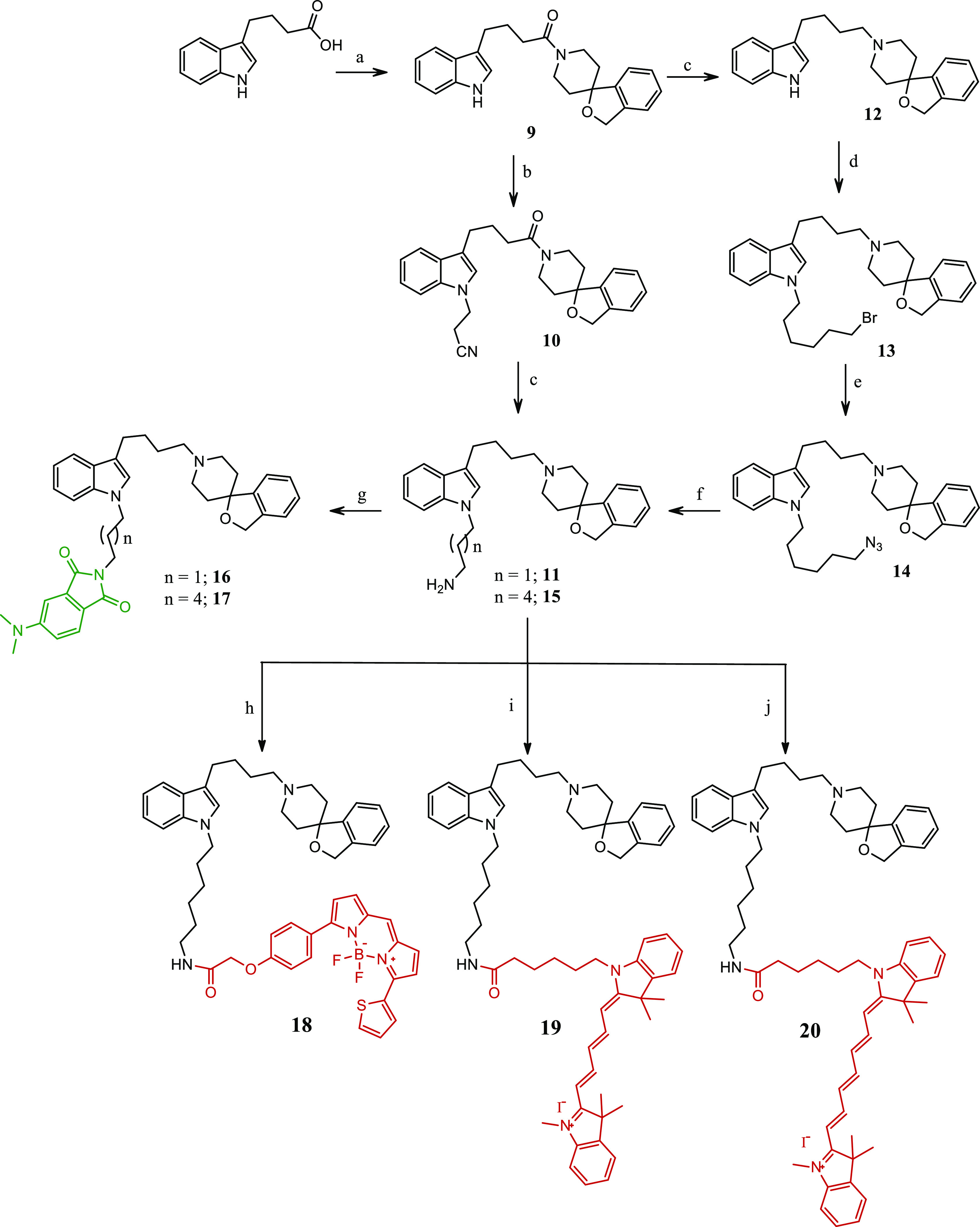
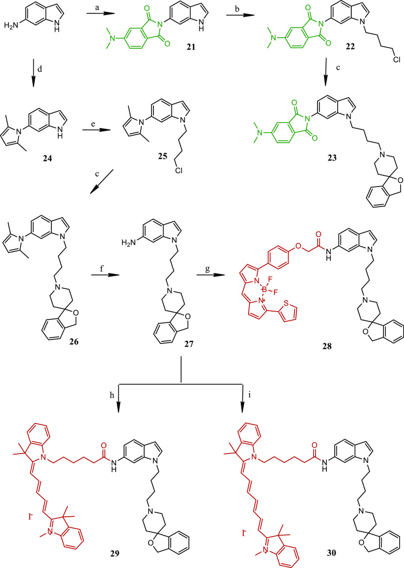
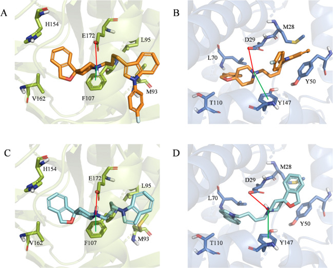
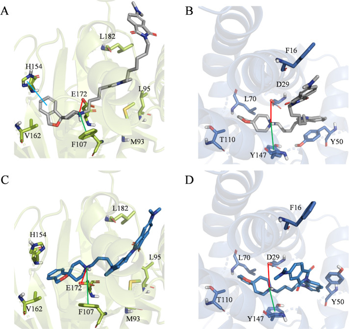
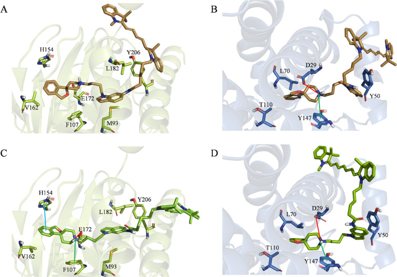
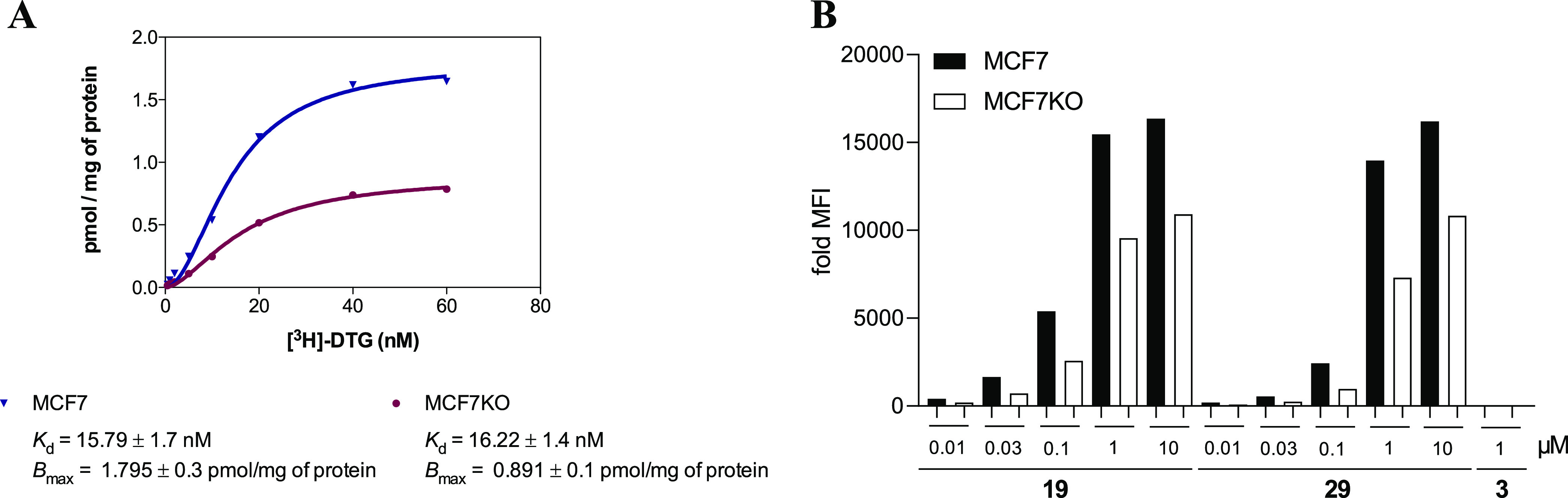
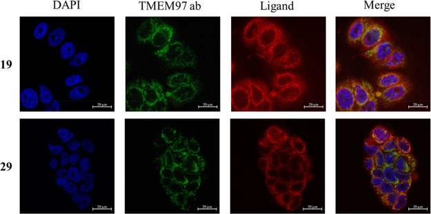
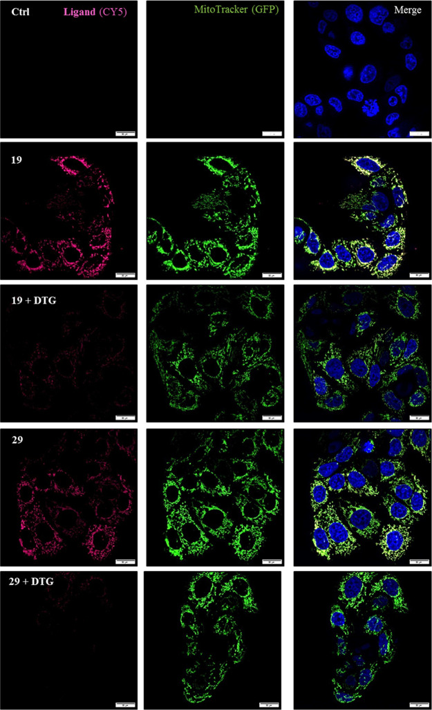
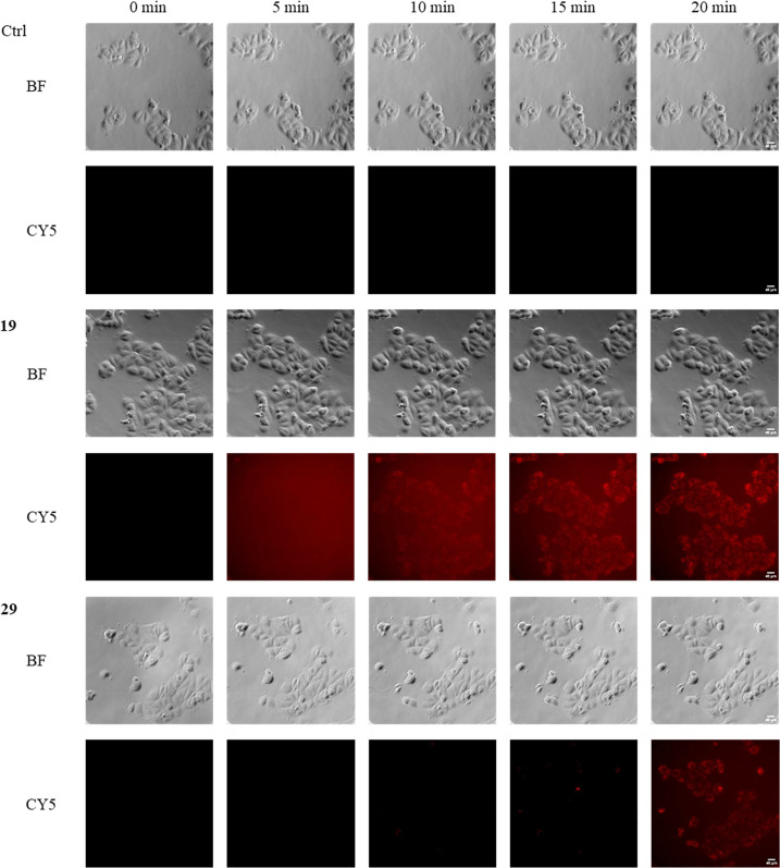
Sigma (σ) receptor subtypes, σ1 and σ2, are targets of wide pharmaceutical interest. The σ2 receptor holds promise for the development of diagnostics and therapeutics against cancer and Alzheimer's disease. Nevertheless, little is known about the mechanisms activated by the σ2 receptor. To contribute to the exploitation of its therapeutic potential, we developed novel specific fluorescent ligands. Indole derivatives bearing the N-butyl-3H-spiro[isobenzofuran-1,4'-piperidine] portion were functionalized with fluorescent tags. Nanomolar-affinity fluorescent σ ligands, spanning from green to red to near-infrared emission, were obtained. Compounds 19 (σ pan affinity) and 292 selective), which displayed the best compromise between pharmacodynamic and photophysical properties, were investigated in flow cytometry, confocal, and live cell microscopy, demonstrating their specificity for the σ2 receptor. To the best of our knowledge, these are the first red-emitting fluorescent σ2 ligands, validated as powerful tools for the study of σ2 receptors via fluorescence-based techniques.


Conflict of interest statement

The authors declare no competing financial interest.

Figures

Figure 1
Figure 1
σ2 receptor reference compounds and fluorescent ligands.
Scheme 1
Scheme 1. Synthetic Pathways for the Synthesis of Final Compounds 1620
Reagents: (a) 3H-spiro[isobenzofuran-1,4′-piperidine], CDI, dry THF, RT, overnight; (b) 3-bromopropanenitrile, KOH, K2CO3, CH3CN, MW, 150 °C, 1 h; (c) BH3·DMS, dry MeOH, reflux, 4 h; (d) 1,6-dibromohexane, TBAB, KOH, dry DMF, RT, 2 h; (e) NaN3, dry DMF, 60 °C, 20 h; (f) PPh3, dry MeOH, 80 °C, 1 h; (g) 4-(dimethylamino)phthalic acid, CDI, dry DMF, RT, overnight; (h) BODIPY-TR-COOH, HATU, DIPEA, CH2Cl2, 30 °C, O/N; (i) Cy-5-COOH, HATU, DIPEA, CH2Cl2, 30 °C, overnight; (j) Cy-7-COOH, HATU, DIPEA, CH2Cl2, 30 °C, overnight.
Scheme 2
Scheme 2. Synthetic Pathways for the Synthesis of Final Compounds 23 and 2830
Reagents: (a) 4-(dimethylamino)phthalic acid, CDI dry DMF, RT, overnight; (b) 1-bromo-4-chloro-butane, t-BuOK, dry DMF, RT, 1 h; (c) 3H-spiro[isobenzofuran-1,4′-piperidine], K2CO3, CH3CN, reflux, overnight; (d) 2,5-hexanedione, dry toluene, reflux, 6 h; (e) 1-bromo-4-chloro-butane, TBAB, KOH, dry DMF, RT, 2 h; (f) NH2OH·HCl, 2:1 EtOH/H2O, 30 °C, overnight; (g) BODIPY-TR-COOH, HATU, DIPEA, CH2Cl2, 30 °C, overnight; (h) Cy-5-COOH HATU, DIPEA, CH2Cl2, 30 °C, overnight; (i) Cy-7-COOH, HATU, DIPEA, CH2Cl2, 30 °C, overnight.
Figure 2
Figure 2
Top-scoring docking poses of (A) siramesine within the binding pocket of σ1 (PDB entry 6DK1), (B) siramesine within the binding pocket of σ2 (PDB entry 7M95), (C) 8 within the binding pocket of σ1 (PDB entry 6DK1), and (D) 8 within the binding pocket of σ2 (PDB entry 7M95). For the sake of clarity, only polar hydrogen atoms are shown. Important residues are rendered as sticks, while the proteins are represented as cartoon. Salt-bridge and cation−π interactions are depicted as red and green lines, respectively.
Figure 3
Figure 3
Top-scoring docking poses of (A) 17 within the binding pocket of σ1 (PDB entry 6DK1), (B) 17 within the binding pocket of σ2 (PDB entry 7M95), (C) 23 within the binding pocket of σ1 (PDB entry 6DK1), and (D) 23 within the binding pocket of σ2 (PDB entry 7M95). For the sake of clarity, only polar hydrogen atoms are shown. Important residues are rendered as sticks, while the proteins are represented as a cartoon. Salt-bridge and cation−π interactions are depicted as red and green lines, respectively.
Figure 4
Figure 4
Top-scoring docking poses of (A) 29 within the binding pocket of σ1 (PDB entry 6DK1), (B) 29 within the binding pocket of σ2 (PDB entry 7M95), (C) 30 within the binding pocket of σ1 (PDB entry 6DK1), and (D) 30 within the binding pocket of σ2 (PDB entry 7M95). For the sake of clarity, only polar hydrogen atoms are shown. Important residues are rendered as sticks, while the proteins are represented as a cartoon. Salt-bridge and cation−π interactions are depicted as red and green lines, respectively.
Figure 5
Figure 5
(A) Saturation binding assay on MCF7 and MCF7KO (silenced in TMEM97/σ2) cells. (B) Flow cytometry. Dose-dependent increase in the mean fluorescence intensity (MFI) in MCF7 and MCF7KO (one representative image of three repetitions) cells upon administration of compound 19, 29, or 3 at the indicated concentrations.
Figure 6
Figure 6
Representative confocal microscopy images showing the co-localization of 19 and 29 with σ2 receptor expression in MCF7 cells. Cells were preincubated with 10 μM selective σ1 receptor agonist (+)-pentazocine for 2 h to mask residual σ1 receptor expression. This was followed by incubation for 1 h with the indicated fluorescent ligands at 5 μM. Then cells were fixed with paraformaldehyde, stained for σ2 receptor expression (TMEM97 ab) as well as nuclei (by DAPI), and analyzed by confocal microscopy. The ligands are colored red (scale bar of 20 μm).
Figure 7
Figure 7
Representative spinning disk images showing the localization of ligand 19 as well as 29 with the mitochondria as well as reduction of the Cy-5 signal of the ligands in the presence of the nonfluorescent σ2 ligand DTG in living MCF7 cells. Cells were preincubated with 10 μM 31 to mask residual σ1 receptor expression with or without 20 μM DTG for 75 min. This was followed by administration of 0.05 μM 19 or 29 for 30 min (red stain in the Cy-5 channel). In addition, mitochondria were stained with 250 nM MitoTracker-Bodipy FL (green stain in the GFP channel) and the nuclei were counterstained with Hoechst 33342 (blue stain in the DAPI channel). The depicted scale bar indicates 20 μm.
Figure 8
Figure 8
Representative microscopically live cell images showing time-dependent accumulation of ligand 19 as well as 29 in MCF7 cells from 0 to 20 min. Cells were preincubated with 10 μM selective σ1 receptor agonist 31 for 2 h to mask residual σ1 receptor expression. This was followed by administration of 19 and 29. For time-lapse experiments, images were collected every 5 min for a period of 4 h. The bright field (BF) and the fluorescence channel Cy-5 for the ligands (red) are shown (scale bar of 40 μm).

Similar articles

Cited by

References

    1. Martin W. R.; Eades C. G.; Thompson J. A.; Huppler R. E.; Gilbert P. E. The effect of morphine- and nalorphine-like drugs in the nondependent and morphine-dependent chronic spinal dog. J. Pharmacol. Exp. Ther. 1976, 197, 517–532. - PubMed
    1. Glennon R. A. The enigmatic sigma receptors. Cent. Nerv. Syst. Agents in Med. Chem. 2009, 9, 159–160. 10.2174/1871524910909030159. - DOI - PubMed
    1. Hanner M.; Moebius F. F.; Flandorfer A.; Knaus H. G.; Striessnig J.; Kempner E.; Glossmann H. Purification, molecular cloning, and expression of the mammalian sigma1-binding site. Proc. Natl. Acad. Sci. U. S. A. 1996, 93, 8072–8077. 10.1073/pnas.93.15.8072. - DOI - PMC - PubMed
    1. Schmidt H. R.; Zheng S.; Gurpinar E.; Koehl A.; Manglik A.; Kruse A. C. Crystal structure of the human σ1 receptor. Nature 2016, 532, 527–530. 10.1038/nature17391. - DOI - PMC - PubMed
    1. Schmidt H. R.; Kruse A. C. The molecular function of σ receptors: Past, present, and future. Trends Pharmacol. Sci. 2019, 40, 636–654. 10.1016/j.tips.2019.07.006. - DOI - PMC - PubMed

Publication types