

Anatomical variants complicating the posterior approaches towards the elbow joint
- PMID: 36920516
- PMCID: PMC10130116
- DOI: 10.1007/s00276-023-03124-9
Anatomical variants complicating the posterior approaches towards the elbow joint
Abstract
Introduction: Anatomical variants observed during the posterior approach to the elbow joint require special attention due to their clinical relevance. We aim to present a compendious review of described variants potentially encountered during the posterior approach towards the elbow joint to the experts in the elbow surgery.
Methods: A narrative review of surgical and anatomical textbooks, as well as search of scientific databases was carried out.
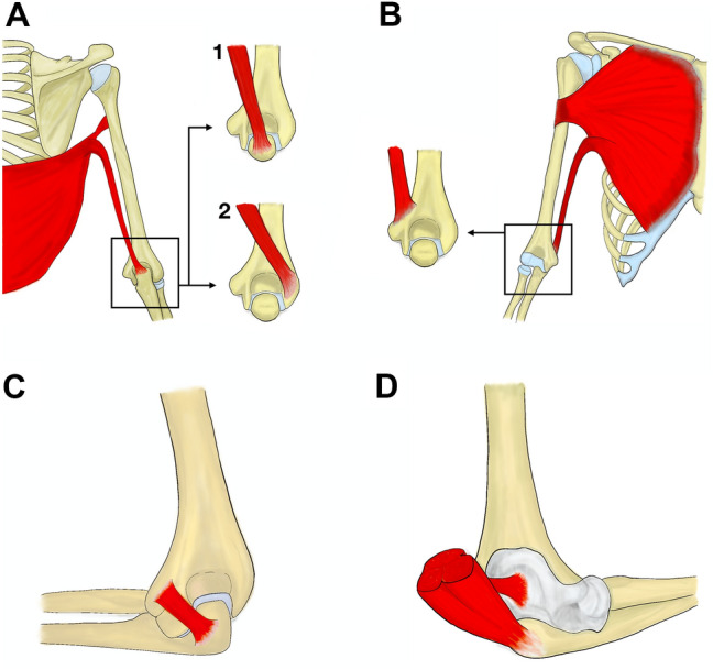
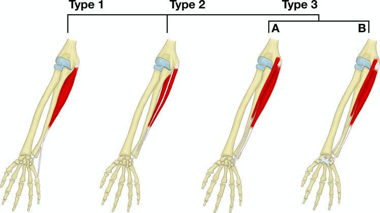
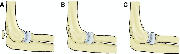
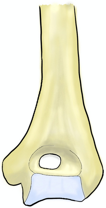
Results: Variability of the subcutaneous nerves is important during incision planning. Accessory muscles such as dorsoepitrochlearis, chondroepitrochlearis, epitrochleoanconeus, subanconeus or supernumerary flexor carpi ulnaris may confuse even the senior surgeon during the dissection and possibly complicate the fracture reduction. Some bony variants such as supratrochlear foramen may lead to fracture or possibly interfere with the osteosynthesis placement. Accessory bones are also present in the region of the elbow joint. Those situated intra-articular may present with symptoms.
Conclusion: Many variants can be encountered in the area of the elbow joint and their knowledge is essential to truly understand its anatomy. The presented review enables easier orientation in the current literature with the aim on the posterior approach towards the elbow joint.
Keywords: Accessory bones; Chondroepitrochlearis; Dorsoepitrochlearis; Epitrochleoanconeus; Posterior approach; Subanconeus; Ulnar nerve; Variability around elbow joint.
© 2023. The Author(s).
Conflict of interest statement
The authors have no conflict of interests to declare that are relevant to the content of this article.
Figures

References
-
- Bando K. Musculus epitrochleo-anconeus. Hirosaki-Igaku. 1956;7:192.
Publication types
MeSH terms
Grants and funding
LinkOut - more resources
Full Text Sources
Medical
