Review
. 2023 Apr;16(4):154-161.
doi: 10.1007/s12178-023-09824-4. Epub 2023 Mar 15.

Discoid Lateral Meniscus

Affiliations
Review

Discoid Lateral Meniscus

Abigail L Campbell et al. Curr Rev Musculoskelet Med. 2023 Apr.

Abstract

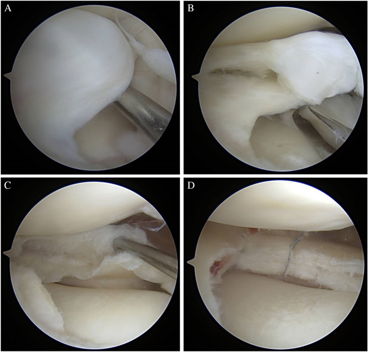
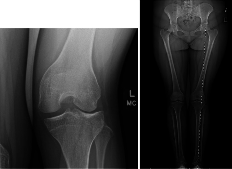
Purpose of review: Discoid lateral meniscus (DLM) is a well-known meniscus variant, and comprises excess and thickened meniscal tissue, altered collagen ultrastructure, and peripheral instability. This article presents a comprehensive review on current knowledge of DLM, focusing on pathology in parallel with surgical techniques and outcomes.

Recent findings: A paradigm shift in surgical management of DLM is taking place as knee surgeons are seeing more patients with long-term sequelae of partial lateral meniscectomy, the standard treatment for DLM for many years. Surgical treatment has evolved alongside the understanding of DLM pathology. A new classification system has been proposed and optimal surgical techniques described in recent years. This article highlights up-to-date evidence and techniques in management of both acute DLM tears and joint restoration following subtotal meniscectomy for DLM. Surgical management of DLM must be tailored to individual pathology, which is variable within the diagnosis of DLM. We present an algorithm for management of DLM and discuss future directions for the understanding and treatment of this debilitating condition.

Keywords: Discoid; Knee preservation; Meniscal repair; Meniscal transplantation.


Conflict of interest statement

Dr Pace is a consultant for Arthrex and JRF Ortho. He serves as a committee member for AOSSM and PRiSM. Dr Mandelbaum and Dr Campbell have no relevant conflicts of interest regarding the subject of this review article.

Figures

Fig. 1
Fig. 1
Anteroposterior radiograph of the left knee of a skeletally mature patient demonstrating all four radiographic findings of DLM: squaring of the lateral femoral condyle, concavity of the lateral tibial plateau, increased lateral joint space, and hypoplasia of the lateral tibial spine
Fig. 2
Fig. 2
MRI T2 coronal × 2 and sagittal images demonstrating a skeletally mature patient with DLM. Increased T2 signal seen within the meniscal tissue as a horizontal strip represents intrasubstance degeneration with myxoid tissue. This is seen most clearly on T2 images due to increased water content due to structural dysregulation within the meniscal tissue. Hypoplasia of the lateral femoral condyle is also appreciated
Fig. 3
Fig. 3
Arthroscopic images of a left knee demonstrating near complete coverage of the lateral plateau followed by saucerization revealing myxoid ID tissue (same knee as MRI images in Fig. 2)
Fig. 4
Fig. 4
A, B, C: Arthroscopic images of a right knee DLM saucerization and repair. Image A: discoid tissue without obvious external tearing. Image B: following saucerization with ID revealed, with similar appearance to horizontal cleavage tear. Image C: following all-inside haybale suture placement
Fig. 5
Fig. 5
AD: arthroscopic images of a right knee. (A) DLM with full coverage of the lateral tibial plateau. (BC) ID revealed as sauceration is performed. (D) all-inside capsular-based anchors being placed with similar compression of leaflets to Fig. 4 images. The difference in this case is the repair technique with capsular-based repair stitches in this figure and all-meniscal suture without capsular inclusion in Fig. 4
Fig. 6
Fig. 6
AP and standing alignment radiographs of an adolescent patient s/p subtotal lateral meniscectomy for DLM, now with 6 degrees valgus deformity

References

    1. Sabbag OD, et al. Incidence and treatment trends of symptomatic discoid lateral menisci: an 18-year population-based study. Orthop J Sports Med. 2018;6:2325967118797886. doi: 10.1177/2325967118797886. - DOI - PMC - PubMed
    1. Grimm NL, et al. Demographics and epidemiology of discoid menisci of the knee: analysis of a large regional insurance database. Orthop J Sports Med. 2020;8:2325967120950669. doi: 10.1177/2325967120950669. - DOI - PMC - PubMed
    1. Rohren EM, Kosarek FJ, Helms CA. Discoid lateral meniscus and the frequency of meniscal tears. Skeletal Radiol. 2001;30:316–320. doi: 10.1007/s002560100351. - DOI - PubMed
    1. Young R. The external semilunar cartilage as a complete disc. London: Williams and Norgate; 1889.
    1. Bellier G, Dupont JY, Larrain M, Caudron C, Carlioz H. Lateral discoid menisci in children. Arthroscopy. 1989;5:52–56. doi: 10.1016/0749-8063(89)90092-3. - DOI - PubMed