Case Reports
. 2023 Mar 16;388(11):1032-1041.
doi: 10.1056/NEJMcpc2211370.

Case 8-2023: A 71-Year-Old Woman with Refractory Hemolytic Anemia

Affiliations
Case Reports

Case 8-2023: A 71-Year-Old Woman with Refractory Hemolytic Anemia

Alfred I Lee et al. N Engl J Med. .
No abstract available


Figures

Figure 1.
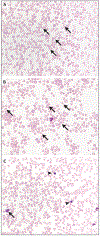
Figure 1.. Peripheral-Blood Specimens on Admission to This Hospital.
Wright–Giemsa staining shows anisopoikilocytosis and schistocytes (Panel A, arrows); in other areas, teardrop cells are present (Panel B, arrows). Immature myeloid precursors (Panel C, arrow) and nucleated red cells (Panel C, arrowheads) can also be seen.
Figure 2.
Figure 2.. Imaging Studies.
An anteroposterior maximum intensity projection image from 18F-fluorodeoxyglucose (FDG) positron-emission tomography–CT shows diffuse, heterogeneous, abnormally increased FDG uptake in the axial and appendicular skeleton (Panel A, arrows). Sagittal images of the skeleton show multiple focal sites of abnormal uptake in the spine (Panel B, arrows), with no correlative lytic or blastic bone lesions on CT (Panel C). Axial images of the pelvis show multiple focal sites of abnormal uptake in the iliac bones and sacrum (Panel D, arrows), with no correlative lytic or blastic bone lesions on CT (Panel E). Overall, this pattern of FDG uptake is seen in patients with osseous metastases or with lymphoproliferative or myeloproliferative processes.
Figure 3.
Figure 3.. Core-Biopsy Specimens of Bone Marrow.
Hematoxylin and eosin staining shows infiltration of atypical nonhematopoietic cells (Panel A, outlined by dashed lines). Immunohistochemical staining shows membranous staining for MNF116 (cytokeratin) (Panel B, in brown) and nuclear staining for estrogen receptor (Panel C, in brown) in the atypical cells, findings that indicate a carcinoma of primary breast origin.

References

    1. Thomas MR, Scully M. How I treat microangiopathic hemolytic anemia in patients with cancer. Blood 2021;137:1310–7. - PMC - PubMed
    1. Zheng XL, Vesely SK, Cataland SR, et al. ISTH guidelines for the diagnosis of thrombotic thrombocytopenic purpura. J Thromb Haemost 2020;18:2486–95. - PMC - PubMed
    1. Bendapudi PK, Hurwitz S, Fry A, et al. Derivation and external validation of the PLASMIC score for rapid assessment of adults with thrombotic microangiopathies: a cohort study. Lancet Haematol 2017;4(4):e157–e164. - PubMed
    1. Cuker A, Cataland SR, Coppo P, et al. Redefining outcomes in immune TTP: an international working group consensus report. Blood 2021;137:1855–61. - PubMed
    1. Jokiranta TS. HUS and atypical HUS. Blood 2017;129:2847–56. - PMC - PubMed

Publication types