. 2023 Mar 28;42(3):112231.
doi: 10.1016/j.celrep.2023.112231. Epub 2023 Mar 14.

Dopamine transporter and synaptic vesicle sorting defects underlie auxilin-associated Parkinson's disease

Affiliations

Dopamine transporter and synaptic vesicle sorting defects underlie auxilin-associated Parkinson's disease

D J Vidyadhara et al. Cell Rep. .

Abstract

Auxilin participates in the uncoating of clathrin-coated vesicles (CCVs), thereby facilitating synaptic vesicle (SV) regeneration at presynaptic sites. Auxilin (DNAJC6/PARK19) loss-of-function mutations cause early-onset Parkinson's disease (PD). Here, we utilized auxilin knockout (KO) mice to elucidate the mechanisms through which auxilin deficiency and clathrin-uncoating deficits lead to PD. Auxilin KO mice display cardinal features of PD, including progressive motor deficits, α-synuclein pathology, nigral dopaminergic loss, and neuroinflammation. Significantly, treatment with L-DOPA ameliorated motor deficits. Unbiased proteomic and neurochemical analyses of auxilin KO brains indicated dopamine dyshomeostasis. We validated these findings by demonstrating slower dopamine reuptake kinetics in vivo, an effect associated with dopamine transporter misrouting into axonal membrane deformities in the dorsal striatum. Defective SV protein sorting and elevated synaptic autophagy also contribute to ineffective dopamine sequestration and compartmentalization, ultimately leading to neurodegeneration. This study provides insights into how presynaptic endocytosis deficits lead to dopaminergic vulnerability and pathogenesis of PD.

Keywords: CP: Neuroscience; L-DOPA; Lewy bodies; axonal deformity; clathrin-coated vesicles; clathrin-mediated endocytosis; dopamine; dorsal striatum; endolysosomal system; substantia nigra; synaptic autophagy; ∝-synuclein.


Conflict of interest statement

Declaration of interests The authors declare no competing interests.

Figures

Figure 1.
Figure 1.. Auxilin KO mice develop progressive motor behavior deficits that are accompanied by nigral DA neuronal loss
(A) Longitudinal open field locomotor behavior tracings of WT and auxilin KO (Aux KO) mice. (B) Distance traveled in open field as a function of age showing a progressive diminishment in locomotion in Aux KO mice (n=1216/genotype). (C) Number of runs performed in 1 min on a balance beam. Performance of Aux KO mice decreased with age, with a significant difference after 9 months. (D) (i) Balance beam performance of individual WT and Aux KO mice at 9–12 months of age before and after treatment with L-DOPA. (ii) Note a significant recovery in Aux KO mice in balance beam performance comparable with that of WT mice post treatment. (E) Representative images showing TH+ SNpc DA neurons in WT and Aux KO midbrain sections at 3 and 9 months of age. Fewer DA neurons (arrows) were present in the SNpc of Aux KO mice at 9 months. Scale bar, 150 μm. (F) Unbiased stereological counting of SNpc DA neurons. A significant (~40%) loss of DA neurons is seen in 9-month-old Aux KO mice (n=56/genotype). Data are presented as mean ± SEM. ns, not significant; **p < 0.01, ***p < 0.001, ****p < 0.0001, $$p < 0.01, $$$p < 0.001, $$$$p < 0.0001. (**comparison between genotypes, $comparison between ages/time points)
Figure 2.
Figure 2.. Aged auxilin KO mice exhibit gliosis and α-synuclein pathology
(A) Representative images of SNpc and SN pars reticulata (SNpr) of WT and Aux KO mice at 3 and 9 months of age (n=56/genotype) immunostained for the astroglial marker GFAP (green) and microglial marker Iba1 (red). Dashed line demarcates SNpc from SNpr. Scale bar, 50 μm. (B) Quantitation of SNpc GFAP-positive cells shows a significant astrogliosis at 9 months. (C) Quantitation of Iba1+ cells shows microgliosis in the SNpc of Aux KO mice at 9 months. (D) Representative images of the SNpc immunostained for pSer129-α-synuclein (green), co-stained with DA marker TH (magenta). Scale bar, 50 μm. (E) Quantitation of pSer129-α-synuclein-positive punctate aggregates in SNpc, which showed an increase in 9-month-old Aux KO mice, but not at 3 months of age. (F) Quantitation of TH staining intensity per DA neuron of SNpc, which showed a moderate decrease in Aux KO mice, suggesting retention of TH phenotype in the surviving neurons. Data are presented as mean ± SEM. *p < 0.05, **p < 0.01.
Figure 3.
Figure 3.. Whole-brain and synaptosome proteomics reveal dopamine degradation dysfunction in young auxilin KO mice
(A) Volcano plot of whole-brain proteome of Aux KO compared with WT mice (age 3 months, n=3mice/genotype). Proteins that exhibit a 1.5-fold change (vertical dotted lines) and a p value of 0.05 (Student’s t test) or less (horizontal dotted line) were considered as significantly changed. (B) Heatmap of significantly changed proteins in whole-brain homogenates of WT and Aux KO mice for all nine technical replicates (3 technical replicates/mouse).Red indicates an increased level (+2) and blue indicates a decreased level (−2). (C) Pathways that are significantly (p<0.05) affected in whole brain of Aux KO mice as determined by IPA. (D) Diagram showing the overlap of significantly affected pathways, where intense red depicts most affected and light red depicts moderately affected pathways. (E) Volcano plot of synaptosome proteome of Aux KO compared with WT mice (age 3 months, n=3mice/genotype). (F) Heatmap of significantly changed proteins in synaptosomes from Aux KO mice in comparison with WT mice for each technical replicate. Red indicates an increased level (+2) and blue indicates a decreased level (−2). (G) Significantly affected pathways due to synaptosome proteomic changes as determined by IPA. (H) Overlap of significantly affected pathways showing highly affected (intense red) and moderately affected (light red) pathways.
Figure 4.
Figure 4.. Dopamine catabolism and dopamine reuptake deficits in young auxilin KO mice
(A) Dopamine levels in the dorsal striatum of WT and Aux KO mice (n=711mice/genotype). (B) DOPAC levels in the dorsal striatum. (C) 3-MT levels in the dorsal striatum. (D) HVA levels in the dorsal striatum. (E) Schematic showing compartmentalization of dopamine and its catabolites in intra- and extra-synaptic space. (F) Schematic showing the location of FSCV recording electrode in the dorsal striatum and the bipolar stimulating electrode in the ventral midbrain of mice under isoflurane anesthesia. (G) Example trace of evoked dopamine release following stimulation of midbrain DA neurons by 30 pulses at a constant 50-Hz frequency in 3-month-old Aux KO and WT mice (scale: y axis, 0.5 μM dopamine; x axis, 2 s). (H) Dopamine release in the dorsal striatum (n=45/genotype). (I) Dopamine reuptake in the dorsal striatum. Reuptake kinetics measured by time taken to clear half the dopamine from its peak levels (t1/2) was significantly delayed in Aux KO mice. (J) Best fits of computational model of dopamine (DA) release (red lines) to averaged FSCV recordings in the dorsal striatum of WT (black dots; R2=0.99, n=4) and Aux KO (green dots; R2=0.99, n=5) mice. Black/green ribbons report SEM. Data are presented as mean ± SEM. *p < 0.05, ***p < 0.001, ****p < 0.0001.
Figure 5.
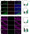
Figure 5.. DAT+ axonal deformities in the dorsal striatum of auxilin KO mice
(A) Representative images of dorsal and ventral striatum (STR) in WT and Aux KO mice (age 3 months), immunostained for DAT and synaptogyrin-3. Note large DAT+ structures in the dorsal STR of Aux KO mice that are absent in the ventral STR. Scale bar, 200 μm. These DAT+ structures were juxtaposed to presynapses (bottom row) as seen by colocalization with synaptogyrin-3 (Aux KO, enlarged, arrowhead), as well as in the soma marked by DAPI staining (Aux KO, enlarged, arrow). Scale bar, 10 μm. (B) Number of DAT+ structures/unit area in the dorsal striatum. (C) Note the absence of DAT+ structures in the ventral striatum. (D) Representative images of ex vivo staining of dichloropane-rhodamine red-X in the dorsal striatum of WT and Aux KO mice (age 3 months). DA axonal projections and presynaptic sites appear as small puncta, whereas axonal deformities appear as large dichloropane-DAT+ structures (arrows). Scale bar, 10 μm. (E) Number of large dichloropane-bound DAT+ structures/unit area in the dorsal striatum, which were significantly higher in Aux KO mice. (F) Number of small dichloropane-bound DAT+ puncta was not altered in Aux KO dorsal striatum in comparison with WT. (G) EM of dorsal striatum of Aux KO mice (age 3 months) showing large axonal whirl-like deformities (arrows), which were present ubiquitously, closer to both synaptic terminals (S) and soma (N, nuclei). Early autophagic vacuole-like structures were also seen in dorsal striatum (arrowheads), closer to axonal whirls. Scale bars, 1 μm and 2 μm. (H) DAT-immunogold labeling of dorsal striatum that mark only DA axonal termini showed dispersed labeling in WT. In Aux KO mice, DAT-immunogold clusters were seen in the dorsal striatum (arrows). Age 3 months. Scale bar, 200 nm. Data are presented as mean ± SEM. *p < 0.05, ***p < 0.001.
Figure 6.
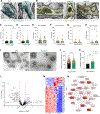
Figure 6.. EM and proteomics revealed imbalance in CCV/SV ratio and SVs with variable membrane composition in auxilin KO mice
(A) Representative EM image of a type I excitatory presynapse with SVs (arrowheads) from dorsal striatum of WT and Aux KO mice (age 3 months). Scale bar, 150 μm. (B) Representative EM image of a type II inhibitory presynapse in the dorsal striatum of WT and Aux KO mice with SVs (arrows) and CCVs (arrows). (i) Aux KO presynapse showing an increase accumulation of CCVs. (ii) Another Aux KO synapse showing CCVs or clathrin cage accumulation (arrows), as well as SV clusters (arrowheads). Scale bar, 150 μm. (C) Number of CCVs in type I synapses. (D) Number of CCVs in type II synapses. (E) Number of SVs in type I synapses. (F) Number of SVs in type II synapses. (G) The CCV/SV ratio in type I synapses of dorsal striatum. (H) The CCV/SV ratio in type II synapses of dorsal striatum. (I) Representative EM images of CCV preparation showing CCVs (arrows) and empty clathrin cages (arrowheads) in WT and Aux KO mice. Scale bar, 100 nm. (J) Diameter of clathrin structures (CCVs + clathrin cages). (K) Proportion of clathrin cages in WT and Aux KO mice. (L) Volcano plot of CCV proteome of Aux KO in comparison with WT mice (n=14mice/experiment,3experiments/genotype). Proteins that were changed 1.5-fold (vertical dotted lines) with a p value of 0.05 (Student’s t test) or less (horizontal dotted line) were considered as significantly changed. (M) Heatmap of significantly changed proteins in Aux KO in comparison with WT mice for each experiment. Red indicates increased expression (+2) and blue indicates decreased expression (−2). (N) Pathways that are significantly affected (p<0.05) in Aux KO mice due to CCV proteome changes, and their overlap. Pathways depicted in intense red are highly affected, whereas those in light red are moderately affected. Data are presented as mean± SEM. *p < 0.05, ***p < 0.001, ****p < 0.0001.
Figure 7.
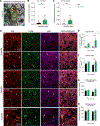
Figure 7.. Synaptic autophagy is enhanced in the dorsal striatum of auxilin KO mice
(A) Representative EM image of a type II synaptic terminal (S) in the dorsal striatum of Aux KO mice showing double-membrane autophagosomes containing CCVs/clathrin cages/missorted SVs (arrows). Scale bar, 150 μm. (B) Autophagosomes per presynaptic terminal in type I synapses. (C) Autophagosomes per presynaptic terminal in type II synapses. (D) Representative images of dorsal striatum immunostained for DA termini marker DAT (red) and autophagosome markers LC3B (green) and p62 (magenta). Note high number of DAT+ DA termini colocalizing with LC3B and p62 (arrows, DAT + LC3B + p62: white) in Aux KO mice, whereas DA termini in WT mice did not show such colocalization (arrowheads). In Aux KO mice, a few large DAT+ structures (axonal whirls) were positive for LC3B and p62 (Aux KO, enlarged, white asterisks) while others were not (Aux KO, enlarged, yellow asterisks). Scale bar, 10 μm. (E) Number of DAT+ termini colocalizing with LC3B and p62 was significantly higher in Aux KO mice. (F) Size of DAT + LC3B + p62-positive puncta was larger in 9-month-old Aux KO compared with WT mice. (G) LC3B expression in dorsal striatum did not show any significant difference between WT and Aux KO mice. (H) p62 expression showed a trend toward higher expression in Aux KO mice, although not significant. Data are presented as mean ± SEM. **p < 0.01, ***p < 0.001.

References

    1. Kordower JH, Olanow CW, Dodiya HB, Chu Y, Beach TG, Adler CH, Halliday GM, and Bartus RT (2013). Disease duration and the integrity of the nigrostriatal system in Parkinson’s disease. Brain 136, 2419–2431. 10.1093/brain/awt192. - DOI - PMC - PubMed
    1. Vidyadhara DJ, Lee JE, and Chandra SS (2019). Role of the endolysosomal system in Parkinson’s disease. J. Neurochem. 150, 487–506. 10.1111/jnc.14820. - DOI - PMC - PubMed
    1. Chanaday NL, Cousin MA, Milosevic I, Watanabe S, and Morgan JR (2019). The synaptic vesicle cycle revisited: new insights into the modes and mechanisms. J. Neurosci. 39, 8209–8216. 10.1523/jneurosci.1158-19.2019. - DOI - PMC - PubMed
    1. Edvardson S, Cinnamon Y, Ta-Shma A, Shaag A, Yim YI, Zenvirt S, Jalas C, Lesage S, Brice A, Taraboulos A, et al. (2012). A deleterious mutation in DNAJC6 encoding the neuronal-specific clathrin-uncoating co-chaperone auxilin, is associated with juvenile parkinsonism. PLoS One 7, e36458. 10.1371/journal.pone.0036458. - DOI - PMC - PubMed
    1. Nalls MA, Pankratz N, Lill CM, Do CB, Hernandez DG, Saad M, DeStefano AL, Kara E, Bras J, Sharma M, et al. (2014). Large-scale meta-analysis of genome-wide association data identifies six new risk loci for Parkinson’s disease. Nat. Genet. 46, 989–993. 10.1038/ng.3043. - DOI - PMC - PubMed

Publication types