. 2023 Mar 17;9(11):eadd8564.
doi: 10.1126/sciadv.add8564. Epub 2023 Mar 15.

Synthetic enforcement of STING signaling in cancer cells appropriates the immune microenvironment for checkpoint inhibitor therapy

Affiliations

Synthetic enforcement of STING signaling in cancer cells appropriates the immune microenvironment for checkpoint inhibitor therapy

Larsen Vornholz et al. Sci Adv. .

Abstract

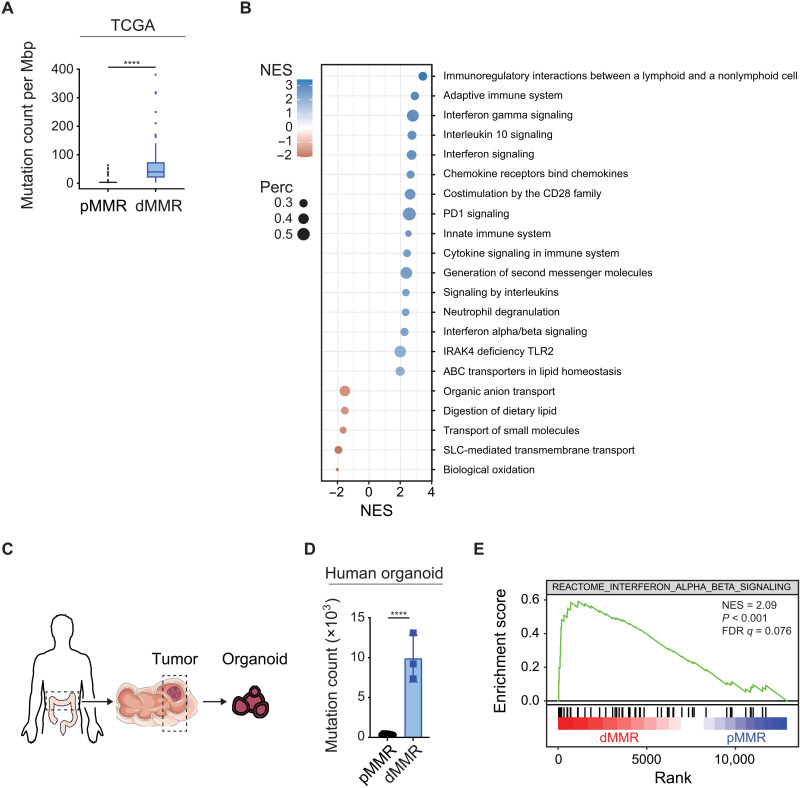
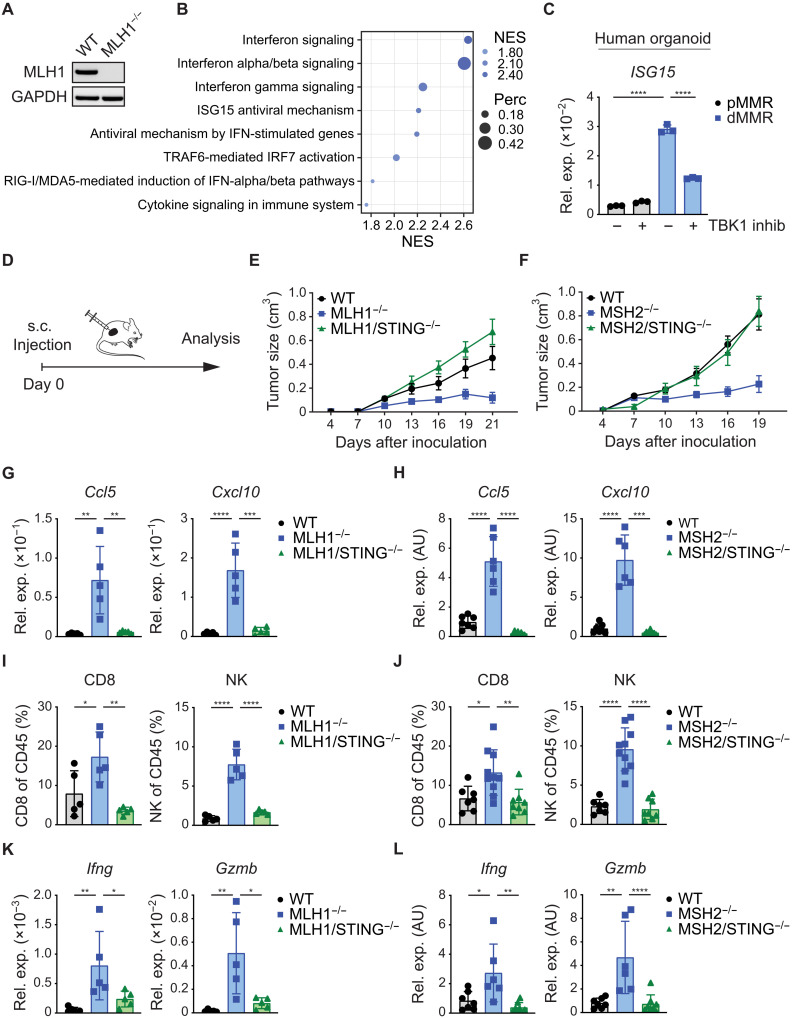
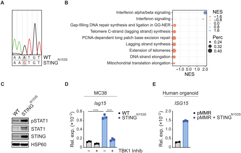
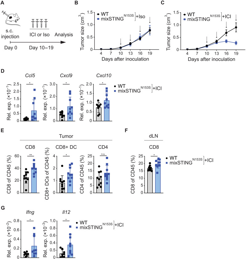
Immune checkpoint inhibitors (ICIs) enhance anticancer immunity by releasing repressive signals into tumor microenvironments (TMEs). To be effective, ICIs require preexisting immunologically "hot" niches for tumor antigen presentation and lymphocyte recruitment. How the mutational landscape of cancer cells shapes these immunological niches remains poorly defined. We found in human and murine colorectal cancer (CRC) models that the superior antitumor immune response of mismatch repair (MMR)-deficient CRC required tumor cell-intrinsic activation of cGAS-STING signaling triggered by genomic instability. Subsequently, we synthetically enforced STING signaling in CRC cells with intact MMR signaling using constitutively active STING variants. Even in MMR-proficient CRC, genetically encoded gain-of-function STING was sufficient to induce cancer cell-intrinsic interferon signaling, local activation of antigen-presenting cells, recruitment of effector lymphocytes, and sensitization of previously "cold" TMEs to ICI therapy in vivo. Thus, our results introduce a rational strategy for modulating cancer cell-intrinsic programs via engineered STING enforcement to sensitize resistant tumors to ICI responsiveness.


Figures

Fig. 1.
Fig. 1.. MMR deficiency drives IFN signaling in CRC.
Exome and RNA-seq data of pMMR and dMMR tumors (A and B) from 524 colorectal and rectal adenocarcinoma patients (TCGA) or (D and E) from patient-derived primary organoids. (A) Mutation count per Megabase pairs (Mbp) of pMMR versus dMMR tumors. (B) GSEA of differentially expressed gene sets from the Reactome database comparing pMMR versus dMMR tumors (+ve NES: dMMR). (C) Schematic representation of organoids from patients. (D) Mutation count of pMMR (n = 9) and dMMR (n = 3) tumor organoids. (E) GSEA of RNA-seq data using the Reactome IFN-α/β signaling gene set comparing dMMR (red) versus pMMR (blue). Student’s t test (A and D) was used to determine significance.
Fig. 2.
Fig. 2.. STING signaling in dMMR CRC mediates immunogenicity.
(A) MLH1 deletion in MC38 tumor cells was confirmed by Western blotting. (B) GSEA of RNA-seq data to identify differentially expressed gene sets of WT versus MLH1−/− MC38 tumor cells by using the Reactome database (+ve NES: MLH1−/−). (C) The relative gene expression of ISG15 in pMMR (PDO#12) versus dMMR (PDO#7) cultured primary organoids was quantified by qPCR 16 hours after TBK1 inhibitor treatment. (D) Schematic representation of the experimental setup in vivo. s.c. subcutaneous. (E) Growth of subcutaneously inoculated WT, MLH1−/−, or MLH1/STING−/− MC38 tumor cells (n = 5) or (F) WT, MSH2−/−, or MSH2/STING−/− MC38 tumor cells (n = 7 to 10) in syngeneic WT C57Bl/6 mice. (G to L) At the end point (MLH1 day 21; MSH2 day 19), subcutaneously grown tumors were explanted for FACS and qPCR analysis. (G and H) Relative gene expression of chemokines (Ccl5, Cxcl10) was quantified by qPCR. (I and J) FACS analyses displaying the percentages (CD8, NK) of live/CD45+ cells. (K and L) The relative gene expression of cytotoxic effector molecules (Gzmb, Ifng) was quantified by qPCR. The data represent n = 3 technical replicates (C). The data are presented as the mean ± SEM (E and F). One-way ANOVA (C and G to L) was used to determine significance. AU, arbitrary units.
Fig. 3.
Fig. 3.. A strategy to genetically enforce STING signaling in cancer cells.
(A) Electropherogram displaying the sequencing result of the PCR-amplified transgene STINGN153S from genomic DNA of MC38 tumor cells. (B) GSEA of RNA-seq data to identify differentially expressed gene sets between WT versus STINGN153S MC38 tumor cells by using the Reactome database with a percentage cutoff of >0.2 (+ve NES: STINGN153S). (C) Phosphorylation of STAT1 in cultured MC38 cells was detected by Western blotting. (D) The relative gene expression of Isg15 in WT versus STINGN153S MC38 tumor cells 16 hours after TBK1 inhibitor treatment was quantified by qPCR. (E) The relative gene expression of ISG15 in pMMR (PDO#12) and STINGN153S-transduced (=pMMR+STINGN153S) cultured primary organoids was quantified by qPCR. The data represent n = 3 independent experiments (D) or n = 2 to 3 technical replicates (E). One-way ANOVA (D) or Student’s t test (E) was used to determine significance.
Fig. 4.
Fig. 4.. Synthetically enforced STING signaling promotes antitumor immunity.
(A) Schematic representation of the experimental setup in vivo. (B) Growth of subcutaneously inoculated WT and STINGN153S MC38 tumor cells in syngeneic WT C57Bl/6 mice (n = 5). (C to F) Subcutaneously grown tumors were explanted on day 17 for qPCR and FACS analysis. (C) The relative gene expression of (C) ISGs (Isg15) and (D) chemokines (Ccl5, Cxcl9, Cxcl10, Cxcl11) was quantified by qPCR. (E) FACS results. Representative dot plots and percentages (CD4, CD8, NK1.1) of live/CD45+ cells are displayed. (F) The relative gene expression of cytotoxic effector molecules (Gzmb, Prf1, Ifng, Tnf) was quantified by qPCR. The data are presented as the mean ± SEM (B). Student’s t test was used to determine significance.
Fig. 5.
Fig. 5.. Tumor cell–intrinsic STING enforcement sensitizes to immune checkpoint inhibitor therapy.
(A) Schematic representation of the experimental setup in vivo. Growth of subcutaneously inoculated WT or mixSTINGN153S MC38 tumors in syngeneic C57Bl/6 mice (B) treated with isotype control (=iso) (n = 4) or (C) treated with anti-PD1/anti-CTLA4 (=ICI) (n = 8 to 10) every 3 days (black arrows). (D to G) Subcutaneously grown tumors that were treated with ICI therapy were explanted on day 21 for qPCR and FACS analysis. The relative gene expression of (D) chemokines (Ccl5, Cxcl9, Cxcl10) was quantified by qPCR. FACS was performed, and the (E) percentages (CD8, CD8+ DCs, CD4) of live/CD45+ cells in the tumor and (F) percentages (CD8) of live/CD45+ cells in the draining lymph node (dLN) are shown. (G) The relative gene expression of cytokines (Ifng, Il12) was quantified by qPCR. The data are presented as the mean ± SEM (B and C). Student’s t test (D to G) was used to determine significance.
Fig. 6.
Fig. 6.. The expression of STINGN153S in a subset of tumor cells reprograms the TME.
CITE-seq analyses of subcutaneously grown WT and mixed STINGN153S MC38 tumors. (A) UMAP plot of annotated clusters displaying the individual clusters. (B) Cellular composition of cell clusters in WT and mixed STINGN153S tumors. (C) Differentially expressed gene sets determined by GSEA by using g:profiler for all GO:BP terms enriched for fewer than 400 genes considering all clusters. (D) Violin plot displaying how strongly the different clusters contribute to the differentially expressed genes observed in (C).

References

    1. C. Robert, A decade of immune-checkpoint inhibitors in cancer therapy. Nat. Commun. 11, 3801 (2020). - PMC - PubMed
    1. D. S. Chen, I. Mellman, Elements of cancer immunity and the cancer-immune set point. Nature 541, 321–330 (2017). - PubMed
    1. J. Galon, D. Bruni, Approaches to treat immune hot, altered and cold tumours with combination immunotherapies. Nat. Rev. Drug Discov. 18, 197–218 (2019). - PubMed
    1. D. T. Le, J. N. Durham, K. N. Smith, H. Wang, B. R. Bartlett, L. K. Aulakh, S. Lu, H. Kemberling, C. Wilt, B. S. Luber, F. Wong, N. S. Azad, A. A. Rucki, D. Laheru, R. Donehower, A. Zaheer, G. A. Fisher, T. S. Crocenzi, J. J. Lee, T. F. Greten, A. G. Duffy, K. K. Ciombor, A. D. Eyring, B. H. Lam, A. Joe, S. P. Kang, M. Holdhoff, L. Danilova, L. Cope, C. Meyer, S. Zhou, R. M. Goldberg, D. K. Armstrong, K. M. Bever, A. N. Fader, J. Taube, F. Housseau, D. Spetzler, N. Xiao, D. M. Pardoll, N. Papadopoulos, K. W. Kinzler, J. R. Eshleman, B. Vogelstein, R. A. Anders, L. A. Diaz Jr., Mismatch repair deficiency predicts response of solid tumors to PD-1 blockade. Science 357, 409–413 (2017). - PMC - PubMed
    1. G.-Y. Lyu, Y.-H. Yeh, Y.-C. Yeh, Y.-C. Wang, Mutation load estimation model as a predictor of the response to cancer immunotherapy. NPJ Genom. Med. 3, 12 (2018). - PMC - PubMed