. 2023 Mar 1;37(5-6):204-217.
doi: 10.1101/gad.350211.122. Epub 2023 Mar 15.

ALOX5-mediated ferroptosis acts as a distinct cell death pathway upon oxidative stress in Huntington's disease

Affiliations

ALOX5-mediated ferroptosis acts as a distinct cell death pathway upon oxidative stress in Huntington's disease

Shujuan Song et al. Genes Dev. .

Abstract

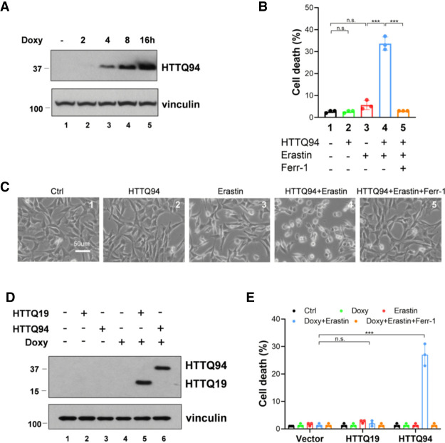
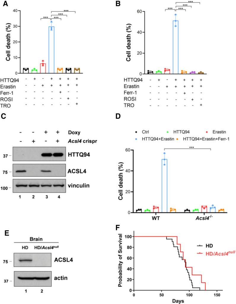
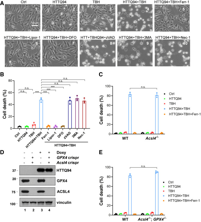
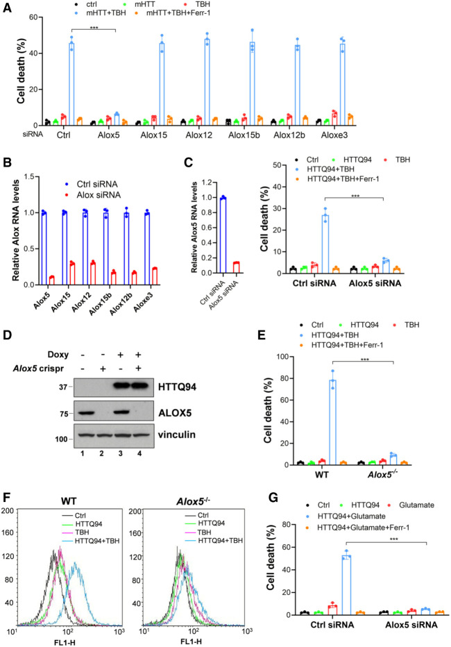
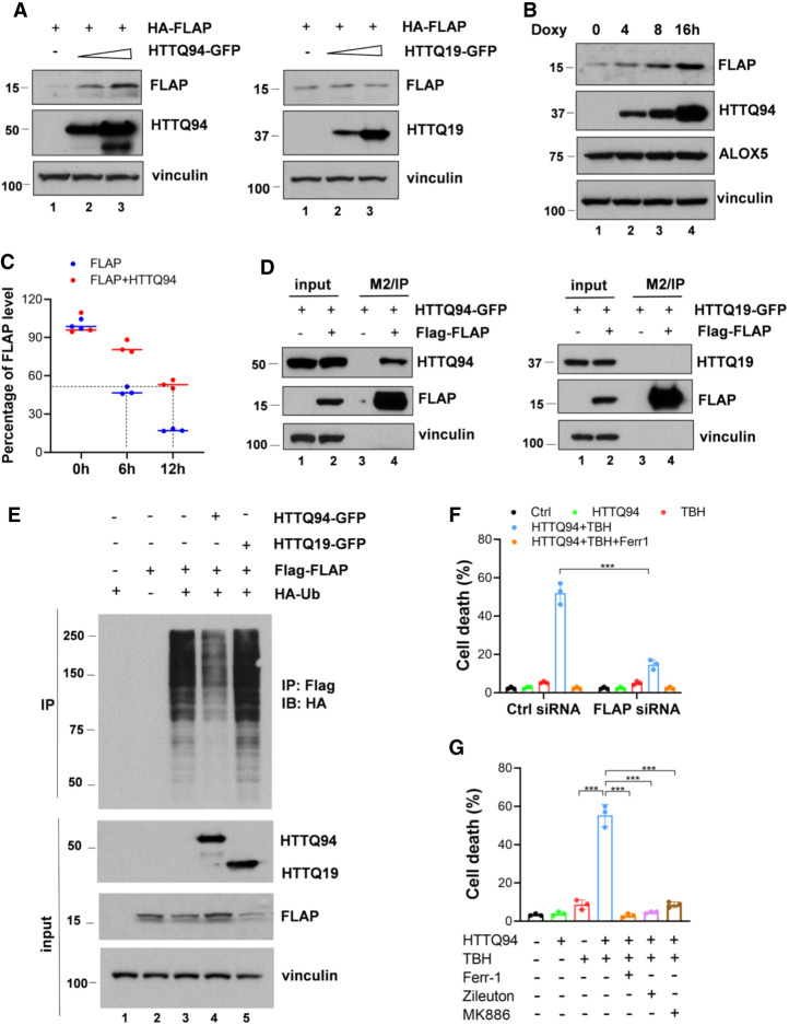
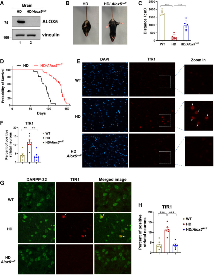
Although it is well established that Huntington's disease (HD) is mainly caused by polyglutamine-expanded mutant huntingtin (mHTT), the molecular mechanism of mHTT-mediated actions is not fully understood. Here, we showed that expression of the N-terminal fragment containing the expanded polyglutamine (HTTQ94) of mHTT is able to promote both the ACSL4-dependent and the ACSL4-independent ferroptosis. Surprisingly, inactivation of the ACSL4-dependent ferroptosis fails to show any effect on the life span of Huntington's disease mice. Moreover, by using RNAi-mediated screening, we identified ALOX5 as a major factor required for the ACSL4-independent ferroptosis induced by HTTQ94. Although ALOX5 is not required for the ferroptotic responses triggered by common ferroptosis inducers such as erastin, loss of ALOX5 expression abolishes HTTQ94-mediated ferroptosis upon reactive oxygen species (ROS)-induced stress. Interestingly, ALOX5 is also required for HTTQ94-mediated ferroptosis in neuronal cells upon high levels of glutamate. Mechanistically, HTTQ94 activates ALOX5-mediated ferroptosis by stabilizing FLAP, an essential cofactor of ALOX5-mediated lipoxygenase activity. Notably, inactivation of the Alox5 gene abrogates the ferroptosis activity in the striatal neurons from the HD mice; more importantly, loss of ALOX5 significantly ameliorates the pathological phenotypes and extends the life spans of these HD mice. Taken together, these results demonstrate that ALOX5 is critical for mHTT-mediated ferroptosis and suggest that ALOX5 is a potential new target for Huntington's disease.

Keywords: ACSL4; ALOX5; GPX4; HTT; ROS; ferroptosis; oxidative stress.


Figures

Figure 1.
Figure 1.
HTTQ94 expression sensitizes neuronal cells and other cell types to ferroptosis. (A) Western blot analysis for HTTQ94 from HTTQ94 tet-on HT-22 cells treated with 0.5 μg/mL doxycycline for 2, 4, 8, and 16 h. (B) HTTQ94 tet-on HT-22 cells were preincubated with 0.5 μg/mL doxycycline for 4 h and then treated with 1 μM erastin for 12 h with/without 2 μM Ferr-1. (C) Representative phase-contrast images of cell death, related to B. (D) Western blot analysis for mutant HTT fragment (HTTQ94) and normal HTT fragment (HTTQ19) expression in the tet-on H1299 cells treated with 0.5 μg/mL doxycycline (tet) for 16 h. (E) Cell death assay. Control, HTTQ94, and HTTQ19 fragment tet-on H1299 cells preincubated with 0.5 μg/mL doxycycline for 16 h were treated with 30 μM erastin for 48 h with/without 2 μM Ferr-1. Cell deaths were calculated from three replicates. Data shown in B and E are the means ± SD. P-values were derived from two-tailed unpaired t-test. (***) P ≤ 0.001, (n.s.) P > 0.05.
Figure 2.
Figure 2.
The role of ACSL4 in mHTT-induced ferroptosis and the life span of the HD mice. (A) Cell death assay. HTTQ94 tet-on HT-22 cells preincubated with 0.5 μg/mL doxycycline for 4 h were treated with 1 μM erastin for 12 h in the presence or absence of 2 μM ferrostatin-1 (Ferr-1) or ACSL4 inhibitors (10 μM rosiglitazone [ROSI] and 10 μM troglitazone [TRO]). (B) Cell death assay. HTTQ94 tet-on SK-N-BE(2)C cells preincubated with 0.5 μg/mL doxycycline for 16 h were treated with 40 μM erastin for 32 h in the presence or absence of 2 μM ferrostatin-1 (Ferr-1), 10 μM rosiglitazone (ROSI), and 10 μM troglitazone(TRO). (C) Western blot analysis for ACSL4 and HTTQ94 in HTTQ94 tet-on SK-N-BE(2)C control and Acsl4-Crispr cells treated with 0.5 μg/mL doxycycline for 16 h. (D) Cell death assay. HTTQ94 tet-on SK-N-BE(2)C control Crispr and Acsl4-Crispr cells were preincubated with 0.5 μg/mL doxycycline for 16 h and then treated with 40 μM erastin for 32 h with/without 2 μM Ferr-1. (E) Western blot analysis for ACSL4 from the cerebral cortex tissues of HD and HD/Acsl4-null mice. (F) Kaplan–Meier survival curves of HD (n = 21 independent mice) and HD/Acsl4-null (n = 7 independent mice) mice. Cell deaths were calculated from three replicates. Data shown in A, B, and D are the means ± SD. P-values were derived from two-tailed unpaired t-test. (***) P ≤ 0.001. In F, P-value (HD vs. HD/Acsl4-null) was calculated using log-rank Mantel–Cox test. P = 0.1356.
Figure 3.
Figure 3.
HTTQ94 is able to induce the ACSL4-independent ferroptosis upon ROS stress. (A) Representative phase-contrast images of cell death from the HTTQ94 tet-on HT-22 cells. HTTQ94 tet-on HT-22 cells were preincubated with 0.5 μg/mL doxycycline for 4 h and then treated with 10 μM TBH for 8 h in the presence or absence of the ferroptosis inhibitors (2 μM ferrostatin-1 [Ferr1], 2 μM liproxstatin-1 [Lipor1], and 100 μM DFO), apoptosis inhibitor (10 μM Z-VADFMK [zVAD]), autophagy inhibitor (2 mM 3-methylademine [3MA]), or necroptosis inhibitor (10 μM necrostatin-1 [Nec1]). (B) Quantification of cell death, related to A. Three replicates were used for each group. (C) HTTQ94 tet-on SK-N-BE(2)C control Crispr and Acsl4Crispr cells were preincubated with 0.5 μg/mL doxycycline for 16 h and then treated with 350 μM TBH for 24 h with/without 2 μM Ferr-1. (D) Western blot analysis of GPX4, ACSL4, and HTTQ94 in HTTQ94 tet-on SK-N-BE(2)C control Crispr and GPX4/Acsl4 double-Crispr cells treated with 0.5 μg/mL doxycycline for 16 h. (E) HTTQ94 tet-on SK-N-BE(2)C control Crispr and GPX4/Acsl4 double-Crispr cells were preincubated with 0.5 μg/mL doxycycline for 16 h and then treated with 350 μM TBH for 24 h with/without 2 μM Ferr-1. Cell deaths were calculated from three replicates; Data shown in B, C, and E are means ± SD. P-values were derived from two-tailed unpaired t-test. (***) P ≤ 0.001, (n.s.) P > 0.05.
Figure 4.
Figure 4.
ALOX5 is required for HTTQ94-mediated ferroptosis induced by ROS stress and glutamate. (A) Cell death assay in HTTQ94 tet-on SK-N-BE(2)C cells with different ALOX knockdowns. Cells were transfected with control siRNA (ctrl) or ALOX family-specific siRNAs, followed by preincubation with 0.5 µg/mL doxycycline for 16 h and then treated with 350 μM TBH for 24 h. (B) qPCR analysis of the knockdown efficiency of ALOX family members in HTTQ94 tet-on SK-N-BE(2)C cells transfected with control siRNA or ALOX family-specific siRNAs. (C) Cell death assay in HTTQ94 tet-on HT-22 cells with ALOX5 knockdown. Cells were transfected with control siRNA (ctrl) or ALOX5-specific siRNA and then preincubated with 0.5 µg/mL doxycycline for 4 h, followed by 10 μM TBH treatment for 8 h. (Left panel) ALOX5 knockdown efficiency. (Right panel) Cell death assay. (D) Western blot analysis of ALOX5 and HTTQ94 in HTTQ94 tet-on SK-N-BE(2)C control Crispr and Alox5-Crispr cells treated with 0.5 μg/mL doxycycline for 16 h. (E) Cell death assay for mHTT tet-on SK-N-BE(2)C control Crispr and Alox5-Crispr cells. Cells were preincubated with 0.5 μg/mL doxycycline for 16 h, followed by incubation with 350 μM TBH for 24 h with/without 2 μM Ferr-1. (F) FACS analysis of lipid ROS production in HTTQ94 tet-on SK-N-BE(2)C control Crispr and Alox5-Crispr cells. Cells were preincubated with 0.5 μg/mL doxycycline for 16 h and then treated with 350 μM TBH for 6 h. Lipid ROS was stained with C11-BODIPY. (G) Cell death assay in HTTQ94 tet-on HT-22 cells. Cells were transfected with control siRNA (ctrl) or ALOX5 siRNA, followed by incubation with 0.5 μg/mL doxycycline and 10 mM glutamate for 16 h in the presence or absence of 2 μM Ferr-1. Cell deaths were calculated from three replicates. Data shown in A, C, E, and G are means ± SD. P-values were derived from two-tailed unpaired t-test. (***) P ≤ 0.001.
Figure 5.
Figure 5.
Mechanistic insight into HTTQ94-induced ALOX5 activation. (A) Western blot analysis of FLAP and HTTQ94 or HTTQ19 in HEK293 cells transfected with an HA-FLAP-expressing plasmid and either an empty vector, HTTQ94-GFP-expressing vector, or HTTQ19-GFP-expressing vector. (B) Western blot analysis of endogenous FLAP and ALOX5 in HTTQ94 tet-on SK-N-BE(2)C cells incubated with 0.5 μg/mL doxycycline for 4, 8, and 16 h. (C) Densitometry quantification of FLAP protein levels calculated using ImageJ software and plotted for half-life determination corresponding to Supplemental Figure S5B. (D) Co-IP of GFP-tagged HTTQ94/HTTQ19 and FLAG-tagged FLAP in HEK293 cells. Cell lysates were immunoprecipitated with anti-FLAG-coupled beads (M2), followed by Western analysis of HTTQ94, HTTQ19, and FLAP. (E) Ubiquitination analysis of FLAP in the presence of HTTQ94 or HTTQ19. HEK293 cells were cotransfected with FLAG-FLAP and HA-ubiquitin (Ub) in the presence of HTTQ94 or HTTQ19. Cell lysates were immunoprecipitated with anti-FLAG-coupled beads (M2), followed by Western blot analysis of HA, FLAP, and HTT proteins. (F) Cell death assay for HTTQ94 tet-on SK-N-BE(2)C cells with FLAP knockdown. Cells were transfected with control siRNA (ctrl) or FLAP-specific siRNA and then preincubated with 0.5 µg/mL doxycycline for 16 h, followed by 350 μM TBH treatment for 24 h. (G) Cell death assay. HTTQ94 tet-on HT-22 cells preincubated with 0.5 μg/mL doxycycline for 4 h were treated with 10 μM TBH for 8 h in the presence or absence of 2 μM Ferr-1, 10 μM zileuton, or 10 μM MK886. Cell deaths were calculated from three replicates. Data shown in F and G are the means ± SD. P-values were derived from two-tailed unpaired t-test. (***) P ≤ 0.001.
Figure 6.
Figure 6.
Loss of ALOX5 ameliorates the phenotypes and significantly extends the life spans of HD mice. (A) Western blot analysis for ALOX5 expression in HD and HD/Alox5-null mouse brains. (B) Representative images of limb clasping from the HD and HD/Alox5-null mice. (C) Open field test. Thirteen-week-old mice were subjected to open field test, and the distance moved was calculated. Six mice were used for each group (HD vs. HD/Alox5-null; P < 0.01). (D) Kaplan–Meier survival curves of HD (n = 21 independent mice) and HD/Alox5-null (n = 25 independent mice) mice. P-value was calculated using log-rank Mantel–Cox test (HD vs. HD/Alox5-null; P < 0.0001). (E,F) TfR1 staining on mouse brain slides. WT, HD-N171-82Q, and HD-N171-82Q/Alox5-null mouse brain paraffin slides were dewaxed, and antigens were retrieved by PH6.0 citric acid solution followed by incubation with TfR1 antibody. (E) Representative images of TfR1 staining (four mice for each group). Nuclei were stained with DAPI. (F) Quantification of TfR1-positive striatal neurons. Data are represented as mean ± SEM. (G) Representative images of TfR1 and DARPP-32 double staining on mouse brain paraffin slides. DARPP-32 was used as a marker for striatal medium spiny neurons (Naia and Rego 2018). (H) Quantification of TfR1-positive striatal neurons, related to G. Data shown in C, F, and H are the means ± SEM. n = 6. P-values were derived from two-tailed unpaired t-test. (***) P ≤ 0.001, (**) P ≤ 0.01.

References

    1. Bates G. 2003. Huntingtin aggregation and toxicity in Huntington's disease. The Lancet 361: 1642–1644. 10.1016/S0140-6736(03)13304-1 - DOI - PubMed
    1. Bates GP, Dorsey R, Gusella JF, Hayden MR, Kay C, Leavitt BR, Nance M, Ross CA, Scahill RI, Wetzel R, et al. 2015. Huntington disease. Nat Rev Dis Primers 1: 15005. 10.1038/nrdp.2015.5 - DOI - PubMed
    1. Bersuker K, Hendricks JM, Li Z, Magtanong L, Ford B, Tang PH, Roberts MA, Tong B, Maimone TJ, Zoncu R, et al. 2019. The CoQ oxidoreductase FSP1 acts parallel to GPX4 to inhibit ferroptosis. Nature 575: 688–692. 10.1038/s41586-019-1705-2 - DOI - PMC - PubMed
    1. Caudle WM, Zhang J. 2009. Glutamate, excitotoxicity, and programmed cell death in Parkinson disease. Exp Neurol 220: 230–233. 10.1016/j.expneurol.2009.09.027 - DOI - PubMed
    1. Chu B, Kon N, Chen D, Li T, Liu T, Jiang L, Song S, Tavana O, Gu W. 2019. ALOX12 is required for p53-mediated tumour suppression through a distinct ferroptosis pathway. Nat Cell Boil 21: 579–591. 10.1038/s41556-019-0305-6 - DOI - PMC - PubMed

Publication types