Review
. 2023 May;53(5):984-1004.
doi: 10.1007/s00247-023-05636-3. Epub 2023 Mar 16.

Endemic mycoses in children in North America: a review of radiologic findings

Affiliations
Review

Endemic mycoses in children in North America: a review of radiologic findings

Abraham P Campbell et al. Pediatr Radiol. 2023 May.

Abstract

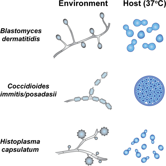
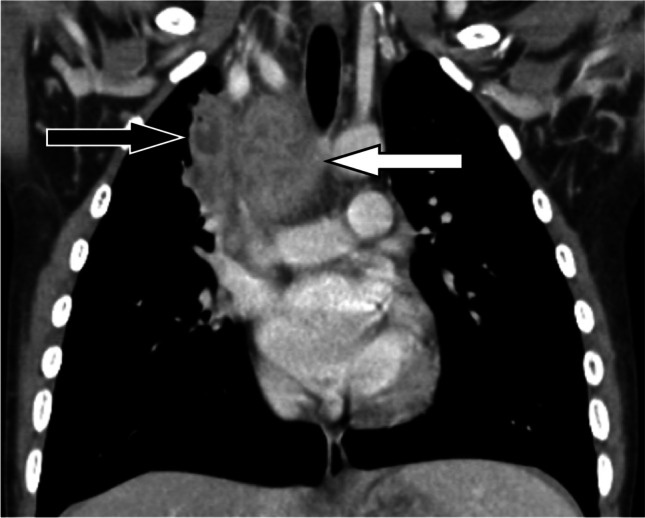
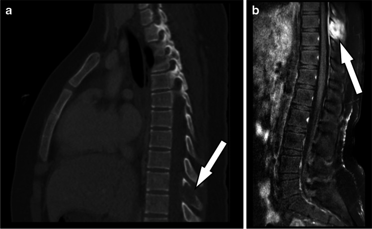
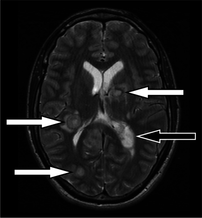
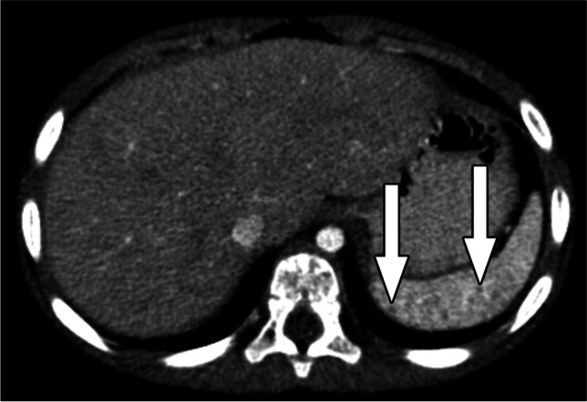
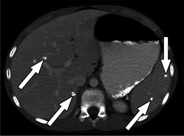
Clinically significant endemic mycoses (fungal infections) in the United States (U.S.) include Blastomyces dermatitidis, Histoplasma capsulatum, and Coccidioides immitis/posadasii. While the majority of infections go clinically unnoticed, symptomatic disease can occur in immunocompromised or hospitalized patients, and occasionally in immune-competent individuals. Clinical manifestations vary widely and their diagnosis may require fungal culture, making the rapid diagnosis a challenge. Imaging can be helpful in making a clinical diagnosis prior to laboratory confirmation, as well as assist in characterizing disease extent and severity. In this review, we discuss the three major endemic fungal infections that occur in the U.S., including mycology, epidemiology, clinical presentations, and typical imaging features with an emphasis on the pediatric population.

Keywords: Blastomycosis; Children; Coccidioidomycosis; Endemic fungal infection; Histoplasmosis; Imaging.


Conflict of interest statement

None

Figures

Fig. 1
Fig. 1
Fungal dimorphic states relative to temperature. Copyright permission granted by original artist, Davina H. Murray, and adapted for the manuscript. Blastomyces exists as septate hyphae with spores in the environment, and as a broad-based budding yeast in the body. Coccidiodes exists as hyphae that link single-cell barrel-shaped spores in the environment, and as spherules in the body that internally contain many endospores that are released upon spherule rupture. Histoplasma exists as septate hyphae with spiky macrospores and smooth microspores in the environment, and as an oval budding yeast in the body
Fig. 2
Fig. 2
Bronchioalveolar lavage (BAL) specimen from a patient with pulmonary blastomycosis. The Grocott methenamine silver (GMS) stain shows numerous Blastomyces yeast forms with characteristic broad-based budding and thick cell walls; scale bar 50 µm
Fig. 3
Fig. 3
Hematoxylin and eosin (H&E) stain showing pulmonary coccidioidomycosis histologic findings. Granulomatous inflammation in pulmonary coccidioidomycosis demonstrating extensive central purulent necrosis (asterisk: collection of neutrophils and cell debris, in a fibrinoid background) surrounded by peripheral palisading/demarcating histiocytes (vague granulomatous lining exemplied by arrows). Scale bar: 440 µm
Fig. 4
Fig. 4
Hematoxylin and eosin (H&E) stain showing Coccidioides spherule with endospores. Pulmonary coccidioidomycosis with a characteristic spherule containing multiple endospores as highlighted by hematoxylin–eosin in a necrotizing background with loss of nuclear staining. Scale bar 25 µm
Fig. 5
Fig. 5
Grocott methenamine silver (GMS) stain in histoplasmosis. GMS highlights Histoplasma organisms in a patient with pulmonary histoplasmosis, showing numerous clustered small yeasts devoid of a capsule. Scale bar 50 µm
Fig. 6
Fig. 6
Hematoxylin and eosin (H&E) stain showing chronic pulmonary histoplasmosis. Representative lung sections show a discrete palisade of epitheloid histiocytes surrounding a centrally necrotic area. Scale bar: 440 µm
Fig. 7
Fig. 7
Hematoxylin and eosin (H&E) stain showing chronic pulmonary histoplasmosis. Hyalinizing granuloma characterized by a well-circumscribed collection of epitheloid histiocytes with concentric layers of hyalinized collagen which eventually may progress into calcification and fibrosis. The Histoplasma organisms cannot be visualized on routine histology and require the use of special stains for identification. Scale bar: 440 µm
Fig. 8
Fig. 8
9-year-old with acute fungal infection due to blastomycosis. Coronal reformatted unenhanced chest CT image shows left upper lobe consolidation (arrow) with air bronchograms, mimicking bacterial community-acquired pneumonia
Fig. 9
Fig. 9
12-year-old boy with acute fungal infection due to blastomycosis. Axial chest CT image with IV contrast material shows right upper lobe consolidation (arrow). Although the morphology of the consolidation is mass-like, the fact that enhancing pulmonary vessels course through the area rather than being displaced by the area suggests consolidated lung parenchyma surrounding bronchovascular bundles rather than a true mass
Fig. 10
Fig. 10
16-year-old girl with acute fungal infection due to coccidioidomycosis. Axial chest CT image with IV contrast material shows left upper lobe consolidation (arrow) with central cavitation and surrounding ground glass opacity
Fig. 11
Fig. 11
17-year-old boy with acute fungal infection due to histoplasmosis. Sagittal reformatted chest CT image with IV contrast material shows left upper lobe consolidation (arrow) with air bronchograms, mimicking bacterial community-acquired pneumonia
Fig. 12
Fig. 12
2-month-old boy with acute fungal infection due to coccidioidomycosis. Axial chest CT image with IV contrast material shows left upper lobe consolidation (white arrows) with air bronchograms, mimicking bacterial community-acquired pneumonia, as well as an enlarged left axillary lymph node (black arrow)
Fig. 13
Fig. 13
15-year-old girl with acute fungal infection due to coccidioidomycosis. Coronal reformatted chest CT image with IV contrast material shows left lower lobe consolidation (arrow) with air bronchograms. The hypoenhancement of the involved lung differentiates infectious consolidation from atelectasis (the majority of the left lower lobe opacity is hypoenhancing, whereas atelectasis should hyperenhance). Heterogeneously enhancing, mediastinal (asterisk) and hilar lymphadenopathy can also be seen
Fig. 14
Fig. 14
15-year-old girl with acute fungal infection due to histoplasmosis. Axial chest CT image with IV contrast material shows a solitary nodule in the left lower lobe (arrow). The ill-defined margins favor an infectious or inflammatory etiology rather than a malignant etiology
Fig. 15
Fig. 15
6-year-old girl with history of juvenile idiopathic arthritis and confirmed histoplasmosis. Axial chest CT image with IV contrast material shows a dominant nodule in the lingula with adjacent bronchovascular nodularity/beading (arrow) suggesting the primary site of infection as well as tiny nodules elsewhere in a miliary pattern suggesting disseminated infection
Fig. 16
Fig. 16
8-year-old girl with sequelae of histoplasmosis. Axial unenhanced chest CT image shows a solitary nodule in the left lower lobe (white arrow) with punctate internal calcifications and associated surrounding satellite nodules, nodularity/beading of the adjacent bronchovascular bundle and pleura (black arrows), peripheral air trapping (arrowhead), and an enlarged left hilar lymph node with punctate calcification (asterisk)
Fig. 17
Fig. 17
17-year-old boy with prior histoplasmosis infection. a Axial chest CT image with IV contrast material shows centrally calcified nodules in the left lower lobe and right upper lobe (arrows). b Axial chest CT image with IV contrast material obtained 7 weeks later for worsened cough after a course of steroids shows new clustered ill-defined nodules around the left lower lobe calcified nodule (arrow) compatible with histoplasmosis reactivation. Enlarged right hilar lymph nodes containing calcifications are also present
Fig. 18
Fig. 18
7-year-old boy with acute fungal infection due to coccidioidomycosis. Axial chest CT image with IV contrast material shows multiple scattered small nodules in the right lung (arrows)
Fig. 19
Fig. 19
17-year-old boy with acute fungal infection due to coccidioidomycosis. Axial chest CT image with IV contrast material shows a dominant cavitary nodule (arrow) in the right lung and several surrounding satellite nodules (arrowheads)
Fig. 20
Fig. 20
16-year-old girl with active histoplasmosis. Coronal reformatted chest CT image with IV contrast material shows enlarged, predominantly hypoenhancing right hilar lymph nodes with septation-like enhancement (arrows) and internal speckled calcifications
Fig. 21
Fig. 21
15-year-old girl with chest pain and active histoplasmosis. a Axial chest CT image with IV contrast material shows peripheral enhancement of an enlarged left peribronchial lymph node (white arrow). Calcifications within this lymph node in the setting of densely calcified lymph nodes elsewhere (black arrow) suggest possible reactivation histoplasmosis, which was confirmed at work-up. b Coronal reformatted chest CT image with IV contrast material shows left lower lobe consolidation and ground-glass opacity around the reactivated lymph node (arrow) as well as clustered small nodules in the lateral basal aspect of the left lower lobe (black arrow), the largest of which is calcified, related to the original infection
Fig. 22
Fig. 22
9-year-old girl with active histoplasmosis. Coronal reformatted chest CT image with IV contrast material shows multiple calcified mediastinal lymph nodes. Both eggshell (white arrow) and dense lymph node calcifications (black arrow) are present
Fig. 23
Fig. 23
4-year-old girl with active histoplasmosis. Axial chest CT image with IV contrast material shows heterogeneously enhancing right paratracheal lymphadenopathy (arrows). There is mass effect on the superior vena cava anteriorly, and the trachea is deviated to the left. Nodal involvement can be bulky and result in substantial mass-effect
Fig. 24
Fig. 24
7-year-old boy with active coccidioidomycosis. Coronal reformatted chest CT image with IV contrast material shows heterogeneously enhancing, conglomerate mediastinal lymphadenopathy (white arrow), including a right upper mediastinal lymph node with discrete central necrosis (black arrow)
Fig. 25
Fig. 25
17-year-old girl with fibrosing mediastinitis due to histoplasmosis infection. Coronal reformatted chest CT image with IV contrast material shows confluent, infiltrative soft tissue involving the mediastinum and both hila that encases and narrows bilateral bronchi (white arrows) and the left upper pulmonary vein (black arrow)
Fig. 26
Fig. 26
2-month-old boy with tenosynovitis and osteomyelitis due to disseminated coccidioidomycosis. a Sagittal T2-weighted MR image of the index finger shows extensive abnormal T2-weighted hyperintense signal in the volar soft tissue of the finger (white arrows) surrounding the flexor tendons, consistent with tenosynovitis, as well as foci of abnormal T2-weighted hyperintense marrow signal in the proximal phalanx, consistent with osteomyelitis (black arrow). b Axial contrast-enhanced T1-weighted MR image shows mild enhancement along the periphery of the area of index finger volar soft tissue T2-weighted hyperintense signal abnormality (white arrow)
Fig. 27
Fig. 27
13-year-old boy with osteomyelitis due to disseminated coccidioidomycosis. a Axial pelvic CT image with IV and oral contrast materials shows destruction of the left ilium (white arrow) with a contiguous large area of mixed phlegmon and abscess containing a small focus of gas extending centrally into the pelvis (black arrows) and superficially into the overlying soft tissues. The left iliac vessels are displaced medially, and the surrounding fat planes are obscured by edema. b Coronal T2-weighted fat-saturated pelvic MR mages shows bone marrow hyperintense signal abnormality in the left ilium consistent with osteomyelitis (white arrows) as well as the large area of mixed phlegmon and abscess in the left iliac fossa seen on CT (black arrows)
Fig. 28
Fig. 28
12-year-old girl with osteomyelitis due to disseminated blastomycosis. Sagittal T2-weighted fat-saturated MR image of the foot shows bone marrow edema throughout the calcaneus (asterisk), an intraosseous abscess (black arrow), and mild surrounding soft tissue edema (white arrows)
Fig. 29
Fig. 29
10-year-old boy with osteomyelitis due to disseminated blastomycosis. Sagittal T2-weighted MR image of the thoracic spine shows T8 vertebral body height loss and marrow edema (arrow)
Fig. 30
Fig. 30
14-year-old girl with osteomyelitis due to disseminated coccidioidomycosis. a Sagittal reformatted chest CT image with IV contrast material shows a “punched-out” lytic lesion in the T9 spinous process (arrow). b Sagittal T1-weighted fat-saturated MR image with IV contrast material shows an enhancing destructive lesion in the T9 spinous process (arrow) with surrounding soft tissue inflammation and minimal epidural extension
Fig. 31
Fig. 31
15-month-old boy with meningitis due to disseminated coccidioidomycosis. Axial contrast-enhanced T1-weighted MR image of the brain shows thick, irregular enhancement of the meninges and basal cisterns (arrows). The partially visualized lateral ventricles are dilated due to obstruction of CSF flow
Fig. 32
Fig. 32
17-year-old boy with brain abscesses due to disseminated histoplasmosis. Axial T2-weighted MR image of the brain shows multiple intraparenchymal abscesses (white arrows) as well as enlargement of the left lateral ventricle choroid plexus consistent with choroid plexitis (black arrow)
Fig. 33
Fig. 33
8-year-old boy with brain involvement from disseminated blastomycosis. Coronal contrast-enhanced T1-weighted fat-saturated MR image of the brain shows scattered punctate foci of enhancement throughout the brain (arrows)
Fig. 34
Fig. 34
3-year-old girl with disseminated histoplasmosis and splenic involvement. Axial abdominal CT image with IV contrast material shows numerous tiny hypoattenuating nodular foci in the spleen (arrows)
Fig. 35
Fig. 35
11-year-old boy remote disseminated histoplasmosis and liver and spleen involvement. Axial abdominal CT image with IV contrast material shows multiple coarse calcifications in the spleen and liver (arrows)
Fig. 36
Fig. 36
17-year-old boy with disseminated histoplasmosis and splenic involvement. Axial abdominal CT image with IV contrast material shows multiple infectious subcapsular splenic fluid collections (arrows)
Fig. 37
Fig. 37
10-year-old boy with disseminated blastomycosis and splenic involvement. Axial abdominal CT image with IV contrast material shows clustered round hypodense lesions in the spleen (arrow).Fine needle aspiration identified budding yeasts
Fig. 38
Fig. 38
14-year-old girl with remote disseminated histoplasmosis and kidney involvement. Axial abdominal CT image with IV contrast material shows a low attenuation, cystic lesion with a thin rim of calcifications in the upper pole of the right kidney (arrow). Additional images from the same CT (not shown) demonstrated splenic calcifications and a calcified nodule in the imaged right lung lower lobe typical of histoplasmosis
Fig. 39
Fig. 39
15-year-old girl with disseminated histoplasmosis and kidney involvement. Axial abdominal CT image with IV contrast material shows small round low attenuation lesions in both kidneys (arrows), the one on the right with a punctate central calcification. Additional images of the abdomen and chest from the same CT (not shown) demonstrated splenic calcifications, bilateral calcified lung nodules, and enlarged, calcified mediastinal lymph nodes typical of histoplasmosis

References

    1. Pfaller MA, Diekema DJ. Epidemiology of invasive mycoses in North America. Crit Rev Microbiol. 2010;36:1–53. doi: 10.3109/10408410903241444. - DOI - PubMed
    1. Salzer HJF, Burchard G, Cornely OA, et al. Diagnosis and management of systemic endemic mycoses causing pulmonary disease. Respiration. 2018;96:283–301. doi: 10.1159/000489501. - DOI - PubMed
    1. Douglas AP, Thursky KA, Worth LJ, et al. FDG PET/CT imaging in detecting and guiding management of invasive fungal infections: a retrospective comparison to conventional CT imaging. Eur J Nucl Med Mol Imaging. 2019;46:166–173. doi: 10.1007/s00259-018-4062-8. - DOI - PubMed
    1. Kunin JR, Blasco LF, Hamid A, et al. Thoracic endemic fungi in the United States: importance of patient location. Radiographics. 2021;41:380–398. doi: 10.1148/rg.2021200071. - DOI - PubMed
    1. Di Mango AL, Zanetti G, Penha D, et al. Endemic pulmonary fungal diseases in immunocompetent patients: an emphasis on thoracic imaging. Expert Rev Respir Med. 2019;13:263–277. doi: 10.1080/17476348.2019.1571914. - DOI - PubMed

MeSH terms