Review
. 2023 Mar 15;8(1):124.
doi: 10.1038/s41392-023-01382-y.

Engineered exosomes from different sources for cancer-targeted therapy

Affiliations
Review

Engineered exosomes from different sources for cancer-targeted therapy

Menghui Zhang et al. Signal Transduct Target Ther. .

Abstract

Exosome is a subgroup of extracellular vesicles, which has been serving as an efficient therapeutic tool for various diseases. Engineered exosomes are the sort of exosomes modified with surface decoration and internal therapeutic molecules. After appropriate modification, engineered exosomes are able to deliver antitumor drugs to tumor sites efficiently and precisely with fewer treatment-related adverse effects. However, there still exist many challenges for the clinical translation of engineered exosomes. For instance, what sources and modification strategies could endow exosomes with the most efficient antitumor activity is still poorly understood. Additionally, how to choose appropriately engineered exosomes in different antitumor therapies is another unresolved problem. In this review, we summarized the characteristics of engineered exosomes, especially the spatial and temporal properties. Additionally, we concluded the recent advances in engineered exosomes in the cancer fields, including the sources, isolation technologies, modification strategies, and labeling and imaging methods of engineered exosomes. Furthermore, the applications of engineered exosomes in different antitumor therapies were summarized, such as photodynamic therapy, gene therapy, and immunotherapy. Consequently, the above provides the cancer researchers in this community with the latest ideas on engineered exosome modification and new direction of new drug development, which is prospective to accelerate the clinical translation of engineered exosomes for cancer-targeted therapy.


Conflict of interest statement

The authors have no financial conflict of interest.

Figures

Fig. 1
Fig. 1
Timeline of exosomes milestones. In 1946, the finding of one clotting factor, similar to the thromboplastic protein, by Chargaff was considered as the beginning of the field of extracellular vesicles (EVs). The EV field emerged the exponential growth in the early 21st century. In 2011, the foundation of the International Society for Extracellular Vesicles (ISEV) promoted the development of exosomes extremely. In 2019, the imaging of exosomes entered the era of single exosomes in vivo
Fig. 2
Fig. 2
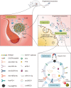
Schematic illustration of the engineered exosomes exhibiting antitumor effects on preclinical models. After intravenous injection, the engineered exosomes carrying the therapeutic molecules arrive at the tumor site under the guide of multiple targeting molecules or a magnetic field. Then the chemotherapeutic drugs, such as PTX, the ncRNAs, such as miR-551-3p, immune molecules, such siPDL1, were released into the tumor microenvironment (TME). Thereafter, the internalization of engineered exosomes led to tumor cell death, such as ferroptosis
Fig. 3
Fig. 3
The common strategies of exosome engineering. a The integration of multiple basic isolation techniques is a common and efficient method to extract high-purity and high-yield exosomes. b The modified strategies comprise biological, immunological, physical, and chemical modification. Each strategy has its own strengths and weaknesses. Therefore, combining different modification strategies is recommended. c The common labeling strategies include lipid dyes (DiR/DiD), fluorescent (GFP), and bioluminescent protein (GLuc-lactadherin and GlucB). However, a ‘one size-fits-all’ reporter does not exist yet. The labeling reporter should be selected based on the practical question and available imaging equipment. d The common administration method of engineered exosomes is intravenous injection in both preclinical and clinical trials. e Based on the labeling reporter, the imaging equipment is distinct. The common tracking technique is MRI
Fig. 4
Fig. 4
Genetically engineered exosomes in various cancers. a In glioma, miR-29a-3p-loaded exosomes inhibit tumor migration and vasculogenic mimicry by targeting ROBO1. b In glioblastoma, MNP@BQR@ANG-EXOsiGPX4 platform delivers siGPX4 into tumor cells and subsequently knockdown GPX4, inducing stronger lipid peroxidation and ferroptosis. c In leukemia, ASOEGFR efficiently target EGFR on the cancer cells. d In osteosarcoma, the internalization of miR317b-5b resulted in changes of tumor progression. And cRGD-Exo-MEG3 achieved stronger anti-OS effects than lncMEG3 alone. e In lung cancer, miRNA-231-Exo strongly inhibited cancer proliferation and migration by interrupting the PTEN/PI3K/AKT signaling pathway. Meanwhile, si-β-Catenin led to loss of β-catenin expression and decreased proliferation. f In prostate cancer, anti-PSMA peptide bind to PSMA receptor on the PC cells, triggering receptor-mediated endocytosis and thereafter intracellular drug release. g In cervical cancer, the exosome-based CRISPR-Cas9 delivery exhibited significant cancer suppression. h In colorectal cancer, si-Survivin knocked down the Survivin gene in tumor cells in vivo. i In PDAC, si-KrasG12D suppressed cancer in multiple mouse models of pancreatic cancer and significantly increased overall survival. j In gastric cancer, CircDIDO1 regulated the level of the signal transducer inhibitor SOSC2 through sponging miR-1307-3p, therefore inhibiting GC progression. k In hepatocellular carcinoma, miR-26a decreased cell migration and proliferation. l In breast cancer, si-VEGF loaded into exosomes exhibited more significant antitumor effects than si-VEGF alone. PC prostate cancer. CRC colorectal cancer. PDAC pancreatic ductal adenocarcinoma. PSMA prostate-specific membrane antigen. GPX4 glutathione peroxidase 4. ASO antisense oligo nucleotide. exoASO-STAT6 ASO targeting STAT6. EGFR epidermal growth factor receptor. cRGD-Exo-MEG3 c(RGDyK)-modified and MEG3-loaded exosomes. LncRNA MEG3 long non-coding RNA maternally expressed gene 3
Fig. 5
Fig. 5
The engineered exosomes remodel the suppressive tumor microenvironment. a The internalization of engineered exosomes not only activates cDC1 and DC2.4, but also trigger a stronger antigen cross-presentation in both B-lymphoblastoid cells and monocyte-derived immature DCs. b Generally, the engineered exosomes reprogram tumor-associated macrophages from M2 to M1. Besides, the SIRPα and CD47 on macrophage could be blocked by the engineered exosomes. Specifically, exoASO-STAT6 could selectively silence STAT6 expression in TAMs. c In TNBC, engineered exosomes could prime and trigger T cells toward killing EGFR-positive TNBC. Additionally, the engineered exosomes could expand and cross-prime TAA-specific CD8 + T cells and activate Th1 cells. d Engineered exosomes induce strong and specific CTL immune response against tumor cells and FAP + CAFs. Furthermore, by releasing IFN-γ and depleting FAP + CAFs, the engineered exosomes could promote tumor ferroptosis

Similar articles

Cited by

References

    1. Chargaff E, West R. The biological significance of the thromboplastic protein of blood. J. Biol. Chem. 1946;166:189–197. doi: 10.1016/S0021-9258(17)34997-9. - DOI - PubMed
    1. Wolf P. The nature and significance of platelet products in human plasma. Br. J. Haematol. 1967;13:269–288. doi: 10.1111/j.1365-2141.1967.tb08741.x. - DOI - PubMed
    1. Nunez EA, Wallis J, Gershon MD. Secretory processes in follicular cells of the bat thyroid. 3. The occurrence of extracellular vesicles and colloid droplets during arousal from hibernation. Am. J. Anat. 1974;141:179–201. doi: 10.1002/aja.1001410203. - DOI - PubMed
    1. Harding C, Heuser J, Stahl P. Receptor-mediated endocytosis of transferrin and recycling of the transferrin receptor in rat reticulocytes. J. Cell Biol. 1983;97:329–339. doi: 10.1083/jcb.97.2.329. - DOI - PMC - PubMed
    1. Pan BT, Johnstone RM. Fate of the transferrin receptor during maturation of sheep reticulocytes in vitro: selective externalization of the receptor. Cell. 1983;33:967–978. doi: 10.1016/0092-8674(83)90040-5. - DOI - PubMed

Publication types